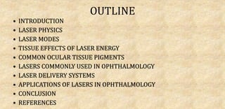
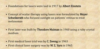
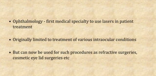
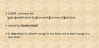
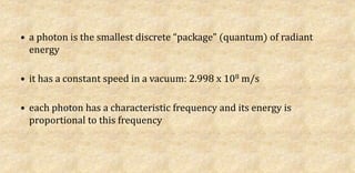
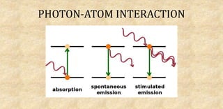
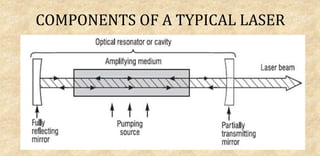
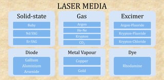
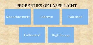
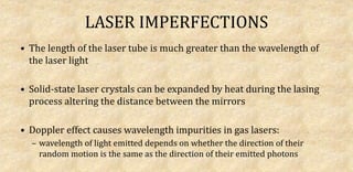
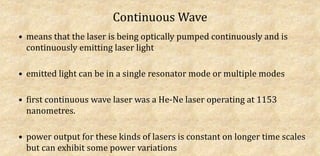
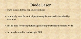
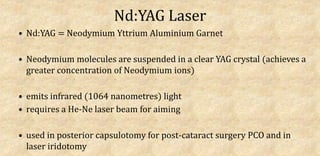
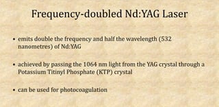
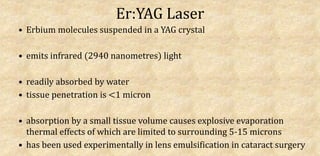
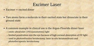
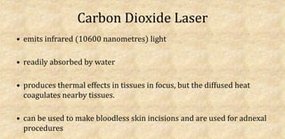
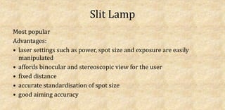
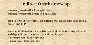
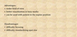
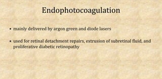
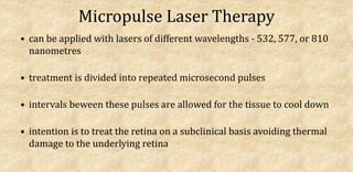
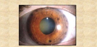
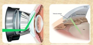
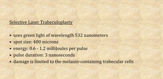
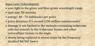
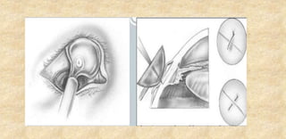
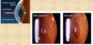
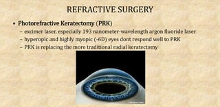
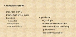
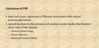
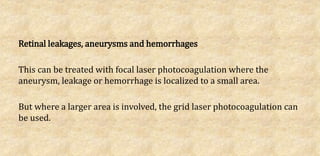
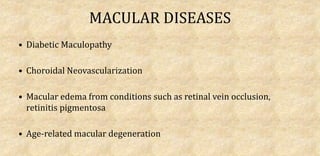
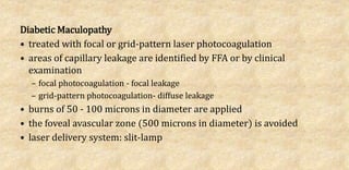
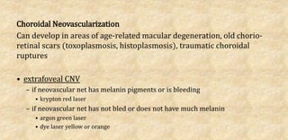
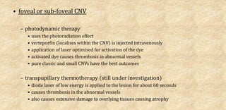
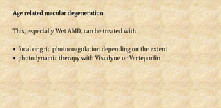
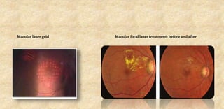
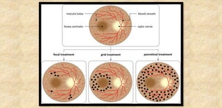
SlideShare a Scribd company logo
1 of 132
BY
DR UGO OSUJI
OUTLINE
• INTRODUCTION
• LASER PHYSICS
• LASER MODES
• TISSUE EFFECTS OF LASER ENERGY
• COMMON OCULAR TISSUE PIGMENTS
• LASERS COMMONLY USED IN OPHTHALMOLOGY
• LASER DELIVERY SYSTEMS
• APPLICATIONS OF LASERS IN OPHTHALMOLOGY
• CONCLUSION
• REFERENCES
• Foundations for lasers were laid in 1917 by Albert Einstein
• Concept of ocular therapy using lasers was formulated by Meyer
Schwikerath who focused sunlight on patients' retinas to treat
melanomas
• First laser was built by Theodore Maiman in 1960 using a ruby crystal
medium
• First medical laser trial was by C. Zweng in 1963
• First clinical laser surgery was by W. Z. Yarn in 1965
• Ophthalmology - first medical specialty to use lasers in patient
treatment
• Originally limited to treatment of various intraocular conditions
• But can now be used for such procedures as refractive surgeries,
cosmetic eye lid surgeries etc
• LASER - acronym for
Light Amplification by Stimuated Emission of Radiation
• coined by Gordon Gould
• to lase means to absorb energy in one form and to emit energy in a
new form
• a photon is the smallest discrete “package” (quantum) of radiant
energy
• it has a constant speed in a vacuum: 2.998 x 10⁸ m/s
• each photon has a characteristic frequency and its energy is
proportional to this frequency
PHOTON-ATOM INTERACTION
COMPONENTS OF A TYPICAL LASER
LASER MEDIA
Solid-state
Ruby
Nd:YAG
Er:YAG
Gas
Argon
He-Ne
Krypton
CO2
Excimer
Argon-Fluoride
Krypton-Fluoride
Krypton-Chloride
Diode
Gallium
Aluminium
Arsenide
Metal Vapour
Copper
Gold
Dye
Rhodamine
HOW LASERS WORK
• lasers use a gain medium that is in a metastable state (an excited state that
has a longer half life than the ordinary excited state but has a shorter half
life than the ground state
• distance between the mirrors must be a multiple of the wavelength of the
light emitted by the medium so that resonance can occur
• during optical pumping, photons are pumped into the optical (resonant)
cavity
• As the photons encounter excited atoms, stimulated emission of photons
occur
• emitted photons travel down the length of the resonator cavity,
exciting other atoms in the process and causing a cascade of
stimulated photon emissions
• these photons travel as waves and as they hit the mirrors at the ends
of the resonator cavity, they are reflected
• because the length of the cavity is a multiple of the wavelength of the
light waves, the reflected waves are in phase with themselves as they
travel back and forth within the cavity
• this causes the waves to reinforce themselves via constructive
interference: resonance
• as more and more stimulated emissions are going on, the
reinforcements keep getting stronger and stronger
• one of the mirrors is only partially reflective and so some of the light
can leave the tube as coherent, mnochromatic and collimated rays:
laser light
PROPERTIES OF LASER LIGHT
Monochromatic Coherent Polarized
Collimated High Energy
LASER IMPERFECTIONS
• The length of the laser tube is much greater than the wavelength of
the laser light
• Solid-state laser crystals can be expanded by heat during the lasing
process altering the distance between the mirrors
• Doppler effect causes wavelength impurities in gas lasers:
– wavelength of light emitted depends on whether the direction of their
random motion is the same as the direction of their emitted photons
Continuous Wave mode
Mode-Locking mode
Q-switching mode
Continuous Wave
• means that the laser is being optically pumped continuously and is
continuously emitting laser light
• emitted light can be in a single resonator mode or multiple modes
• first continuous wave laser was a He-Ne laser operating at 1153
nanometres.
• power output for these kinds of lasers is constant on longer time scales
but can exhibit some power variations
Mode locking
• lasers do not produce light of a single frequency or wavelength
• they produce light that span a narrow range of frequencies:
bandwidth
• a laser's bandwidth depends on:
– the material of the laser's gain medium
– the distance between the mirrors in the optical (resonant) cavity
• as the light waves in the laser cavity bounce from one mirror to the
other, constructive and destructive interference
• constructive interference causes the formation of standing waves
between the mirrors
• together, these standing waves form a discrete set of frequencies:
longitudinal modes
• these longitudinal modes are self-regenerating and continue to
oscillate within the resonant cavity
• each laser type has a particular number of longitudinal modes that it can
support
• if each of these longitudinal modes is allowed to oscillate independently,
they interfere randomly with each other causing either fluctuations in
intensity or causing a near-constant output of intensity
• but if these modes are made to oscillate in phase with each other, they all
can be made to constructively interfere with each other periodically to
produce an intense burst or pulse of laser light - this is called mode-locking
• pulse duration ranges from picoseconds to femtoseconds
Q-switching
• lasers have a property called Quality factor or Q factor
• it is the ratio of the inital energy stored in a resonator to the energy lost
in one cycle of oscillation
• higher Q factor means lower rate of energy loss per oscillation so the
oscillations continue for longer periods
• the reverse is the case for lower Q factor
• in Q switching for lasers, initally, when the laser is being pumped, the Q
factor is reduced to such a level that energy losses are kept high enough
to prevent the lasing process from starting
• the pumped-in energy accumulates in the gain medium
• when the energy in the gain medium reaches a maximum level, it is said
to be gain saturated
• at this point, the Q factor is suddenly increased such that the lasing
process commences very rapidly and builds up and produces laser light
of very high energy and extremely high peak power in a short pulse
• Q switching causes lower pulse repetition and has much longer pulse
durations than mode locking
• pulse durations are in the nanoseconds range
laser
effects
photothermal
photocoagulation
photovaporisation
photochemical
photoradiation
photoablation
photoionising photodisruption
PHOTOTHERMAL EFFECTS
produced at shorter exposure times (ranging from microseconds to a
minute) and higher power densities (ranging from 10 W/cm2 to 106
W/cm2)
photocoagulation (occurs at temperatures >60oC)
photovaporisation (occurs at temperatures of 100oC)
Photocoagulation
• most commonly used thermal laser-tissue effect in ophthalmic surgery
• tissue pigments absorb the laser light and convert it to heat
• the heat raises the temperature of the target tissue high enough to
coagulate and denature the cellular components
• laser wavelengths greater than 500 nanometres are preferred
• examples of photocoagulation lasers
– argon
– krypton
– diode (810 nanometers)
– frequency doubled Nd:YAG
Photovaporisation
• these lasers use long-wavelength infrared heat beams
• they are easily absorbed by water and do not enter the interior of the
eye as 90% of the energy is absorbed within a depth of 200 microns of
the surface to which it is applied
• the light is absorbed by the target tissue causing vaporisation of both
intra- and extra-cellular water
• CO2 laser, at wavelength of 10, 600 nanometres, uses this effect
PHOTOCHEMICAL EFFECTS
occurs with long exposure times (ranging from seconds to
continuously) and very low power densities (typically 1 W/cm2)
photoradiation
photoablation
Photoradiation
• a photosensitizing agent (eg, verteporfin, riboflavin, rose bengal) is first
injected intravenously or instilled within the tissue to be treated
• target tissue takes up the agent concentrates it and is sensitized
• exposure of the sensitized tissue to laser light causes activation of the
photsensitizer
• chemical reaction occurs that will cause formation of new chemical bonds
within the tissue or cause thrombosis and closure of vessels
• PhotoDynamic Therapy (PDT) and Corneal Collagen Cross-linking employ
this effect
Photoablation
• occurs when sufficient laser energy is delivered to a tissue in such a
short time that no heat is transferred
• the energy will be sufficient to directly split molecules - break their
covalent bonds
• long-chain polymers in the tissue are broken up into smaller volatile
fragments which evaporate/vaporise
• because no heat is transferred, there is no thermal damage to adjacent
tissues
• Excimer lasers that generate laser light in the UV range are suitable for
photoablation
PHOTOIONISING EFFECTS
uses high energy laser pulses to strip ions from atoms
photodisruption
Photodisruption
• these lasers emit a very high pulse of energy (with wavelength of
about 1064 nanometres) within a few nanoseconds
• when focused on a spot of about 15 - 25 microns in diameter, this
high energy pulse exceeds the critical energy density of the tissue
• this raises the temperature of that spot in focus so high (10, 000 K)
that electrons are stripped from atoms resulting in a mixture of ions
and electrons known as plasma
• the heat makes the plasma expand with short momentary pressures
as high as 150, 000 psi and produces a cutting effect on the tissue
• the plasma size is small and it has little total energy therefore it
produces very little effect away from the point of focus
• in this process, it is mechanical effect of the expanding plasma that
produces the visible cutting effect and not the laser light itself
• Nd:YAG laser uses this effect
Melanin
• found mainly in RPE and Uvea
• absorbs mainly wavelengths between 400 - 700 nanometres
• the longer the wavelength, the more the melanin is penetrated
• diode lasers using wavelength of 810 nanometres penetrate deep into
the choroid
Xanthophyll
• located in the inner and outer plexiform layers and is abundant at the
macula
• protects photoreceptors from damage from short wavelength light
• is damaged by light in the blue spectrum and so blue light is avoided
in macular laser proedures
• argon green is preferred to argon blue in macular photocoagulation
Haemoglobin
• absorption varies according to oxygen saturation
• absorbs yellow, green and blue wavelengths very well
• absorbs red light poorly
• as such, macular lasers can damage retinal vessels
WAVELENGTHS OF LASER TYPES
Argon Blue-Green Gas Laser
• emits 70% blue (488 nanometres) and 30% green (514 nanometres)
light
• older models were large and had a water cooling system to dissipate
generated heat
• newer models are smaller, have air cooling systems and emit
primarily green light
• mainly used for retinal photocoagulation (well-absorbed by melanin
and haemoglobin)
• argon green can be used in macular photocoagulation (blue light is
well absorbed by xanthophyll)
Helium-Neon Laser
• low power gas laser
• emits red (632.8 nanometres) light
• used an aiming beam for lasers that emit invisible light
Diode Laser
• emits infrared (810 nanometres) light
• commonly used for retinal photocoagulation (well-absorbed by
melanin)
• can be used for cyclophotocoagulation (penetrates the sclera well)
• can also be used in endoscopic DCR
Nd:YAG Laser
• Nd:YAG = Neodymium Yttrium Aluminium Garnet
• Neodymium molecules are suspended in a clear YAG crystal (achieves a
greater concentration of Neodymium ions)
• emits infrared (1064 nanometres) light
• requires a He-Ne laser beam for aiming
• used in posterior capsulotomy for post-cataract surgery PCO and in
laser iridotomy
Frequency-doubled Nd:YAG Laser
• emits double the frequency and half the wavelength (532
nanometres) of Nd:YAG
• achieved by passing the 1064 nm light from the YAG crystal through a
Potassium Titinyl Phosphate (KTP) crystal
• can be used for photocoagulation
Er:YAG Laser
• Erbium molecules suspended in a YAG crystal
• emits infrared (2940 nanometres) light
• readily absorbed by water
• tissue penetration is <1 micron
• absorption by a small tissue volume causes explosive evaporation
thermal effects of which are limited to surrounding 5-15 microns
• has been used experimentally in lens emulsification in cataract surgery
Excimer Laser
• Excimer = excited dimer
• Two atoms form a molecule in their excited state but dissociate in their
ground state
• A common example in clinical use is the Argon-Flouride dimer laser
– emits ultraviolet (193 nanometres) light
– limited penetration into the eye because of high corneal absorption of UV light
– used in photorefractive keratectomy, laser in-situ keratomileusis and
phototherapeutic keratectomy
Carbon Dioxide Laser
• emits infrared (10600 nanometres) light
• readily absorbed by water
• produces thermal effects in tissues in focus, but the diffused heat
coagulates nearby tissues.
• can be used to make bloodless skin incisions and are used for adnexal
procedures
Slit Lamp
Most popular
Advantages:
• laser settings such as power, spot size and exposure are easily
manipulated
• affords binocular and stereoscopic view for the user
• fixed distance
• accurate standardisation of spot size
• good aiming accuracy
Indirect Ophthalmoscope
• commonly used with a fibreoptic cable
• commonly used with argon or diode lasers
• used to treat peripheral retinal pathologies such as peripheral retinal
breaks and ROP
• spot size is altered by the dioptric power of the condensing lens used
and depending on the refractive status of the eye
– hyperopic eyes - smaller spot size
– myopic eyes - larger spot size
Advantages:
• wider field of view
• better visualization in hazy media
• can be used with patient in the supine position
Disadvantages
• difficulty focusing
• difficulty standardising spot size
Endophotocoagulation
• mainly delivered by argon green and diode lasers
• used for retinal detachment repairs, extrusion of subretinal fluid, and
proliferative diabetic retinopathy
Micropulse Laser Therapy
• can be applied with lasers of different wavelengths - 532, 577, or 810
nanometres
• treatment is divided into repeated microsecond pulses
• intervals beween these pulses are allowed for the tissue to cool down
• intention is to treat the retina on a subclinical basis avoiding thermal
damage to the underlying retina
LENSES USED FOR LASER DELIVERY
Contact Lenses for PRP
Contact Lenses for Focal/Grid Laser
THERAPEUTIC
DIAGNOSTIC
THERAPEUTIC
 Anterior Segment
 Posterior Segment
ANTERIOR SEGMENT
 Glaucoma
 Cataract
 Refractive Surgeries
GLAUCOMA
Angle-Closure Glaucoma
• Laser Iridotomy: made more effective with the use of Abraham
contact lens (+66D focusing button) and Wise Iridotomy-
Sphincterotomy lens (+103D focusing button)
– Argon Laser
• preferred for heavily pigmented and thick irides
• beam focused on the far peripheral iris fibers
• cut parallel to the limbus
• with multiple shots at high energy level with exposures lasting 10 - 20
milliseconds
– Q-switched Nd:YAG laser
• uses a single point method
• 5 - 10 milliJoules per shot in a single shot burst is applied to the peripheral iris
• the iridotomy can be enlarged by cutting the far peripheral iris fibres in a line
parallel to the limbus with multiple shots at 1 - 2 milliJoules.
Laser Peripheral Iridoplasty
• done when the cornea is too cloudy (from edema) to allow for laser
iridotomy
• a ring of contraction burns are applied to the peripheral iris using a
standard iridotomy lens
• burns of about 200 milliWatts, about 0.5 seconds, and large spot size
of about 500 microns
• leads to contraction of the iris stroma near the angle pulling the angle
open
• this causes fall in IOP allowing for laser iridotomy
Open Angle Glaucoma
Laser Trabeculoplasty
• performed if medical therapy is not adequate for IOP control
• 100 or more non-perforating laser burns are placed circumferentially
360 degrees around the trabecular meshwork with the aid of a
goniolens
• this shrinks the collagen in the TM, pulls the trabecular layers apart
leading to reopening of the intertrabecular spaces and the Schlemm
canal
• one major complication is the immediate rise in IOP for 1 - 4 hours in
33% of treated eyes (prevented by instillation of apraclonidine
drops) and 1 - 3 weeks IOP elevation in 2% of treated eyes
• some surgeons reduce the severity of this IOP rise by applying 50
laser burns to 180 degrees of the TM initially (the rest is left for later
treatment)
• Advanced Glaucoma Intervention Study proposed that initial
treatment in black patients with advanced glaucoma should be laser
trabeculoplasty while in white patients with advanced glaucoma
should be trabeculectomy
• trabeculoplasty is most effective in patients with Pseudoexfoliation
and Pigmentary Glaucoma
• in most other patients, its effect is just for about 1 to 2 years
Selective Laser Trabeculoplasty
• uses green light of wavelength 532 nanometers
• spot size: 400 microns
• energy: 0.6 - 1.2 milliJoules per pulse
• pulse duration: 3 nanoseconds
• damage is limited to the melanin-containing trabecular cells
Argon Laser Trabeculoplasty
• uses light in the green and blue-green wavelength range
• spot size: 50 microns
• energy: 40 - 70 milliJoules per pulse
• pulse duration: 0.1 second (100 million nanoseconds)
• damage is not limited to the melanin-containing trabecular
cells but extends to the trabecular beams and other
extracellular tissues in the angle
• slowly being replaced in recent times by the frequency
doubled Nd:YAG lasers
Multipulse Laser Trabeculoplasty
• uses light of wavelength 810 nanometers
• generated by a diode laser
• spot size: 300 microns
• energy: 0.6 milliJoules
• pulse duration: 0.2 seconds (200 million nanoseconds)
divided into 100 microsecond pulses at a duty cycle of 15%
• does not destroy the melanin-containing trabecular cells but
generates enough energy to injure the cells and cause TM
remodelling
Cyclophotocoagulation
• used for glaucoma that is refractory to usual operative procedures
• involves direct destruction of the ciliary processes
• Initially done by diathermy, later by cryosurgery and now with lasers
• can be done by ab externo or ab interno approach
• ab externo technique (through intact conjunctiva and sclera) was
developed by Beckman using high-energy ruby lasers
• currently performed using the thermal mode Nd:YAG laser or the
diode laser contact-delivered with a fibre-optic probe
• laser endocyclophotocoagulation (ab interno) done by passing a
fibreoptic probe through the pars plana during vitrectomy
Laser Suture Lysis
• done in the early post-trabeculectomy period
• to increase degree of drainage and possibly achieve longer-term IOP
reduction
• short laser pulses are focused on the partial-thickness scleral flap
sutures through a clear conjunctiva to cut them
• visibility of the sutures is aided by compressing overlying tissues with
the Hoskins suture lens
• argon laser can be used but if hemorrhage is present, krypton red or
diode infrared laser is used
CATARACT SURGERY
Femtosecond lasers
• corneal incisions
• capsulorrhexis
• initial fragmentation of the crystalline lens
• astigmatism-relieving incisions
Advantages of
femtosecond lasers
Greater precision
Greater incision integrity
Reduced phaco energy
Improved refractive outcomes due to
more precise capsulorrhexis
Disadvantages of
femtosecond lasers
Higher cost
Longer operating time
Steep learning curve
Difficulties with challenging cases
(patients with small pupils)
Posterior Capsulotomy
• done in posterior capsule opacification following cataract surgery
• Q-switched Nd:YAG laser is used
• laser pulses are focused (with the aid of a condensing contact lens)
just posterior to the posterior capsule to produce a central
capsulotomy
Capsule Contraction Syndrome (Capsulophimosis)
• fibrosis of the margins of anterior capsulotomy may lead to
contracture and obscuration of the visual axis
• this fibrosis can be released with radial incisions made with Q-
switched Nd:YAG laser
Anterior Vitreolysis
• done with Q-switched Nd:YAG laser with the aid of condensing corneal
contact lens
• used to clear vitreous remnants in the AC from incomplete clearance of
vitreous due to vitreous loss on account of trauma or surgery
• topical miotics constrict the pupil thereby tightening the vitreous strands
for easier cutting
• concussions to the cornea and iris should be minimized by using multiple
shots at optical breakdown levels
REFRACTIVE SURGERY
• Photorefractive Keratectomy (PRK)
– excimer laser, especially 193 nanometer-wavelength argon fluoride laser
– hyperopic and highly myopic (-6D) eyes dont respond well to PRK
– PRK is replacing the more traditional radial keratectomy
• Laser In-Situ Keratomileusis (LASIK)
– better than PRK in that it preserves the Bowman layer, provides faster visual
recovery and less patient discomfort
– has higher risk of long-term complications
• Laser Subepithelial Keratomileusis (LASEK)
– combines the benefits of both PRK and LASIK
• Phototherapeutic keratectomy (PTK)
– excimer lasers are used
– to remove superficial corneal opacities such as in band keratopathy
– to treat superficial corneal diseases such as recurrent corneal erosions
• Modern excimer lasers have
– smaller spot size
– an eye tracking system
– wavefront custom ablation
improve accuracy of treatment and reduce incidence of
spherical aberration
POSTERIOR SEGMENT
 Retinal Tears and Detachments
 Vascular Diseases
 Macular Diseases
 Neoplasms
RETINAL TEARS and DETACHMENT
• These are treated by a procedure known as Laser Retinopexy which
involves laser photocoagulation mostly using argon lasers
• Laser burns are placed around the retinal tear or the detaching retina
in one or two rows:
– to prevent the tear from enlarging
– to spot-weld the neurosensory retina to the underlying RPE at the laser spots
to prevent retinal detachment (rhegmatogenous) from developing
RETINAL VASCULAR DISEASES
• Diabetic Retinopathy
• Central Retinal Artery and Vein Occlusion
• Retinopathy of Prematurity
• Eale’s Disease
• Central Serous Chorioretinopathy
• Retinal Aneurysms
Some of these conditions lead to retinal neovascularization and, if they
persist long enough, can predispose to
– Retinal detachment
– Neovascularization at the disc
– Neovascularization at other retinal sites
– Neovascularization in other locations such as the iris and the angles
– Neovascular glaucoma
Others can lead to leakages and retinal and vitreous hemorrhages if not
treated on time
Retinal Neovascularization
• treated with pan-retinal photocoagulation
• It is aimed at applying laser burns to the ischemic areas of the retina
making them atrophic thereby reducing the hypoxia that drives
neovascularization
• burns of 200 - 500 microns in diameter, separated by 1-1.5 burn
diameter, are applied in pulses to the whole retina except the area
within the temporal vascular arcades
• for ROP, the laser burns are limited to the avascular areas of the retina
• pulse duration: 100ms
• at least 2000 burns are applied - can be up to 6000 burns or more
• power: 200 - 250mW
• laser delivery system: indirect ophthalmoscope and +20D lens
• usually performed in 3 sessions that are spaced about 1 to 2
weeks apart
– 1st session - inferior and nasal retina
– 2nd session - temporal retina
– 3rd session - superior retina
• recurrent or recalcitrant neovascularisation might need to be
retreated - local anaesthesia
• PRP can be done as a prophylaxis for iris neovascularization and can
also be used to cause regression of the early stages of iris
neovascularization
• PRP can be done as prophylaxis for neovascular glaucoma - most
effective in the presence of iris neovascularisation but before the
development of neovascular glaucoma (may be difficult to achieve
this timing clinically)
• PRP in early neovascular glaucoma can cause regression of angle
fibrosis - allowing for other methods of glaucoma control
• in established neovascular glaucoma - associated with miosis, corneal
edema, and hyphema, only cyclophotocoagulation can be done
• CRVO associated with RAPD, VA <6/60 and multiple cotton wool
spots are highly suggestive of retinal ischemia and should warrant
prophylactic PRP (to prevent retinal neovascularization)
Complications of PRP
• induction of PVD
• inadvertent foveal burns
• transient
– headaches
– blurry vision
– macular oedema
• persistent
– nyctalopia
– defective accommodation
– reduced contrast sensitivity
– photophobia
– reduced visual fields
Limitations of PRP
• does not cause regression of fibrosis associated with retinal
neovascularization
• cannot be done in the presence of unclear ocular media that hinders
clear view of the fundus:
– vitreous hemorrhage
– dense cataracts
– advanced corneal edema
Retinal leakages, aneurysms and hemorrhages
This can be treated with focal laser photocoagulation where the
aneurysm, leakage or hemorrhage is localized to a small area.
But where a larger area is involved, the grid laser photocoagulation can
be used.
Macular laser grid Macular focal laser treatment: before and after
MACULAR DISEASES
• Diabetic Maculopathy
• Choroidal Neovascularization
• Macular edema from conditions such as retinal vein occlusion,
retinitis pigmentosa
• Age-related macular degeneration
Diabetic Maculopathy
• treated with focal or grid-pattern laser photocoagulation
• areas of capillary leakage are identified by FFA or by clinical
examination
– focal photocoagulation - focal leakage
– grid-pattern photocoagulation- diffuse leakage
• burns of 50 - 100 microns in diameter are applied
• the foveal avascular zone (500 microns in diameter) is avoided
• laser delivery system: slit-lamp
Choroidal Neovascularization
Can develop in areas of age-related macular degeneration, old chorio-
retinal scars (toxoplasmosis, histoplasmosis), traumatic choroidal
ruptures
• extrafoveal CNV
– if neovascular net has melanin pigments or is bleeding
• krypton red laser
– if neovascular net has not bled or does not have much melanin
• argon green laser
• dye laser yellow or orange
• foveal or sub-foveal CNV
– photodynamic therapy
• uses the photoradiation effect
• verteporfin (localises within the CNV) is injected intravenously
• application of laser optimised for activation of the dye
• activated dye causes thrombosis in abnormal vessels
• pure classic and small CNVs have the best outcomes
– transpupillary thermotherapy (still under investigation)
• diode laser of low energy is applied to the lesion for about 60 seconds
• causes thrombosis in the abnormal vessels
• also causes extensive damage to overlying tissues causing atrophy
Age related macular degeneration
This, especially Wet AMD, can be treated with
• focal or grid photocoagulation depending on the extent
• photodynamic therapy with Visudyne or Verteporfin
Macular laser grid Macular focal laser treatment: before and after
NEOPLASMS
• Vascular tumors such as
– Retinal capillary hemangiomas
– Choroidal hemangioma
• Solid tumors such as choroidal melanoma
Vascular Tumors
• For choroidal hemangiomas associated with serous retinal
detachment, entire tumor surface can be treated with laser burns
• For choroidal hemangiomas around the macular area or in the
peripapillary region, photodynamic therapy can be used.
• For small capillary hemangiomas, laser burns can be applied directly
to the surface of the lesions.
• For larger retinal capillary hemangiomas, the feeder vessels are
treated with lasers and the lesions shrink subsequently with loss of
blood supply
Solid tumors
Choroidal melanoma is the commonest primary intraocular malignancy
For tumors with basal diameter of < 15mm and thickness of <5mm,
treatment can be done with argon laser photocoagulation
OTHER POSTERIOR SEGMENT APPLICATIONS
• Pre-macular hemorrhage (especially when the hemorrhage > 3 disc
diameters in size) secondary to diabetic retinopathy, Valsalva
retinopathy, Terson syndrome, ruptured retinal aneurysm:
– YAG laser hyaloidotomy
• Optic disc pit associated with serous macular detachment:
– Laser photocoagulation along the temporal margin of the disc
ADNEXAL
Cosmetic Eyelid Surgeries
 Lid Tumours
COSMETIC EYELID SURGERY
• repeated 1 millisecond pulses from a CO2 laser on exposed wrinkled
eyelid skin
• leads to evaporation of the epidermis and contraction of the dermal
collagen
• epidermal regeneration leads to a tightened skin without the wrinkles
LID TUMOURS
• lid tumors can be managed by vaporisation
• CO2 lasers can be used to cause the photovaporisation effect
• both benign and malignant tumors can be treated
• limitations
– scarring
– lack of specimen for histology
– inability to assess tumour margins
DIAGNOSTIC
CONFOCAL SCANNING LASER OPHTHALMOSCOPY
• rapidly scanning tiny laser spot is focused on the part of the eye to be
examined.
• reflected light from the laser spot is imaged through a pinhole in a
detector thereby suppressing all reflections except those from the
focal plane
• these reflections are combined and processed by a computer program
to produce precise and reproducible 3D images
• used to evaluate and follow glaucomatous changes in the ONH.
• can also be used to image the cornea, lens and macula
• can be used to perform flourescein and indocyanine green angiography
• can be used to perform microperimetry
• Argon blue (488 nanometres), Argon green (514 nanometres), Helium-
Neon red (633 nanometres) and diode infrared (780 nanometres) can
all be used.
SCANNING LASER POLARIMETRY
• measures thickness of the retinal nerve fibre layer
• polarised light (780 nanometres) is projected onto the retina which
passes through the RNFL to deeper retinal layers from where it is
reflected
• as RNFL fibres are arranged in a parallel manner, it acts as a
birefringerent medium and changes the state of the polarised light
passing through it
• the magnitude of this change of polarisation (retardation) correlates
with RNFL thickness
CONFOCAL SCANNING LASER TOMOGRAPHY
• produces a topographic image of the retina and optic nerve head
• diode (670 nanometres) laser light is focused on the retina
• only light reflected from the layer of the retina in the focal plane of the
laser light is imaged in pixels
• 32 images are acquired totally.
• the first image is from the the region parallel to the retinal surface
just anterior to the blood vessels emerging from the cup
• subsequent images are acquired by gradually advancing the focal
plane of the laser beam towards the lamina cribrosa
• a computer reconstructs these pixels into a 3D picture which can be
used to evaluate optic disc damage in glaucoma
LASER INTERFEROMETRY
• interferometers project laser light from 2 sources onto the retina
• the laser light commonly used is the Helium-Neon laser
• these 2 laser light beams meet, produce an interference which is seen as a
sine wave grating
• distance between the light sources can be reduced to reduce the spatial
frequency of the sine wave grating
• this allows estimation of the eye's potential visual acuity despite presence
of a refractive error or a cataract that prevents macular visibility
LASER DOPPLER FLOWMETRY
• used to detect blood flow through the ciliary body and the retinal blood
vessels
• uses the Doppler principle (change in wave frequency due to relative
motion between its source and the observer)
• moving blood cells reflect laser light at a different frequency than that of
the incident beam
• the greater the difference in frequency the greater the blood flow velocity
CONCLUSION
• Lasers are increasingly becoming an indispensable part of the
ophthalmic practice worldwide
• A good knowledge of the theory and practice of the use of lasers will
be a very important tool in the arsenal of the ophthalmic surgeon
BIBLIOGRAPHY
• Riordan-Eva P., Whitcher J. P. (2003) Vaughan & Asbury's General Ophthalmology,
16th edition.
• Salmon J. F. (2020). Kanski's Clinical Ophthalmology, 9th edition.
• AlBoushi A. F., Abouammoh M. A. (2021). Lasers (Surgery),
https://www.eyewiki.aao.org/Lasers_(surgery).
• Elkington A. R., Frank H. J., Greaney M. J. (1999). Clinical Optics, 3rd edition.
• American Academy of Ophthalmology (2019). Clinical Optics: 2019 - 2020 Basic and
Clinical Science Course.
• Singh A., Retinal Lasers, https://www.slideshare.net, 2018
• Paschotta R., Q-switching, https://www.rp-photonics.com/q_switching.html
• Paschotta R., Mode-locking, https://www.rp-photonics.com/mode_locking.html
• Wikipedia, Mode-locking, https://www.wikipedia.org/wiki/Mode-locking

More Related Content

What's hot (20)

Tonometry and tonography
Tonometry and tonographyTonometry and tonography
Tonometry and tonography
 
Stops pupils and ports
Stops pupils and portsStops pupils and ports
Stops pupils and ports
 
Electrooculogram- EOG
Electrooculogram- EOG  Electrooculogram- EOG
Electrooculogram- EOG
 
Lens shapes.pptx
Lens shapes.pptxLens shapes.pptx
Lens shapes.pptx
 
Techniques of retinoscopy
Techniques of retinoscopyTechniques of retinoscopy
Techniques of retinoscopy
 
Accessories of trial set
Accessories of trial setAccessories of trial set
Accessories of trial set
 
Fermet's principle
Fermet's principleFermet's principle
Fermet's principle
 
Lensometer
LensometerLensometer
Lensometer
 
Keratometry & autorefraction
Keratometry & autorefractionKeratometry & autorefraction
Keratometry & autorefraction
 
Ophthalmic prisms
Ophthalmic prismsOphthalmic prisms
Ophthalmic prisms
 
Gonioscopy
GonioscopyGonioscopy
Gonioscopy
 
Lensometry.
Lensometry.Lensometry.
Lensometry.
 
Lens power measurement
Lens power measurementLens power measurement
Lens power measurement
 
Aberrations
AberrationsAberrations
Aberrations
 
WORTH FOUR DOT TEST.pptx
WORTH FOUR DOT TEST.pptxWORTH FOUR DOT TEST.pptx
WORTH FOUR DOT TEST.pptx
 
Oct
OctOct
Oct
 
The Schematic, Reduced Eye, Axes and Angle of the eye.pptx
The Schematic, Reduced Eye, Axes and Angle of the eye.pptxThe Schematic, Reduced Eye, Axes and Angle of the eye.pptx
The Schematic, Reduced Eye, Axes and Angle of the eye.pptx
 
Vergence
VergenceVergence
Vergence
 
Radiometry and Photometry by Sumayya Naseem
Radiometry and Photometry by Sumayya NaseemRadiometry and Photometry by Sumayya Naseem
Radiometry and Photometry by Sumayya Naseem
 
Retinal laser therapy
Retinal laser therapyRetinal laser therapy
Retinal laser therapy
 

Similar to Lasers in Ophthalmology

Lasers in ophthalmology - Dr. Parag Apte
Lasers in ophthalmology - Dr. Parag ApteLasers in ophthalmology - Dr. Parag Apte
Lasers in ophthalmology - Dr. Parag Apteparag apte
 
PRINCIPLES OF LASERS copy.pptx
PRINCIPLES OF LASERS copy.pptxPRINCIPLES OF LASERS copy.pptx
PRINCIPLES OF LASERS copy.pptxLavanyaCool
 
LASERS IN CONSERVATIVE DENTISTRY AND ENDODONTICS new.pptx
LASERS IN CONSERVATIVE DENTISTRY AND ENDODONTICS new.pptxLASERS IN CONSERVATIVE DENTISTRY AND ENDODONTICS new.pptx
LASERS IN CONSERVATIVE DENTISTRY AND ENDODONTICS new.pptxCmenonMenon
 
Lasers in Oncosurgery
Lasers in OncosurgeryLasers in Oncosurgery
Lasers in OncosurgeryDr KAMBLE
 
Lasers in oral surgery
Lasers in oral surgeryLasers in oral surgery
Lasers in oral surgeryTejal Ragji
 
Lasers in Ophthalmology.pptx
Lasers in Ophthalmology.pptxLasers in Ophthalmology.pptx
Lasers in Ophthalmology.pptxZaid Azhar
 
Laser in Ophthalmology and types of Laser
Laser in Ophthalmology and types of LaserLaser in Ophthalmology and types of Laser
Laser in Ophthalmology and types of LaserAsif469093
 
Lasers in oral and maxillofacial surgery
Lasers in oral and maxillofacial surgery Lasers in oral and maxillofacial surgery
Lasers in oral and maxillofacial surgery Jeff Zacharia
 
LINEAR ACCELERATOR PRINCIPAL AND WORKING
LINEAR ACCELERATOR PRINCIPAL AND WORKINGLINEAR ACCELERATOR PRINCIPAL AND WORKING
LINEAR ACCELERATOR PRINCIPAL AND WORKINGKrishan Murari Yadav
 
Laser characteristics as applied to medicine and biology
Laser characteristics as applied to medicine and biologyLaser characteristics as applied to medicine and biology
Laser characteristics as applied to medicine and biologykaroline Enoch
 
Lasers for mpctc
Lasers for mpctcLasers for mpctc
Lasers for mpctcKyaw Tun
 
basics in laser
basics in laserbasics in laser
basics in laserchinnu04
 
LASERS IN vitreoRETINAaaaaaaaaa2023.pptx
LASERS IN vitreoRETINAaaaaaaaaa2023.pptxLASERS IN vitreoRETINAaaaaaaaaa2023.pptx
LASERS IN vitreoRETINAaaaaaaaaa2023.pptxMadhuri521470
 

Similar to Lasers in Ophthalmology (20)

Lasers in ophthalmology - Dr. Parag Apte
Lasers in ophthalmology - Dr. Parag ApteLasers in ophthalmology - Dr. Parag Apte
Lasers in ophthalmology - Dr. Parag Apte
 
PRINCIPLES OF LASERS copy.pptx
PRINCIPLES OF LASERS copy.pptxPRINCIPLES OF LASERS copy.pptx
PRINCIPLES OF LASERS copy.pptx
 
LASERS IN CONSERVATIVE DENTISTRY AND ENDODONTICS new.pptx
LASERS IN CONSERVATIVE DENTISTRY AND ENDODONTICS new.pptxLASERS IN CONSERVATIVE DENTISTRY AND ENDODONTICS new.pptx
LASERS IN CONSERVATIVE DENTISTRY AND ENDODONTICS new.pptx
 
Lasers in Oncosurgery
Lasers in OncosurgeryLasers in Oncosurgery
Lasers in Oncosurgery
 
Lasers in urology
Lasers in urologyLasers in urology
Lasers in urology
 
Laser
LaserLaser
Laser
 
Laser
LaserLaser
Laser
 
Laser Basics
Laser BasicsLaser Basics
Laser Basics
 
Lasers
LasersLasers
Lasers
 
Lasers in oral surgery
Lasers in oral surgeryLasers in oral surgery
Lasers in oral surgery
 
Lasers in Ophthalmology.pptx
Lasers in Ophthalmology.pptxLasers in Ophthalmology.pptx
Lasers in Ophthalmology.pptx
 
Laser in Ophthalmology and types of Laser
Laser in Ophthalmology and types of LaserLaser in Ophthalmology and types of Laser
Laser in Ophthalmology and types of Laser
 
Spectroscopy
Spectroscopy   Spectroscopy
Spectroscopy
 
Lasers in oral and maxillofacial surgery
Lasers in oral and maxillofacial surgery Lasers in oral and maxillofacial surgery
Lasers in oral and maxillofacial surgery
 
LINEAR ACCELERATOR PRINCIPAL AND WORKING
LINEAR ACCELERATOR PRINCIPAL AND WORKINGLINEAR ACCELERATOR PRINCIPAL AND WORKING
LINEAR ACCELERATOR PRINCIPAL AND WORKING
 
Laser characteristics as applied to medicine and biology
Laser characteristics as applied to medicine and biologyLaser characteristics as applied to medicine and biology
Laser characteristics as applied to medicine and biology
 
Lasers for mpctc
Lasers for mpctcLasers for mpctc
Lasers for mpctc
 
basics in laser
basics in laserbasics in laser
basics in laser
 
Laser
Laser Laser
Laser
 
LASERS IN vitreoRETINAaaaaaaaaa2023.pptx
LASERS IN vitreoRETINAaaaaaaaaa2023.pptxLASERS IN vitreoRETINAaaaaaaaaa2023.pptx
LASERS IN vitreoRETINAaaaaaaaaa2023.pptx
 

Recently uploaded

Call Girls Service Bellary Road Just Call 7001305949 Enjoy College Girls Service
Call Girls Service Bellary Road Just Call 7001305949 Enjoy College Girls ServiceCall Girls Service Bellary Road Just Call 7001305949 Enjoy College Girls Service
Call Girls Service Bellary Road Just Call 7001305949 Enjoy College Girls Servicenarwatsonia7
 
Russian Call Girls in Pune Riya 9907093804 Short 1500 Night 6000 Best call gi...
Russian Call Girls in Pune Riya 9907093804 Short 1500 Night 6000 Best call gi...Russian Call Girls in Pune Riya 9907093804 Short 1500 Night 6000 Best call gi...
Russian Call Girls in Pune Riya 9907093804 Short 1500 Night 6000 Best call gi...Miss joya
 
Low Rate Call Girls Ambattur Anika 8250192130 Independent Escort Service Amba...
Low Rate Call Girls Ambattur Anika 8250192130 Independent Escort Service Amba...Low Rate Call Girls Ambattur Anika 8250192130 Independent Escort Service Amba...
Low Rate Call Girls Ambattur Anika 8250192130 Independent Escort Service Amba...narwatsonia7
 
Hi,Fi Call Girl In Mysore Road - 7001305949 | 24x7 Service Available Near Me
Hi,Fi Call Girl In Mysore Road - 7001305949 | 24x7 Service Available Near MeHi,Fi Call Girl In Mysore Road - 7001305949 | 24x7 Service Available Near Me
Hi,Fi Call Girl In Mysore Road - 7001305949 | 24x7 Service Available Near Menarwatsonia7
 
Russian Call Girls in Bangalore Manisha 7001305949 Independent Escort Service...
Russian Call Girls in Bangalore Manisha 7001305949 Independent Escort Service...Russian Call Girls in Bangalore Manisha 7001305949 Independent Escort Service...
Russian Call Girls in Bangalore Manisha 7001305949 Independent Escort Service...narwatsonia7
 
Call Girls Doddaballapur Road Just Call 7001305949 Top Class Call Girl Servic...
Call Girls Doddaballapur Road Just Call 7001305949 Top Class Call Girl Servic...Call Girls Doddaballapur Road Just Call 7001305949 Top Class Call Girl Servic...
Call Girls Doddaballapur Road Just Call 7001305949 Top Class Call Girl Servic...narwatsonia7
 
Aspirin presentation slides by Dr. Rewas Ali
Aspirin presentation slides by Dr. Rewas AliAspirin presentation slides by Dr. Rewas Ali
Aspirin presentation slides by Dr. Rewas AliRewAs ALI
 
Bangalore Call Girls Marathahalli 📞 9907093804 High Profile Service 100% Safe
Bangalore Call Girls Marathahalli 📞 9907093804 High Profile Service 100% SafeBangalore Call Girls Marathahalli 📞 9907093804 High Profile Service 100% Safe
Bangalore Call Girls Marathahalli 📞 9907093804 High Profile Service 100% Safenarwatsonia7
 
Russian Call Girl Brookfield - 7001305949 Escorts Service 50% Off with Cash O...
Russian Call Girl Brookfield - 7001305949 Escorts Service 50% Off with Cash O...Russian Call Girl Brookfield - 7001305949 Escorts Service 50% Off with Cash O...
Russian Call Girl Brookfield - 7001305949 Escorts Service 50% Off with Cash O...narwatsonia7
 
VIP Mumbai Call Girls Hiranandani Gardens Just Call 9920874524 with A/C Room ...
VIP Mumbai Call Girls Hiranandani Gardens Just Call 9920874524 with A/C Room ...VIP Mumbai Call Girls Hiranandani Gardens Just Call 9920874524 with A/C Room ...
VIP Mumbai Call Girls Hiranandani Gardens Just Call 9920874524 with A/C Room ...Garima Khatri
 
Call Girls Horamavu WhatsApp Number 7001035870 Meeting With Bangalore Escorts
Call Girls Horamavu WhatsApp Number 7001035870 Meeting With Bangalore EscortsCall Girls Horamavu WhatsApp Number 7001035870 Meeting With Bangalore Escorts
Call Girls Horamavu WhatsApp Number 7001035870 Meeting With Bangalore Escortsvidya singh
 
High Profile Call Girls Jaipur Vani 8445551418 Independent Escort Service Jaipur
High Profile Call Girls Jaipur Vani 8445551418 Independent Escort Service JaipurHigh Profile Call Girls Jaipur Vani 8445551418 Independent Escort Service Jaipur
High Profile Call Girls Jaipur Vani 8445551418 Independent Escort Service Jaipurparulsinha
 
Call Girls Service Chennai Jiya 7001305949 Independent Escort Service Chennai
Call Girls Service Chennai Jiya 7001305949 Independent Escort Service ChennaiCall Girls Service Chennai Jiya 7001305949 Independent Escort Service Chennai
Call Girls Service Chennai Jiya 7001305949 Independent Escort Service ChennaiNehru place Escorts
 
Call Girls Service in Bommanahalli - 7001305949 with real photos and phone nu...
Call Girls Service in Bommanahalli - 7001305949 with real photos and phone nu...Call Girls Service in Bommanahalli - 7001305949 with real photos and phone nu...
Call Girls Service in Bommanahalli - 7001305949 with real photos and phone nu...narwatsonia7
 
Sonagachi Call Girls Services 9907093804 @24x7 High Class Babes Here Call Now
Sonagachi Call Girls Services 9907093804 @24x7 High Class Babes Here Call NowSonagachi Call Girls Services 9907093804 @24x7 High Class Babes Here Call Now
Sonagachi Call Girls Services 9907093804 @24x7 High Class Babes Here Call NowRiya Pathan
 
CALL ON ➥9907093804 🔝 Call Girls Baramati ( Pune) Girls Service
CALL ON ➥9907093804 🔝 Call Girls Baramati ( Pune)  Girls ServiceCALL ON ➥9907093804 🔝 Call Girls Baramati ( Pune)  Girls Service
CALL ON ➥9907093804 🔝 Call Girls Baramati ( Pune) Girls ServiceMiss joya
 
Call Girls Service In Shyam Nagar Whatsapp 8445551418 Independent Escort Service
Call Girls Service In Shyam Nagar Whatsapp 8445551418 Independent Escort ServiceCall Girls Service In Shyam Nagar Whatsapp 8445551418 Independent Escort Service
Call Girls Service In Shyam Nagar Whatsapp 8445551418 Independent Escort Serviceparulsinha
 
Housewife Call Girls Hoskote | 7001305949 At Low Cost Cash Payment Booking
Housewife Call Girls Hoskote | 7001305949 At Low Cost Cash Payment BookingHousewife Call Girls Hoskote | 7001305949 At Low Cost Cash Payment Booking
Housewife Call Girls Hoskote | 7001305949 At Low Cost Cash Payment Bookingnarwatsonia7
 
VIP Call Girls Pune Vani 9907093804 Short 1500 Night 6000 Best call girls Ser...
VIP Call Girls Pune Vani 9907093804 Short 1500 Night 6000 Best call girls Ser...VIP Call Girls Pune Vani 9907093804 Short 1500 Night 6000 Best call girls Ser...
VIP Call Girls Pune Vani 9907093804 Short 1500 Night 6000 Best call girls Ser...Miss joya
 

Recently uploaded (20)

Call Girls Service Bellary Road Just Call 7001305949 Enjoy College Girls Service
Call Girls Service Bellary Road Just Call 7001305949 Enjoy College Girls ServiceCall Girls Service Bellary Road Just Call 7001305949 Enjoy College Girls Service
Call Girls Service Bellary Road Just Call 7001305949 Enjoy College Girls Service
 
Russian Call Girls in Pune Riya 9907093804 Short 1500 Night 6000 Best call gi...
Russian Call Girls in Pune Riya 9907093804 Short 1500 Night 6000 Best call gi...Russian Call Girls in Pune Riya 9907093804 Short 1500 Night 6000 Best call gi...
Russian Call Girls in Pune Riya 9907093804 Short 1500 Night 6000 Best call gi...
 
Low Rate Call Girls Ambattur Anika 8250192130 Independent Escort Service Amba...
Low Rate Call Girls Ambattur Anika 8250192130 Independent Escort Service Amba...Low Rate Call Girls Ambattur Anika 8250192130 Independent Escort Service Amba...
Low Rate Call Girls Ambattur Anika 8250192130 Independent Escort Service Amba...
 
Hi,Fi Call Girl In Mysore Road - 7001305949 | 24x7 Service Available Near Me
Hi,Fi Call Girl In Mysore Road - 7001305949 | 24x7 Service Available Near MeHi,Fi Call Girl In Mysore Road - 7001305949 | 24x7 Service Available Near Me
Hi,Fi Call Girl In Mysore Road - 7001305949 | 24x7 Service Available Near Me
 
Russian Call Girls in Bangalore Manisha 7001305949 Independent Escort Service...
Russian Call Girls in Bangalore Manisha 7001305949 Independent Escort Service...Russian Call Girls in Bangalore Manisha 7001305949 Independent Escort Service...
Russian Call Girls in Bangalore Manisha 7001305949 Independent Escort Service...
 
Call Girls Doddaballapur Road Just Call 7001305949 Top Class Call Girl Servic...
Call Girls Doddaballapur Road Just Call 7001305949 Top Class Call Girl Servic...Call Girls Doddaballapur Road Just Call 7001305949 Top Class Call Girl Servic...
Call Girls Doddaballapur Road Just Call 7001305949 Top Class Call Girl Servic...
 
Aspirin presentation slides by Dr. Rewas Ali
Aspirin presentation slides by Dr. Rewas AliAspirin presentation slides by Dr. Rewas Ali
Aspirin presentation slides by Dr. Rewas Ali
 
Bangalore Call Girls Marathahalli 📞 9907093804 High Profile Service 100% Safe
Bangalore Call Girls Marathahalli 📞 9907093804 High Profile Service 100% SafeBangalore Call Girls Marathahalli 📞 9907093804 High Profile Service 100% Safe
Bangalore Call Girls Marathahalli 📞 9907093804 High Profile Service 100% Safe
 
Russian Call Girl Brookfield - 7001305949 Escorts Service 50% Off with Cash O...
Russian Call Girl Brookfield - 7001305949 Escorts Service 50% Off with Cash O...Russian Call Girl Brookfield - 7001305949 Escorts Service 50% Off with Cash O...
Russian Call Girl Brookfield - 7001305949 Escorts Service 50% Off with Cash O...
 
sauth delhi call girls in Bhajanpura 🔝 9953056974 🔝 escort Service
sauth delhi call girls in Bhajanpura 🔝 9953056974 🔝 escort Servicesauth delhi call girls in Bhajanpura 🔝 9953056974 🔝 escort Service
sauth delhi call girls in Bhajanpura 🔝 9953056974 🔝 escort Service
 
VIP Mumbai Call Girls Hiranandani Gardens Just Call 9920874524 with A/C Room ...
VIP Mumbai Call Girls Hiranandani Gardens Just Call 9920874524 with A/C Room ...VIP Mumbai Call Girls Hiranandani Gardens Just Call 9920874524 with A/C Room ...
VIP Mumbai Call Girls Hiranandani Gardens Just Call 9920874524 with A/C Room ...
 
Call Girls Horamavu WhatsApp Number 7001035870 Meeting With Bangalore Escorts
Call Girls Horamavu WhatsApp Number 7001035870 Meeting With Bangalore EscortsCall Girls Horamavu WhatsApp Number 7001035870 Meeting With Bangalore Escorts
Call Girls Horamavu WhatsApp Number 7001035870 Meeting With Bangalore Escorts
 
High Profile Call Girls Jaipur Vani 8445551418 Independent Escort Service Jaipur
High Profile Call Girls Jaipur Vani 8445551418 Independent Escort Service JaipurHigh Profile Call Girls Jaipur Vani 8445551418 Independent Escort Service Jaipur
High Profile Call Girls Jaipur Vani 8445551418 Independent Escort Service Jaipur
 
Call Girls Service Chennai Jiya 7001305949 Independent Escort Service Chennai
Call Girls Service Chennai Jiya 7001305949 Independent Escort Service ChennaiCall Girls Service Chennai Jiya 7001305949 Independent Escort Service Chennai
Call Girls Service Chennai Jiya 7001305949 Independent Escort Service Chennai
 
Call Girls Service in Bommanahalli - 7001305949 with real photos and phone nu...
Call Girls Service in Bommanahalli - 7001305949 with real photos and phone nu...Call Girls Service in Bommanahalli - 7001305949 with real photos and phone nu...
Call Girls Service in Bommanahalli - 7001305949 with real photos and phone nu...
 
Sonagachi Call Girls Services 9907093804 @24x7 High Class Babes Here Call Now
Sonagachi Call Girls Services 9907093804 @24x7 High Class Babes Here Call NowSonagachi Call Girls Services 9907093804 @24x7 High Class Babes Here Call Now
Sonagachi Call Girls Services 9907093804 @24x7 High Class Babes Here Call Now
 
CALL ON ➥9907093804 🔝 Call Girls Baramati ( Pune) Girls Service
CALL ON ➥9907093804 🔝 Call Girls Baramati ( Pune)  Girls ServiceCALL ON ➥9907093804 🔝 Call Girls Baramati ( Pune)  Girls Service
CALL ON ➥9907093804 🔝 Call Girls Baramati ( Pune) Girls Service
 
Call Girls Service In Shyam Nagar Whatsapp 8445551418 Independent Escort Service
Call Girls Service In Shyam Nagar Whatsapp 8445551418 Independent Escort ServiceCall Girls Service In Shyam Nagar Whatsapp 8445551418 Independent Escort Service
Call Girls Service In Shyam Nagar Whatsapp 8445551418 Independent Escort Service
 
Housewife Call Girls Hoskote | 7001305949 At Low Cost Cash Payment Booking
Housewife Call Girls Hoskote | 7001305949 At Low Cost Cash Payment BookingHousewife Call Girls Hoskote | 7001305949 At Low Cost Cash Payment Booking
Housewife Call Girls Hoskote | 7001305949 At Low Cost Cash Payment Booking
 
VIP Call Girls Pune Vani 9907093804 Short 1500 Night 6000 Best call girls Ser...
VIP Call Girls Pune Vani 9907093804 Short 1500 Night 6000 Best call girls Ser...VIP Call Girls Pune Vani 9907093804 Short 1500 Night 6000 Best call girls Ser...
VIP Call Girls Pune Vani 9907093804 Short 1500 Night 6000 Best call girls Ser...
 

Lasers in Ophthalmology

  • 2. OUTLINE • INTRODUCTION • LASER PHYSICS • LASER MODES • TISSUE EFFECTS OF LASER ENERGY • COMMON OCULAR TISSUE PIGMENTS • LASERS COMMONLY USED IN OPHTHALMOLOGY • LASER DELIVERY SYSTEMS • APPLICATIONS OF LASERS IN OPHTHALMOLOGY • CONCLUSION • REFERENCES
  • 3.
  • 4. • Foundations for lasers were laid in 1917 by Albert Einstein • Concept of ocular therapy using lasers was formulated by Meyer Schwikerath who focused sunlight on patients' retinas to treat melanomas • First laser was built by Theodore Maiman in 1960 using a ruby crystal medium • First medical laser trial was by C. Zweng in 1963 • First clinical laser surgery was by W. Z. Yarn in 1965
  • 5. • Ophthalmology - first medical specialty to use lasers in patient treatment • Originally limited to treatment of various intraocular conditions • But can now be used for such procedures as refractive surgeries, cosmetic eye lid surgeries etc
  • 6. • LASER - acronym for Light Amplification by Stimuated Emission of Radiation • coined by Gordon Gould • to lase means to absorb energy in one form and to emit energy in a new form
  • 7.
  • 8. • a photon is the smallest discrete “package” (quantum) of radiant energy • it has a constant speed in a vacuum: 2.998 x 10⁸ m/s • each photon has a characteristic frequency and its energy is proportional to this frequency
  • 10. COMPONENTS OF A TYPICAL LASER
  • 12. HOW LASERS WORK • lasers use a gain medium that is in a metastable state (an excited state that has a longer half life than the ordinary excited state but has a shorter half life than the ground state • distance between the mirrors must be a multiple of the wavelength of the light emitted by the medium so that resonance can occur • during optical pumping, photons are pumped into the optical (resonant) cavity • As the photons encounter excited atoms, stimulated emission of photons occur
  • 13. • emitted photons travel down the length of the resonator cavity, exciting other atoms in the process and causing a cascade of stimulated photon emissions • these photons travel as waves and as they hit the mirrors at the ends of the resonator cavity, they are reflected • because the length of the cavity is a multiple of the wavelength of the light waves, the reflected waves are in phase with themselves as they travel back and forth within the cavity
  • 14. • this causes the waves to reinforce themselves via constructive interference: resonance • as more and more stimulated emissions are going on, the reinforcements keep getting stronger and stronger • one of the mirrors is only partially reflective and so some of the light can leave the tube as coherent, mnochromatic and collimated rays: laser light
  • 15. PROPERTIES OF LASER LIGHT Monochromatic Coherent Polarized Collimated High Energy
  • 16. LASER IMPERFECTIONS • The length of the laser tube is much greater than the wavelength of the laser light • Solid-state laser crystals can be expanded by heat during the lasing process altering the distance between the mirrors • Doppler effect causes wavelength impurities in gas lasers: – wavelength of light emitted depends on whether the direction of their random motion is the same as the direction of their emitted photons
  • 17.
  • 18. Continuous Wave mode Mode-Locking mode Q-switching mode
  • 19. Continuous Wave • means that the laser is being optically pumped continuously and is continuously emitting laser light • emitted light can be in a single resonator mode or multiple modes • first continuous wave laser was a He-Ne laser operating at 1153 nanometres. • power output for these kinds of lasers is constant on longer time scales but can exhibit some power variations
  • 20. Mode locking • lasers do not produce light of a single frequency or wavelength • they produce light that span a narrow range of frequencies: bandwidth • a laser's bandwidth depends on: – the material of the laser's gain medium – the distance between the mirrors in the optical (resonant) cavity
  • 21. • as the light waves in the laser cavity bounce from one mirror to the other, constructive and destructive interference • constructive interference causes the formation of standing waves between the mirrors • together, these standing waves form a discrete set of frequencies: longitudinal modes • these longitudinal modes are self-regenerating and continue to oscillate within the resonant cavity
  • 22. • each laser type has a particular number of longitudinal modes that it can support • if each of these longitudinal modes is allowed to oscillate independently, they interfere randomly with each other causing either fluctuations in intensity or causing a near-constant output of intensity • but if these modes are made to oscillate in phase with each other, they all can be made to constructively interfere with each other periodically to produce an intense burst or pulse of laser light - this is called mode-locking • pulse duration ranges from picoseconds to femtoseconds
  • 23. Q-switching • lasers have a property called Quality factor or Q factor • it is the ratio of the inital energy stored in a resonator to the energy lost in one cycle of oscillation • higher Q factor means lower rate of energy loss per oscillation so the oscillations continue for longer periods • the reverse is the case for lower Q factor
  • 24. • in Q switching for lasers, initally, when the laser is being pumped, the Q factor is reduced to such a level that energy losses are kept high enough to prevent the lasing process from starting • the pumped-in energy accumulates in the gain medium • when the energy in the gain medium reaches a maximum level, it is said to be gain saturated
  • 25. • at this point, the Q factor is suddenly increased such that the lasing process commences very rapidly and builds up and produces laser light of very high energy and extremely high peak power in a short pulse • Q switching causes lower pulse repetition and has much longer pulse durations than mode locking • pulse durations are in the nanoseconds range
  • 26.
  • 28. PHOTOTHERMAL EFFECTS produced at shorter exposure times (ranging from microseconds to a minute) and higher power densities (ranging from 10 W/cm2 to 106 W/cm2) photocoagulation (occurs at temperatures >60oC) photovaporisation (occurs at temperatures of 100oC)
  • 29. Photocoagulation • most commonly used thermal laser-tissue effect in ophthalmic surgery • tissue pigments absorb the laser light and convert it to heat • the heat raises the temperature of the target tissue high enough to coagulate and denature the cellular components • laser wavelengths greater than 500 nanometres are preferred • examples of photocoagulation lasers – argon – krypton – diode (810 nanometers) – frequency doubled Nd:YAG
  • 30. Photovaporisation • these lasers use long-wavelength infrared heat beams • they are easily absorbed by water and do not enter the interior of the eye as 90% of the energy is absorbed within a depth of 200 microns of the surface to which it is applied • the light is absorbed by the target tissue causing vaporisation of both intra- and extra-cellular water • CO2 laser, at wavelength of 10, 600 nanometres, uses this effect
  • 31. PHOTOCHEMICAL EFFECTS occurs with long exposure times (ranging from seconds to continuously) and very low power densities (typically 1 W/cm2) photoradiation photoablation
  • 32. Photoradiation • a photosensitizing agent (eg, verteporfin, riboflavin, rose bengal) is first injected intravenously or instilled within the tissue to be treated • target tissue takes up the agent concentrates it and is sensitized • exposure of the sensitized tissue to laser light causes activation of the photsensitizer • chemical reaction occurs that will cause formation of new chemical bonds within the tissue or cause thrombosis and closure of vessels • PhotoDynamic Therapy (PDT) and Corneal Collagen Cross-linking employ this effect
  • 33. Photoablation • occurs when sufficient laser energy is delivered to a tissue in such a short time that no heat is transferred • the energy will be sufficient to directly split molecules - break their covalent bonds • long-chain polymers in the tissue are broken up into smaller volatile fragments which evaporate/vaporise • because no heat is transferred, there is no thermal damage to adjacent tissues • Excimer lasers that generate laser light in the UV range are suitable for photoablation
  • 34. PHOTOIONISING EFFECTS uses high energy laser pulses to strip ions from atoms photodisruption
  • 35. Photodisruption • these lasers emit a very high pulse of energy (with wavelength of about 1064 nanometres) within a few nanoseconds • when focused on a spot of about 15 - 25 microns in diameter, this high energy pulse exceeds the critical energy density of the tissue • this raises the temperature of that spot in focus so high (10, 000 K) that electrons are stripped from atoms resulting in a mixture of ions and electrons known as plasma
  • 36. • the heat makes the plasma expand with short momentary pressures as high as 150, 000 psi and produces a cutting effect on the tissue • the plasma size is small and it has little total energy therefore it produces very little effect away from the point of focus • in this process, it is mechanical effect of the expanding plasma that produces the visible cutting effect and not the laser light itself • Nd:YAG laser uses this effect
  • 37.
  • 38.
  • 39. Melanin • found mainly in RPE and Uvea • absorbs mainly wavelengths between 400 - 700 nanometres • the longer the wavelength, the more the melanin is penetrated • diode lasers using wavelength of 810 nanometres penetrate deep into the choroid
  • 40. Xanthophyll • located in the inner and outer plexiform layers and is abundant at the macula • protects photoreceptors from damage from short wavelength light • is damaged by light in the blue spectrum and so blue light is avoided in macular laser proedures • argon green is preferred to argon blue in macular photocoagulation
  • 41. Haemoglobin • absorption varies according to oxygen saturation • absorbs yellow, green and blue wavelengths very well • absorbs red light poorly • as such, macular lasers can damage retinal vessels
  • 42.
  • 44. Argon Blue-Green Gas Laser • emits 70% blue (488 nanometres) and 30% green (514 nanometres) light • older models were large and had a water cooling system to dissipate generated heat • newer models are smaller, have air cooling systems and emit primarily green light • mainly used for retinal photocoagulation (well-absorbed by melanin and haemoglobin) • argon green can be used in macular photocoagulation (blue light is well absorbed by xanthophyll)
  • 45. Helium-Neon Laser • low power gas laser • emits red (632.8 nanometres) light • used an aiming beam for lasers that emit invisible light
  • 46. Diode Laser • emits infrared (810 nanometres) light • commonly used for retinal photocoagulation (well-absorbed by melanin) • can be used for cyclophotocoagulation (penetrates the sclera well) • can also be used in endoscopic DCR
  • 47. Nd:YAG Laser • Nd:YAG = Neodymium Yttrium Aluminium Garnet • Neodymium molecules are suspended in a clear YAG crystal (achieves a greater concentration of Neodymium ions) • emits infrared (1064 nanometres) light • requires a He-Ne laser beam for aiming • used in posterior capsulotomy for post-cataract surgery PCO and in laser iridotomy
  • 48. Frequency-doubled Nd:YAG Laser • emits double the frequency and half the wavelength (532 nanometres) of Nd:YAG • achieved by passing the 1064 nm light from the YAG crystal through a Potassium Titinyl Phosphate (KTP) crystal • can be used for photocoagulation
  • 49. Er:YAG Laser • Erbium molecules suspended in a YAG crystal • emits infrared (2940 nanometres) light • readily absorbed by water • tissue penetration is <1 micron • absorption by a small tissue volume causes explosive evaporation thermal effects of which are limited to surrounding 5-15 microns • has been used experimentally in lens emulsification in cataract surgery
  • 50. Excimer Laser • Excimer = excited dimer • Two atoms form a molecule in their excited state but dissociate in their ground state • A common example in clinical use is the Argon-Flouride dimer laser – emits ultraviolet (193 nanometres) light – limited penetration into the eye because of high corneal absorption of UV light – used in photorefractive keratectomy, laser in-situ keratomileusis and phototherapeutic keratectomy
  • 51. Carbon Dioxide Laser • emits infrared (10600 nanometres) light • readily absorbed by water • produces thermal effects in tissues in focus, but the diffused heat coagulates nearby tissues. • can be used to make bloodless skin incisions and are used for adnexal procedures
  • 52.
  • 53. Slit Lamp Most popular Advantages: • laser settings such as power, spot size and exposure are easily manipulated • affords binocular and stereoscopic view for the user • fixed distance • accurate standardisation of spot size • good aiming accuracy
  • 54. Indirect Ophthalmoscope • commonly used with a fibreoptic cable • commonly used with argon or diode lasers • used to treat peripheral retinal pathologies such as peripheral retinal breaks and ROP • spot size is altered by the dioptric power of the condensing lens used and depending on the refractive status of the eye – hyperopic eyes - smaller spot size – myopic eyes - larger spot size
  • 55. Advantages: • wider field of view • better visualization in hazy media • can be used with patient in the supine position Disadvantages • difficulty focusing • difficulty standardising spot size
  • 56. Endophotocoagulation • mainly delivered by argon green and diode lasers • used for retinal detachment repairs, extrusion of subretinal fluid, and proliferative diabetic retinopathy
  • 57. Micropulse Laser Therapy • can be applied with lasers of different wavelengths - 532, 577, or 810 nanometres • treatment is divided into repeated microsecond pulses • intervals beween these pulses are allowed for the tissue to cool down • intention is to treat the retina on a subclinical basis avoiding thermal damage to the underlying retina
  • 58. LENSES USED FOR LASER DELIVERY
  • 60. Contact Lenses for Focal/Grid Laser
  • 61.
  • 64. ANTERIOR SEGMENT  Glaucoma  Cataract  Refractive Surgeries
  • 65. GLAUCOMA Angle-Closure Glaucoma • Laser Iridotomy: made more effective with the use of Abraham contact lens (+66D focusing button) and Wise Iridotomy- Sphincterotomy lens (+103D focusing button) – Argon Laser • preferred for heavily pigmented and thick irides • beam focused on the far peripheral iris fibers • cut parallel to the limbus • with multiple shots at high energy level with exposures lasting 10 - 20 milliseconds
  • 66. – Q-switched Nd:YAG laser • uses a single point method • 5 - 10 milliJoules per shot in a single shot burst is applied to the peripheral iris • the iridotomy can be enlarged by cutting the far peripheral iris fibres in a line parallel to the limbus with multiple shots at 1 - 2 milliJoules.
  • 67.
  • 68.
  • 69. Laser Peripheral Iridoplasty • done when the cornea is too cloudy (from edema) to allow for laser iridotomy • a ring of contraction burns are applied to the peripheral iris using a standard iridotomy lens • burns of about 200 milliWatts, about 0.5 seconds, and large spot size of about 500 microns • leads to contraction of the iris stroma near the angle pulling the angle open • this causes fall in IOP allowing for laser iridotomy
  • 70.
  • 71. Open Angle Glaucoma Laser Trabeculoplasty • performed if medical therapy is not adequate for IOP control • 100 or more non-perforating laser burns are placed circumferentially 360 degrees around the trabecular meshwork with the aid of a goniolens • this shrinks the collagen in the TM, pulls the trabecular layers apart leading to reopening of the intertrabecular spaces and the Schlemm canal
  • 72. • one major complication is the immediate rise in IOP for 1 - 4 hours in 33% of treated eyes (prevented by instillation of apraclonidine drops) and 1 - 3 weeks IOP elevation in 2% of treated eyes • some surgeons reduce the severity of this IOP rise by applying 50 laser burns to 180 degrees of the TM initially (the rest is left for later treatment)
  • 73. • Advanced Glaucoma Intervention Study proposed that initial treatment in black patients with advanced glaucoma should be laser trabeculoplasty while in white patients with advanced glaucoma should be trabeculectomy • trabeculoplasty is most effective in patients with Pseudoexfoliation and Pigmentary Glaucoma • in most other patients, its effect is just for about 1 to 2 years
  • 74.
  • 75. Selective Laser Trabeculoplasty • uses green light of wavelength 532 nanometers • spot size: 400 microns • energy: 0.6 - 1.2 milliJoules per pulse • pulse duration: 3 nanoseconds • damage is limited to the melanin-containing trabecular cells
  • 76. Argon Laser Trabeculoplasty • uses light in the green and blue-green wavelength range • spot size: 50 microns • energy: 40 - 70 milliJoules per pulse • pulse duration: 0.1 second (100 million nanoseconds) • damage is not limited to the melanin-containing trabecular cells but extends to the trabecular beams and other extracellular tissues in the angle • slowly being replaced in recent times by the frequency doubled Nd:YAG lasers
  • 77.
  • 78. Multipulse Laser Trabeculoplasty • uses light of wavelength 810 nanometers • generated by a diode laser • spot size: 300 microns • energy: 0.6 milliJoules • pulse duration: 0.2 seconds (200 million nanoseconds) divided into 100 microsecond pulses at a duty cycle of 15% • does not destroy the melanin-containing trabecular cells but generates enough energy to injure the cells and cause TM remodelling
  • 79. Cyclophotocoagulation • used for glaucoma that is refractory to usual operative procedures • involves direct destruction of the ciliary processes • Initially done by diathermy, later by cryosurgery and now with lasers • can be done by ab externo or ab interno approach
  • 80. • ab externo technique (through intact conjunctiva and sclera) was developed by Beckman using high-energy ruby lasers • currently performed using the thermal mode Nd:YAG laser or the diode laser contact-delivered with a fibre-optic probe • laser endocyclophotocoagulation (ab interno) done by passing a fibreoptic probe through the pars plana during vitrectomy
  • 81.
  • 82. Laser Suture Lysis • done in the early post-trabeculectomy period • to increase degree of drainage and possibly achieve longer-term IOP reduction • short laser pulses are focused on the partial-thickness scleral flap sutures through a clear conjunctiva to cut them • visibility of the sutures is aided by compressing overlying tissues with the Hoskins suture lens • argon laser can be used but if hemorrhage is present, krypton red or diode infrared laser is used
  • 83.
  • 84. CATARACT SURGERY Femtosecond lasers • corneal incisions • capsulorrhexis • initial fragmentation of the crystalline lens • astigmatism-relieving incisions
  • 85. Advantages of femtosecond lasers Greater precision Greater incision integrity Reduced phaco energy Improved refractive outcomes due to more precise capsulorrhexis Disadvantages of femtosecond lasers Higher cost Longer operating time Steep learning curve Difficulties with challenging cases (patients with small pupils)
  • 86. Posterior Capsulotomy • done in posterior capsule opacification following cataract surgery • Q-switched Nd:YAG laser is used • laser pulses are focused (with the aid of a condensing contact lens) just posterior to the posterior capsule to produce a central capsulotomy
  • 87.
  • 88. Capsule Contraction Syndrome (Capsulophimosis) • fibrosis of the margins of anterior capsulotomy may lead to contracture and obscuration of the visual axis • this fibrosis can be released with radial incisions made with Q- switched Nd:YAG laser
  • 89. Anterior Vitreolysis • done with Q-switched Nd:YAG laser with the aid of condensing corneal contact lens • used to clear vitreous remnants in the AC from incomplete clearance of vitreous due to vitreous loss on account of trauma or surgery • topical miotics constrict the pupil thereby tightening the vitreous strands for easier cutting • concussions to the cornea and iris should be minimized by using multiple shots at optical breakdown levels
  • 90. REFRACTIVE SURGERY • Photorefractive Keratectomy (PRK) – excimer laser, especially 193 nanometer-wavelength argon fluoride laser – hyperopic and highly myopic (-6D) eyes dont respond well to PRK – PRK is replacing the more traditional radial keratectomy
  • 91. • Laser In-Situ Keratomileusis (LASIK) – better than PRK in that it preserves the Bowman layer, provides faster visual recovery and less patient discomfort – has higher risk of long-term complications • Laser Subepithelial Keratomileusis (LASEK) – combines the benefits of both PRK and LASIK
  • 92. • Phototherapeutic keratectomy (PTK) – excimer lasers are used – to remove superficial corneal opacities such as in band keratopathy – to treat superficial corneal diseases such as recurrent corneal erosions • Modern excimer lasers have – smaller spot size – an eye tracking system – wavefront custom ablation improve accuracy of treatment and reduce incidence of spherical aberration
  • 93. POSTERIOR SEGMENT  Retinal Tears and Detachments  Vascular Diseases  Macular Diseases  Neoplasms
  • 94. RETINAL TEARS and DETACHMENT • These are treated by a procedure known as Laser Retinopexy which involves laser photocoagulation mostly using argon lasers • Laser burns are placed around the retinal tear or the detaching retina in one or two rows: – to prevent the tear from enlarging – to spot-weld the neurosensory retina to the underlying RPE at the laser spots to prevent retinal detachment (rhegmatogenous) from developing
  • 95.
  • 96.
  • 97. RETINAL VASCULAR DISEASES • Diabetic Retinopathy • Central Retinal Artery and Vein Occlusion • Retinopathy of Prematurity • Eale’s Disease • Central Serous Chorioretinopathy • Retinal Aneurysms
  • 98. Some of these conditions lead to retinal neovascularization and, if they persist long enough, can predispose to – Retinal detachment – Neovascularization at the disc – Neovascularization at other retinal sites – Neovascularization in other locations such as the iris and the angles – Neovascular glaucoma Others can lead to leakages and retinal and vitreous hemorrhages if not treated on time
  • 99. Retinal Neovascularization • treated with pan-retinal photocoagulation • It is aimed at applying laser burns to the ischemic areas of the retina making them atrophic thereby reducing the hypoxia that drives neovascularization
  • 100. • burns of 200 - 500 microns in diameter, separated by 1-1.5 burn diameter, are applied in pulses to the whole retina except the area within the temporal vascular arcades • for ROP, the laser burns are limited to the avascular areas of the retina • pulse duration: 100ms • at least 2000 burns are applied - can be up to 6000 burns or more • power: 200 - 250mW • laser delivery system: indirect ophthalmoscope and +20D lens
  • 101. • usually performed in 3 sessions that are spaced about 1 to 2 weeks apart – 1st session - inferior and nasal retina – 2nd session - temporal retina – 3rd session - superior retina • recurrent or recalcitrant neovascularisation might need to be retreated - local anaesthesia
  • 102.
  • 103. • PRP can be done as a prophylaxis for iris neovascularization and can also be used to cause regression of the early stages of iris neovascularization • PRP can be done as prophylaxis for neovascular glaucoma - most effective in the presence of iris neovascularisation but before the development of neovascular glaucoma (may be difficult to achieve this timing clinically)
  • 104. • PRP in early neovascular glaucoma can cause regression of angle fibrosis - allowing for other methods of glaucoma control • in established neovascular glaucoma - associated with miosis, corneal edema, and hyphema, only cyclophotocoagulation can be done • CRVO associated with RAPD, VA <6/60 and multiple cotton wool spots are highly suggestive of retinal ischemia and should warrant prophylactic PRP (to prevent retinal neovascularization)
  • 105. Complications of PRP • induction of PVD • inadvertent foveal burns • transient – headaches – blurry vision – macular oedema • persistent – nyctalopia – defective accommodation – reduced contrast sensitivity – photophobia – reduced visual fields
  • 106. Limitations of PRP • does not cause regression of fibrosis associated with retinal neovascularization • cannot be done in the presence of unclear ocular media that hinders clear view of the fundus: – vitreous hemorrhage – dense cataracts – advanced corneal edema
  • 107. Retinal leakages, aneurysms and hemorrhages This can be treated with focal laser photocoagulation where the aneurysm, leakage or hemorrhage is localized to a small area. But where a larger area is involved, the grid laser photocoagulation can be used.
  • 108. Macular laser grid Macular focal laser treatment: before and after
  • 109. MACULAR DISEASES • Diabetic Maculopathy • Choroidal Neovascularization • Macular edema from conditions such as retinal vein occlusion, retinitis pigmentosa • Age-related macular degeneration
  • 110. Diabetic Maculopathy • treated with focal or grid-pattern laser photocoagulation • areas of capillary leakage are identified by FFA or by clinical examination – focal photocoagulation - focal leakage – grid-pattern photocoagulation- diffuse leakage • burns of 50 - 100 microns in diameter are applied • the foveal avascular zone (500 microns in diameter) is avoided • laser delivery system: slit-lamp
  • 111. Choroidal Neovascularization Can develop in areas of age-related macular degeneration, old chorio- retinal scars (toxoplasmosis, histoplasmosis), traumatic choroidal ruptures • extrafoveal CNV – if neovascular net has melanin pigments or is bleeding • krypton red laser – if neovascular net has not bled or does not have much melanin • argon green laser • dye laser yellow or orange
  • 112. • foveal or sub-foveal CNV – photodynamic therapy • uses the photoradiation effect • verteporfin (localises within the CNV) is injected intravenously • application of laser optimised for activation of the dye • activated dye causes thrombosis in abnormal vessels • pure classic and small CNVs have the best outcomes – transpupillary thermotherapy (still under investigation) • diode laser of low energy is applied to the lesion for about 60 seconds • causes thrombosis in the abnormal vessels • also causes extensive damage to overlying tissues causing atrophy
  • 113. Age related macular degeneration This, especially Wet AMD, can be treated with • focal or grid photocoagulation depending on the extent • photodynamic therapy with Visudyne or Verteporfin
  • 114. Macular laser grid Macular focal laser treatment: before and after
  • 115.
  • 116. NEOPLASMS • Vascular tumors such as – Retinal capillary hemangiomas – Choroidal hemangioma • Solid tumors such as choroidal melanoma
  • 117. Vascular Tumors • For choroidal hemangiomas associated with serous retinal detachment, entire tumor surface can be treated with laser burns • For choroidal hemangiomas around the macular area or in the peripapillary region, photodynamic therapy can be used. • For small capillary hemangiomas, laser burns can be applied directly to the surface of the lesions. • For larger retinal capillary hemangiomas, the feeder vessels are treated with lasers and the lesions shrink subsequently with loss of blood supply
  • 118. Solid tumors Choroidal melanoma is the commonest primary intraocular malignancy For tumors with basal diameter of < 15mm and thickness of <5mm, treatment can be done with argon laser photocoagulation
  • 119. OTHER POSTERIOR SEGMENT APPLICATIONS • Pre-macular hemorrhage (especially when the hemorrhage > 3 disc diameters in size) secondary to diabetic retinopathy, Valsalva retinopathy, Terson syndrome, ruptured retinal aneurysm: – YAG laser hyaloidotomy • Optic disc pit associated with serous macular detachment: – Laser photocoagulation along the temporal margin of the disc
  • 121. COSMETIC EYELID SURGERY • repeated 1 millisecond pulses from a CO2 laser on exposed wrinkled eyelid skin • leads to evaporation of the epidermis and contraction of the dermal collagen • epidermal regeneration leads to a tightened skin without the wrinkles
  • 122. LID TUMOURS • lid tumors can be managed by vaporisation • CO2 lasers can be used to cause the photovaporisation effect • both benign and malignant tumors can be treated • limitations – scarring – lack of specimen for histology – inability to assess tumour margins
  • 124. CONFOCAL SCANNING LASER OPHTHALMOSCOPY • rapidly scanning tiny laser spot is focused on the part of the eye to be examined. • reflected light from the laser spot is imaged through a pinhole in a detector thereby suppressing all reflections except those from the focal plane • these reflections are combined and processed by a computer program to produce precise and reproducible 3D images
  • 125. • used to evaluate and follow glaucomatous changes in the ONH. • can also be used to image the cornea, lens and macula • can be used to perform flourescein and indocyanine green angiography • can be used to perform microperimetry • Argon blue (488 nanometres), Argon green (514 nanometres), Helium- Neon red (633 nanometres) and diode infrared (780 nanometres) can all be used.
  • 126. SCANNING LASER POLARIMETRY • measures thickness of the retinal nerve fibre layer • polarised light (780 nanometres) is projected onto the retina which passes through the RNFL to deeper retinal layers from where it is reflected • as RNFL fibres are arranged in a parallel manner, it acts as a birefringerent medium and changes the state of the polarised light passing through it • the magnitude of this change of polarisation (retardation) correlates with RNFL thickness
  • 127. CONFOCAL SCANNING LASER TOMOGRAPHY • produces a topographic image of the retina and optic nerve head • diode (670 nanometres) laser light is focused on the retina • only light reflected from the layer of the retina in the focal plane of the laser light is imaged in pixels • 32 images are acquired totally.
  • 128. • the first image is from the the region parallel to the retinal surface just anterior to the blood vessels emerging from the cup • subsequent images are acquired by gradually advancing the focal plane of the laser beam towards the lamina cribrosa • a computer reconstructs these pixels into a 3D picture which can be used to evaluate optic disc damage in glaucoma
  • 129. LASER INTERFEROMETRY • interferometers project laser light from 2 sources onto the retina • the laser light commonly used is the Helium-Neon laser • these 2 laser light beams meet, produce an interference which is seen as a sine wave grating • distance between the light sources can be reduced to reduce the spatial frequency of the sine wave grating • this allows estimation of the eye's potential visual acuity despite presence of a refractive error or a cataract that prevents macular visibility
  • 130. LASER DOPPLER FLOWMETRY • used to detect blood flow through the ciliary body and the retinal blood vessels • uses the Doppler principle (change in wave frequency due to relative motion between its source and the observer) • moving blood cells reflect laser light at a different frequency than that of the incident beam • the greater the difference in frequency the greater the blood flow velocity
  • 131. CONCLUSION • Lasers are increasingly becoming an indispensable part of the ophthalmic practice worldwide • A good knowledge of the theory and practice of the use of lasers will be a very important tool in the arsenal of the ophthalmic surgeon
  • 132. BIBLIOGRAPHY • Riordan-Eva P., Whitcher J. P. (2003) Vaughan & Asbury's General Ophthalmology, 16th edition. • Salmon J. F. (2020). Kanski's Clinical Ophthalmology, 9th edition. • AlBoushi A. F., Abouammoh M. A. (2021). Lasers (Surgery), https://www.eyewiki.aao.org/Lasers_(surgery). • Elkington A. R., Frank H. J., Greaney M. J. (1999). Clinical Optics, 3rd edition. • American Academy of Ophthalmology (2019). Clinical Optics: 2019 - 2020 Basic and Clinical Science Course. • Singh A., Retinal Lasers, https://www.slideshare.net, 2018 • Paschotta R., Q-switching, https://www.rp-photonics.com/q_switching.html • Paschotta R., Mode-locking, https://www.rp-photonics.com/mode_locking.html • Wikipedia, Mode-locking, https://www.wikipedia.org/wiki/Mode-locking