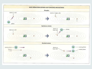
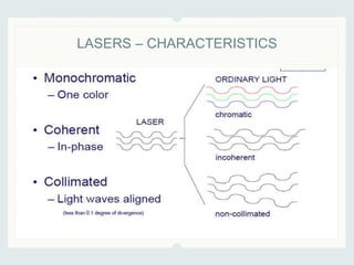
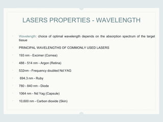
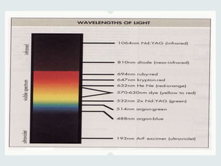
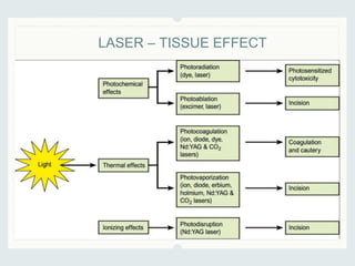
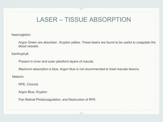
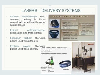
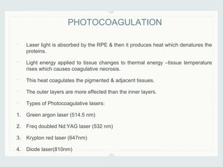
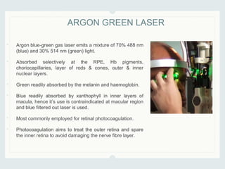
2. • LASER is an acronym that stands for
• Light
• Amplification
• by Stimulated
• Emission
• of Radiation
LASERS - DEFINITION
3. • Light acts as electromagnetic waves, emitting radiant energy in tiny package
called ‘quanta’/photon. Each photon has a characteristic frequency and its
energy is proportional to its frequency.
• Three basic ways for photons and atoms to interact:
1. Absorption
2. Spontaneous Emission
3. Stimulated Emission
LASERS - INTRO
6. • The basic laser cavity consists of an active medium in a resonant cavity with
two mirrors placed at opposite ends.
• One of the mirrors allows partial transmission of laser light out of the laser
cavity, toward the target tissue.
• A pump source introduces energy into the active medium and excites a
number of atoms.
• In this manner, amplified, coherent, and collimated light energy is released as
laser energy through the mirror that partially transmits.
• The various lasers differ mainly in the characteristics of the active medium
and the way this active medium is pumped.
LASERS – MECHANISM OF EMISSION
8. • Wavelength: choice of optimal wavelength depends on the absorption spectrum of the target
tissue
• PRINCIPAL WAVELENGTHS OF COMMONLY USED LASERS
• 193 nm - Excimer (Cornea)
• 488 - 514 nm - Argon (Retina)
• 532nm - Frequency doubled Nd:YAG
• 694.3 nm - Ruby
• 780 - 840 nm - Diode
• 1064 nm - Nd Yag (Capsule)
• 10,600 nm - Carbon dioxide (Skin)
LASERS PROPERTIES - WAVELENGTH
9.
10. • This is the amount of power delivered to a unit area of tissue.
• To prevent creating very intense burn decrease in the spot size should be
accompanied by decrease in power.
• Power may be increased by delivering the same energy over a shorter time.
This is achieved by Q-switching to produce bursts of power.
LASER PROPERTIES - POWER
11. • Continuous Wave (CW) Laser: It delivers the energy in a continuous
stream of photons.
• Pulsed Lasers: Produce energy pulses of a few tens of micro to few
mili second.
• Q Switched Lasers: Deliver energy pulses of extremely short duration
(nano second).
• Mode-locked Lasers: Emits a train of short duration pulses
(picoseconds) to femtoseconds
LASER – MODES
12. Transverse electromagnetic modes
Comparison of cross section of the laser beam at different points along its path
reveals that it is very slightly divergent, and that it is more intense at certain points.
This mode is important in photodisruption (e.g. YAG capsulotomy).
Fundamental mode
• At the point of focus, energy is most. concentrated at the centre of the laser
beam and diminishes peripherally in a distribution described by a Gaussian
curve
LASER – MODES
• Newer YAG laser designs increase the distribution
of energy towards the centre of the beam to create
a smaller point of focus and produce the same
effect whilst delivering less energy.
13. Q-switching mode
• Q-switching is a mechanism whereby a shutter
is placed in front of one of the two mirrors in
the laser tube between which the oscillation of
the beam normally occurs
• This maximises the energy state of the laser
medium by limiting energy loss to spontaneous
emission alone
• Opening the shutter allows oscillation to occur
and produce a single pulsed surge of
stimulated emission with a duration of 2–30
nanoseconds.
• Various shutters are used, including rotating
mirrors, dyes and electrooptic switches.
LASER – MODES
14. Mode locking mode
• Mode locking is a refinement of Q-switching which synchronises the
variouswavelengths so that periodically they are in phase and summate as a
train of very high energy pulses
• A pulse lasts about 30 picoseconds and produces up to 100 times as much
power for the same energy compared with Q-switching.
LASER – MODES
17. Haemoglobin:
• Argon Green are absorbed , Krypton yellow. These lasers are found to be useful to coagulate the
blood vessels.
Xanthophyll:
• Present in inner and outer plexiform layers of macula.
• Maximum absorption is blue. Argon blue is not recommended to treat macular lesions.
Melanin:
• RPE, Choroid
• Argon Blue, Krypton
• Pan Retinal Photocoagulation, and Destruction of RPE
LASER – TISSUE ABSORPTION
18. • Slit-lamp biomicroscope: most
common, delivery is trans-
corneal, with or without the aid of
contact lenses
• Indirect ophthalmoscope:
condensing lens ,trans-corneal
• Endolaser probes: fiber-optic
probes used within the eye
• Exolaser probes: fiber-optic
probes used trans-sclerally
LASERS – DELIVERY SYSTEMS
19. • Laser light is absorbed by the RPE & then it produces heat which denatures the
proteins.
• Light energy applied to tissue changes to thermal energy –tissue temperature
rises which causes coagulative necrosis.
• This heat coagulates the pigmented & adjacent tissues.
• The outer layers are more effected than the inner layers.
• Types of Photocoagulative lasers:
1. Green argon laser (514.5 nm)
2. Freq doubled Nd:YAG laser (532 nm)
3. Krypton red laser (647nm)
4. Diode laser(810nm)
PHOTOCOAGULATION
20. • Argon blue-green gas laser emits a mixture of 70% 488 nm
(blue) and 30% 514 nm (green) light.
• Absorbed selectively at the RPE, Hb pigments,
choriocapillaries, layer of rods & cones, outer & inner
nuclear layers.
• Green readily absorbed by the melanin and haemoglobin.
• Blue readily absorbed by xanthophyll in inner layers of
macula, hence it’s use is contraindicated at macular region
and blue filtered out laser is used.
• Most commonly employed for retinal photocoagulation.
• Photocoagulation aims to treat the outer retina and spare
the inner retina to avoid damaging the nerve fibre layer.
ARGON GREEN LASER
21. • 532 nm radiation
• Produces a pea green beam.
• Often termed as “green laser”/ktp laser as potassium titinyl
phosphate crystal may also be used.
• Highly absorbed by Hb & the melanin pigment.
• It coagulates from choriocapillaries to ONL.
• It causes coagulation with least energy transmission & shows
considerable safety in macular treatment.
DOUBLE FREQUENCY YAG LASER
22. • Melanin absorbs it readily.
• It is not absorbed by xanthophylls & Hb
& thus it is particularly suitable for
macular photocoagulation.
• It coagulates deeper into the RPE &
choroids. It has insignificant effect on
the vascular system of retina.
• It is less absorbed & more highly
transmitted through RPE
KRYPTON RED LASER (647NM)
23. • The diode lasers emit a wavelength of 810 nm infrared in continuous wave
mode. The laser energy is generated by a semiconductor diode chip.
• Diode lasers are efficient, generate little excess heat and are portable.
• In the eye, diode laser light is absorbed only by melanin and consequently is
most commonly used for retinal. photocoagulation. Low scattering of this
wavelength ensures good penetration of the ocular media and of oedematous
retina.
• The 810 nm wavelength also penetrates the sclera. Thus, even if the retina
isobscured from view through the pupil, photocoagulation may still be
performed by placing the delivering probe on the surface of the eye.
• The transparency of the sclera to diode laser also allows photocycloablation of
the ciliary body in 'end stage' glaucoma.
• Diode photocoagulation of vascular structures (e.g. neovascular membranes
and tumours) is enhanced by intravenous indocyanine green dye with an
absorption peak of 800–810 nm.
• Diode laser light has been used endoscopically to create a
dacryocystorhinostomy (DCR).
DIODE LASER
24. • The energy produced is released in a very short time.
• The laser beam is focused, concentrating the power into small area
• It produces a spark & an acoustic wave—which disrupts the tissue.
• ex: Nd:YAG laser.
PHOTODISRUPTIVE
25. • Neodymium-doped yttrium aluminum garnet is a crystal that is used as a lasing
medium for solid-state lasers
• Nd:YAG lasers typically emit light with a wavelength of 1064 nm, in the infrared
region.
• It is a powerful continuous wave (CW) laser which is usually Q-switched when
used to treat the eye.
• The 1064 nm wavelength is invisible and requires a He–Ne laser red aiming
beam. Before use on a patient's eye, the operator must ensure that the laser
beam and the aiming beam are focused at the same point.
Applications
• Correct posterior capsular opacification
• Peripheral iridotomy in patients with angle-closure glaucoma
• Frequency-doubled Nd:YAG lasers (wavelength 532 nm) are used for PRP.
YAG
26. • Breaks the chemical bonds that hold tissue together essentially vaporizing the
tissue, e.g. Photorefractive Keratectomy, Argon Fluoride (ArF) Excimer Laser.
PHOTOABLATIVE
27. • The excimer laser derives its name from 'excited dimer', two atoms forming a
molecule in the excited
• state but which dissociate in the ground state.
• Excimer lasers in clinical use employ an argon–fluorine (Ar–F) dimer laser
medium to emit 193 nm ultraviolet (UV) radiation. High absorption of UV by the
cornea limits its penetration.
• The delivery of a relatively high level of energy to a small volume of tissue causes
tissue removal (i.e. ablation). The ablation depth may be precisely determined.
• The excimer laser is therefore ideally suited to photorefractive keratectomy (PRK)
and laser intrastromal keratomileusis (Femto-LASIK) to reshape the corneal
surface as well as phototherapeutic keratectomy (PTK) to remove abnormal
corneal surface tissue.
EXCIMER LASER
28. • It is a conversion of chemical from one form to another by light.
• Also called Photodynamic Therapy
• Photochemical reaction following visible/infrared light particularly after
administration of exogenous chromophore.
Commonly used photosensitizers:
• Hematoporphyrin
• Benzaporphyrin Derivatives
• e.g. Treatment of ocular tumour and CNV by the use of verteporfin –a drug that
is chemically inert but is activated by light , after which it destroys neovascular
tissue.
PHOTOACTIVATION/ PHOTOCHEMICAL
30. • Class-I: inherently safe, usually because the light is contained in an enclosure,
for example in CD players.
• Class-II: safe during normal use; the blink reflex of the eye will prevent damage.
Usually up to 1 mW power, for example laser pointers.
• Class-IIIA: usually up to 5 mW and involve a small risk of eye damage within the
time of the blink reflex. Staring into such a beam for several seconds is likely to
cause damage to a spot on the retina.
• Class –IIIB: can cause immediate eye damage upon exposure.
• Class-IV: lasers can burn skin, and in some cases, even scattered light can
cause eye and/or skin damage. Many industrial and scientific lasers are in this
class.
LASER SAFETY
31. LASER SAFETY REGULATION:
• Patient safety is ensured by correct positioning.
• Danger to the surgeon is avoided by safety filter
system.
• Safety of observers and assistants.
LASER SAFETY
32. • Confocal Scanning: Imaging system and optical system focus at same point, Ar
green. He-Ne Red and diode infrared lasers are used to view surface of cornea,
optic nerve head and used to perform fluoroscein angiography.
• Scanning laser Polarimetry: Measures thickness of RNFL by projecting a spot of
polarized laser light onto the retina.
• Confocal Tomography: Laser light used to produce a topographic map of the optic
nerve head.
• Laser inferometry/ microperimetry: Determine light sensistivty of small areas of
retina
• Laser Doppler flowmetry: Measuring Retinal capillary blood flow using incident laser
light on moving RBCs.
LASERS IN INVESTIGATIONS