Downloaded 880 times
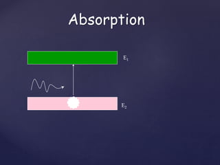
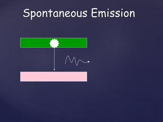
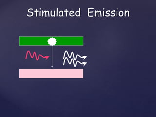
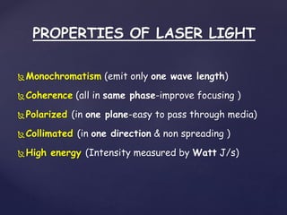
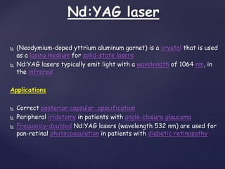
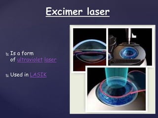
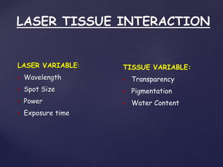
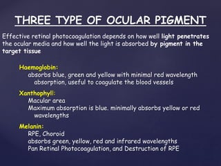
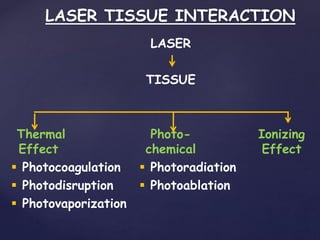
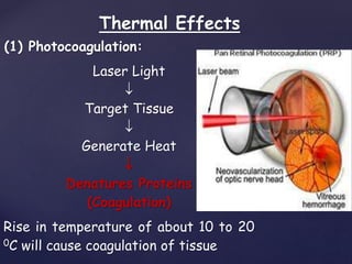
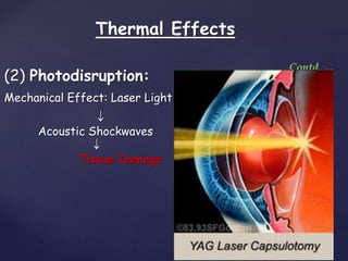
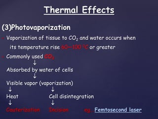


The document discusses lasers used in ophthalmology. It begins by defining what a laser is in terms of its acronym parts. It then covers laser physics including absorption, spontaneous emission, and stimulated emission. It describes different types of lasers used in ophthalmology like Nd:YAG, excimer, and diode lasers. Applications covered include treatments for glaucoma, cataracts, retinal diseases, and refractive errors. Mechanisms of laser tissue interaction like photocoagulation and photodisruption are also summarized.


