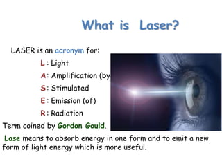
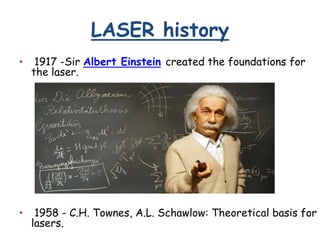
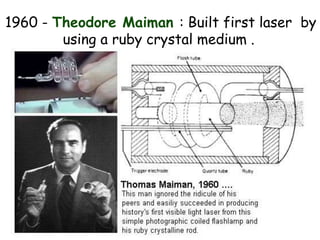
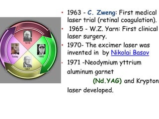
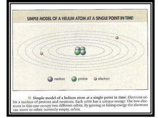
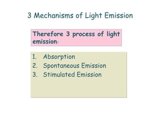
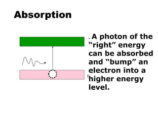
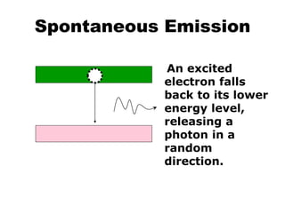
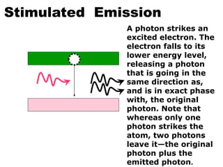
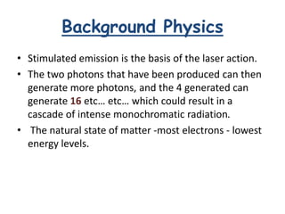
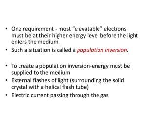
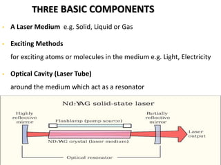
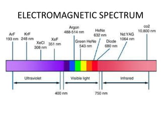
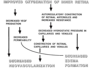
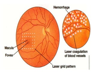
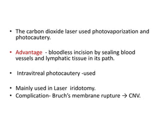
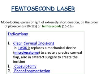
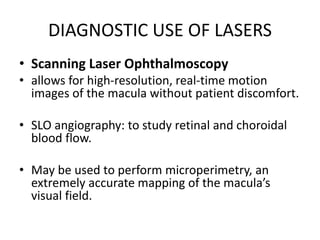
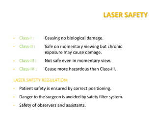
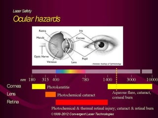
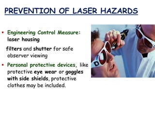
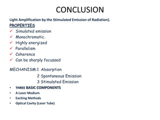
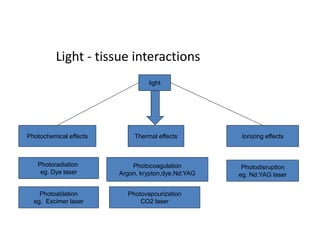
2. Objectives
• What is Laser ?
• LASER history..
• LASER Properties.
• How LASER is produced ?
• Effects of laser.
• Application of LASERs in Ophthalmology.
• LASER Safety.
3. What is Laser?
LASER is an acronym for:
L : Light
A: Amplification (by)
S: Stimulated
E : Emission (of)
R : Radiation
Term coined by Gordon Gould.
Lase means to absorb energy in one form and to emit a new
form of light energy which is more useful.
4. LASER history
• 1917 -Sir Albert Einstein created the foundations for
the laser.
• 1958 - C.H. Townes, A.L. Schawlow: Theoretical basis for
lasers.
5. 1960 - Theodore Maiman : Built first laser by
using a ruby crystal medium .
6. • 1963 - C. Zweng: First medical
laser trial (retinal coagulation).
• 1965 - W.Z. Yarn: First clinical
laser surgery.
• 1970- The excimer laser was
invented in by Nikolai Basov
• 1971 -Neodymium yttrium
aluminum garnet
(Nd.YAG) and Krypton
laser developed.
7. Lasers have many important applications.
• Common consumer devices such as DVD players, laser
printers, and barcode scanners.
• laser surgery and various skin treatments,
• Industry - cutting and welding materials.
• Military and law
enforcement devices marking
targets and measuring
range and speed.
• Laser lighting displays
8. Laser physics
• Laser light differs from “ordinary” light in
several important ways.
• These differences are a direct result of the
manner in which laser light is generated
Fig. 1. “White” light is composed of all wavelengths, traveling in all
directions.
Fig. 2. Ordinary monochromatic light consists of light of
approximately the same wavelength. However, the light is not traveling
in one direction.
9. PROPERTIES OF LASER LIGHT
• Monochromatic (emit only one wave length)
• Coherence (all in step with one another-improve
• focusing )
• Polarized (in one plane-easy to pass through media)
• Collimated (in one direction & non spreading )
• High energy (Intensity measured by Watt J/s)
10. LASER Vs. LIGHT
LASER LIGHT
Simulated emission
Monochromatic.
Highly energized
Parallelism
Coherence
Can be sharply focussed.
Spontaneous emission.
Polychromatic.
Poorly energized.
Highly divergence
Not coherent
Can not be sharply
focussed.
11. How LASER is produced ?
Light is a form of energy at which the human eye is sensitive
12. LASER PHYSICS
• Light as electromagnetic waves, emitting radiant
energy in tiny package called ‘quanta’/photon.
• Each photon has a characteristic frequency and its
energy is proportional to its frequency.
• When light is passed through certain kinds of
materials, the photons may excite electrons around
certain atoms into the next higher energy level
13.
14. 3 Mechanisms of Light Emission
1. Absorption
2. Spontaneous Emission
3. Stimulated Emission
Therefore 3 process of light
emission:
15. Absorption
. A photon of the
“right” energy
can be absorbed
and “bump” an
electron into a
higher energy
level.
E2
17. Stimulated Emission
A photon strikes an
excited electron. The
electron falls to its
lower energy level,
releasing a photon
that is going in the
same direction as,
and is in exact phase
with, the original
photon. Note that
whereas only one
photon strikes the
atom, two photons
leave it—the original
photon plus the
emitted photon.
18. Background Physics
• Stimulated emission is the basis of the laser action.
• The two photons that have been produced can then
generate more photons, and the 4 generated can
generate 16 etc… etc… which could result in a
cascade of intense monochromatic radiation.
• The natural state of matter -most electrons - lowest
energy levels.
19. • One requirement - most “elevatable” electrons
must be at their higher energy level before the light
enters the medium.
• Such a situation is called a population inversion.
• To create a population inversion-energy must be
supplied to the medium
• External flashes of light (surrounding the solid
crystal with a helical flash tube)
• Electric current passing through the gas
20. THREE BASIC COMPONENTS
• A Laser Medium e.g. Solid, Liquid or Gas
• Exciting Methods
for exciting atoms or molecules in the medium e.g. Light, Electricity
• Optical Cavity (Laser Tube)
around the medium which act as a resonator
21. The Resonator-
• The energy of the emitted laser beam - increased still further
by causing the light beam to traverse the material multiple
times.
• This is accomplished by placing a mirror over each end of the
crystal or gas tube so that the distance between them is an
even multiple of the laser light’s wavelength.
• The coherent light beam is reflected back and forth becoming
more and more intense.
If the “exciting” energy is supplied in brief pulses, laser
output of higher energy levels (in pulses) can be
obtained.
22.
23.
24. LASER INSTRUMENTATION
Three Main Components –
• Console: laser medium and tube, power
supply and laser control system.
• Control Panel: dials or push buttons for
controlling various parameters & standby
switch as a safety measure.
• Delivery System
25. Delivery systems
• Transpupillary: - Slit lamp
- Laser Indirect
Ophthalmoscopy
• Trans scleral : - Contact
- Non contact
• Endophotocoagulation.
26. Slit lamp biomicroscopic laser delivery
• Most commonly employed mode for anterior
and posterior segment.
• ADVANTAGES:
• Binocular and stereoscopic view.
• Fixed distance.
• Standardization of spot size is more accurate.
• Aiming accuracy is good.
27. Laser indirect ophthalmoscope.
• Advantages :
• Wider field(ability to reach periphery).
• Better visualization and laser application in hazy
medium.
• Ability to treat in supine position.
• Disadvantage : difficulty in focusing.
• Difficulty to standardize spot size.
• Expensive.
• Un co-operative patient.
• Learning curve.
28. MODES OF LASER OPERATION
• Continuous Wave (CW) Laser: deliver - energy in a continuous stream
of photons.
• Pulsed Lasers: Produce energy pulses of a few tens of micro to few
mili second.
• Q Switches Lasers: Deliver energy pulses of extremely short duration
(nano second).
• A Mode-locked Lasers: Emits a train of short duration pulses
(picoseconds).
• Fundamental System: Optical condition in which only one type of
wave is oscillating in the laser cavity.
• Multimode system: Large number of waves, each in a slight different
direction ,oscillate in laser cavity.
29. CLASSIFICATION OF LASER
• Solid State
Ruby
Nd.Yag
Erbium.YAG
• Gas
Ion
Argon
Krypton
He-Neon
CO2
• Metal Vapour
Cu
Gold
Dye
Rhodamine
Excimer
Argon Fluoride
Krypton Fluoride
Krypton Chloride
Diode
Gallium-Aluminum Arsenide
(GaAlAs)
32. THREE TYPE OF OCULAR PIGMENT
Effective retinal photocoagulation depends –
• how well light penetrates the ocular media
• how well the light is absorbed by pigment in the target tissue
• Haemoglobin:
Argon Green are absorbed - useful to coagulate the blood
vessels.
• Xanthophyll:
Present in inner and outer plexiform layers of macula.
Maximum absorption is blue. Argon blue is not recommended
to treat macular lesions.
• Melanin:
RPE, Choroid
Argon Blue, Krypton
Pan Retinal Photocoagulation, and Destruction of RPE
39. Thermal Effects
(1) Photocoagulation:
Laser Light
Target Tissue
Generate Heat -Tissue temp.:increases
from 37 deg c to 50 deg. C or higher
Denatures Proteins (Coagulation)
Risein temperature of about 10 to 20
0C will cause coagulation of tissue.
40. Panretinal photocoagulation (PRP) ctd
1. Proliferative diabetic retinopathy with high risk
characteristics
2. Neovascularisation of iris
3. Severe non proliferative diabetic retinopathy associated
with-poor compliance for follow up-before cataract
surgery-renal failure-one eyed patient and-pregnancy
4. central retinal vein occlusion, branch retinal vein
occlusion,
5. sickle retinopathy,
6. Eales disease and IRVAN (idiopathic retinal vasculitis,
aneurysms, and neuroretinitis )
Indications
45. • The carbon dioxide laser used photovaporization and
photocautery.
• Advantage - bloodless incision by sealing blood
vessels and lymphatic tissue in its path.
• Intravitreal photocautery -used
• Mainly used in Laser iridotomy.
• Complication- Bruch’s membrane rupture → CNV.
46. FEMTOSECOND LASER
Mode-locking -pulses of light of extremely short duration, on the order
of picoseconds (10−12s) or femtoseconds (10−15s).
Indications
1. Clear Corneal Incisions
in LASIK it replaces a mechanical device
(microkeratome) to create a precise corneal
flap, also in cataract surgery to create the
incision
1. Capsulotomy
2. Phacofragmentation
47. Photochemical effcts
Photoablation:
• Breaks the chemical bonds that hold tissue
together essentially vaporizing the tissue, e.g.
Photorefractive Keratectomy, Argon Fluoride
(ArF) Excimer Laser.
Contd. …
Is a form of ultraviolet laser
• Used in
delicate surgeries such
as eye surgery eg ;LASIK
48. PHOTOCHEMICAL EFFECT
Photoradiation OR Photodynamic therapy (PDT)
• 42°C and 52°C temp
• Photoradiation by red (630 nm)- cytotoxic free radicals in a tumor previously
sensitized by a hematoporphyrin-derived uptake.
• The most common - PDT.
• This new treatment modality combines low-power diode lasers (689 nm) with
an infusion of verteporfin to ablate subretinal neovascular membranes and
treatment of various tumors.
• The advantage -over conventional photocoagulation -lower energy levels result
in much less damage to adjacent normal tissue
E.G. Treatment of Ocular tumours and CNV
49. Photodisruption:
Extremely high irradiance delivered - very short exposure - small spot
electrons - energized from molecules - target tissue (microscopically
localized temp. rise from 37 deg. c to 1500degc)
collection of free electrons and ions – plasma
Rapid expansion of the plasma creates acoustic and shock waves
combines with latent tissue stress, incise the target tissue
Tissue damage
Contd. …
50. Nd:YAG laser
• (neodymium-doped yttrium aluminum garnet) is a crystal that is used
as a lasing medium for solid-state lasers.
• Nd:YAG lasers typically emit light with a wavelength of 1064nm, in
the infrared.
Applications
• Correct posterior capsular opacification
• Peripheral iridotomy in patients with acute angle-closure glaucoma.
• Frequency-doubled Nd:YAG lasers (wavelength 532 nm) are used for
pan-retinal photocoagulation in patients with diabetic retinopathy.
51. [
-
The most common laser
source used is the Nd:YAG
1064-nm.
Methods of pulsing and
Nd:YAG laser:-
1)Q- switching
2)mode- locking
65. DIAGNOSTIC USE OF LASERS
• Scanning Laser Ophthalmoscopy
• allows for high-resolution, real-time motion
images of the macula without patient discomfort.
• SLO angiography: to study retinal and choroidal
blood flow.
• May be used to perform microperimetry, an
extremely accurate mapping of the macula’s
visual field.
66. LASER SAFETY
• Class-I : Causing no biological damage.
• Class-II : Safe on momentary viewing but chronic
exposure may cause damage.
• Class-III : Not safe even in momentary view.
• Class-IV : Cause more hazardous than Class-III.
LASER SAFETY REGULATION:
• Patient safety is ensured by correct positioning.
• Danger to the surgeon is avoided by safety filter system.
• Safety of observers and assistants.
70. Complications(contd..)
• Ischaemic papillitis.
• Paracentral visual field loss and scotoma.
• Photocoagulation scar enlargement.
• Subretinal fibrosis.
• Iatrogenic choroidal neovascularisation.
• Accidental foveal burns.
71. PREVENTION OF LASER HAZARDS
Engineering Control Measure:
laser housing
filters and shutter for safe
observer viewing
Personal protective devices, like
protective eye wear or goggles
with side shields, protective
clothes may be included.
76. REFERENCES
• YANOFF AND DUKER OPHTHALMOLOGY- 3rd
edition.
• LASERS IN OPTHALMOLOGY
• A practical guide-AIIMS.
• LASER SURGERY OF THE POSTERIOR
SEGMENT- Steven M. Bloom