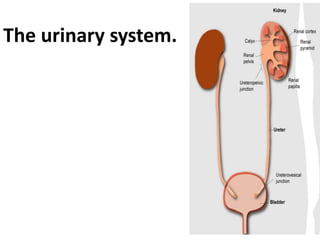
3. The kidneys, ureters, urinary bladder and urethra are the
main components of the urinary system.
A function of the urinary system that immediately comes
to mind is the excretion of waste products from the body.
This is only one of many functions of the system.
Others are elimination of foreign substances and
regulation of the amount of water in the body,
-control of the concentration of most compounds in the
extracellular fluid .
4. Most of these tasks are performed in the kidneys.
Functionally the processes can be divided into two steps,
each of which have their anatomical correlate:
•filtration - glomeruli of the kidney
•selective resorption and excretion - tubular system of
the kidney
In addition, the kidney also functions as an endocrine
organ. Fibrocytes in the cortex release the hormone
erythropoietin, which stimulates the formation of red
blood cells. Modified fibrocytes of the medulla secrete
prostaglandins which are able to decrease blood
pressure.
5. Kidney
Glomeruli and the tubular system are both part of the
basic functional unit of the kidney, the nephron.
The Glomerulus (or renal corpuscle)
The Glomerulus is the round (~0.2 mm in diameter)
blind beginning of the nephron. It is invaginated by a
tuft of capillaries at the vascular pole of the
Glomerulus. The tuft of capillaries and other cells in
contact with them form the anatomical Glomerulus.
Substances which leave the capillaries enter the renal
tubule at the urinary pole of the Glomerulus.
6. The anatomical glomerulus is enclosed by two layers of
epithelium, Bowman's capsule. Cells of the outer or
parietal layer of Bowman's capsule form a simple
squamous epithelium. Cells of the inner layer, podocytes in
the visceral layer, are extremely complex in shape. Small
foot-like processes, pedicles, of their cytoplasm form a
fenestrated epithelium around the fenestrated capillaries
of the glomerulus. The openings between the pedicles are
called filtration slits. They are spanned by a thin
membrane, the filtration slit membrane.
7. -Mesangial cells in the glomerulus form the connective
tissue that gives structural support to podocytes and
vessels.
Blood pressure is the driving force in the formation of about
125 ml of glomerular filtrate per minute. About 124 ml of
the glomerular filtrate is reabsorbed in the tubules of the
nephron.
- Between the podocytes and the endothelial cells of the
capillaries we find a comparatively thick basal lamina, which
can be subdivided into an outer lamina rara externa, a
middle lamina densa and an inner lamina rara interna. The
basal lamina and the slit membranes form the glomerular
filtration barrier, which prevents some large molecules from
entering the capsular space between the outer and inner
epithelial layers of Bowman's capsule.
8.
9. Tubules of the Nephron
The tubular system can be divided into proximal and
distal tubules, which in turn have convoluted and
straight portions. Intermediate tubules connect the
proximal and distal tubules. Running from the cortex of
the kidney towards the medulla (descending), then
turning and running back towards the cortex
(ascending), the tubules form the loop of Henle.
10. The proximal tubule is the longest section of the
nephron (about 14 mm). The convoluted part of the
proximal tubules coils close to the glomerulus in the
cortex. The diameter of proximal tubules is ~65 µm.
Their walls are formed by a low columnar epithelium.
The eosinophilic cells of the epithelium have a wide
brush border (long microvilli ) and are active in
endocytosis. They almost completely reabsorb
substances of nutritional value from the glomerular
filtrate (glucose, amino acids, protein, vitamins etc. -
11. The straight portion of the proximal tubule merges
with the intermediate tubule (thin segment of the
loop of Henle).
A flattened, only ~1-2 µm high epithelium forms the
intermediate tubule, which is only ~15 µm wide.
Descending parts of the straight proximal and
intermediate tubules are permeable to water but not
to solutes
12. The thin segment of Henle's loop leads into the straight
part of the distal tubule, which is formed by low cuboidal
cells without a brush border. A few short microvilli are
present, but they are difficult to see in the light
microscope. The diameter of the tubule expands to ~35
µm. Epithelial cells in the ascending parts of the
intermediate and straight distal tubules cells transport
chloride (active) and sodium ions (passive) out of the
tubular lumen into the surrounding peritubular space.
The epithelium can not be penetrated by water.
Consequently, the transport of ions over the epithelium
sets up a gradient in osmotic pressure, which serves as
driving force in the further concentration of the urine.
13. The straight portion of the distal tubule contacts the
glomerulus forming the macula densa.
Thereafter, the distal tubule forms its convoluted portion
(about 5 mm long).
Cells in the distal tubule are sensitive to the hormone
aldosterone, which is produced in the zona glomerulosa
of the adrenal glands.
Aldosterone stimulates the active reabsorption of
sodium ions and the excretion of potassium ions.
14.
15.
16. The Juxtaglomerular Apparatus
As mentioned above, the distal tubule contacts the
glomerulus forming a specialized section of tubular
epithelium, the macula densa. At the point of contact
with the glomerulus, the distal tubule is always in close
contact with the efferent and afferent arterioles of the
glomerulus.
Other parts of the juxtaglomerular apparatus are
extraglomerular mesangial cells and the juxtaglomerular
cells surrounding the afferent arteriole (modified
smooth muscle cells), which produce and secrete renin
17. Different theories exist that try to explain the
interactions between the cells that eventually lead to the
release of renin. One of them, the baroreceptor theory,
assumes that the juxtaglomerular cells function as
stretch receptors (high blood pressure would inhibit the
release of renin). Another theory, the macula densa
theory, claims that the secretion of renin is regulated by
the composition of the fluid in the distal tubule and/or
the afferent arteriole (low sodium would increase in the
release of renin).
18. Excretory Passages
The minor calyces merge to form major calyces within
the kidney, which in turn merge to form the renal
pelvis (still within the kidney). The urine flows through
these structures to the ureter and is channeled to the
bladder.
The basic structure of all these components is the
same. The mucosa is lined with a transitional
epithelium , which occurs exclusively in the urinary
system. The epithelium is virtually impenetrable to any
components of the urine , which consequently does
not change in composition as it passes through the
excretory passages.
19. The lamina propria consists mainly of dense
connective tissue, with many bundles of coarse
collagenous fibres. The muscularis usually consists of
an inner longitudinal and outer circular layer of
smooth muscle cells . In lower parts of the ureter and
the bladder an additional outer longitudinal layer of
muscles is added to the first two
20.
21.
22. The bladder is finally emptied through the urethra.
Initially, the urethra is lined by a transitional epithelium in
males and females. In males, it is replaced by a
pseudostratified or stratified columnar epithelium below
the openings of the ejaculatory ducts into the urethra. The
distal parts of the female urethra and the distal end of the
male urethra are lined by a stratified squamous
epithelium. The lamina propria contains loose connective
tissue. Smooth muscle cells in the muscularis are mainly
oriented longitudinally. They are surrounded, in the middle
part of the urethra (below the prostate in males), by
striated muscle cells of the sphincter urethrae.