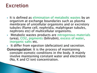
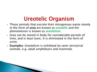
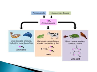
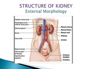
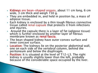
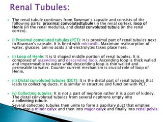
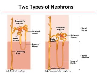
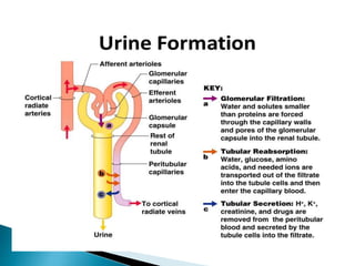
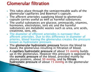
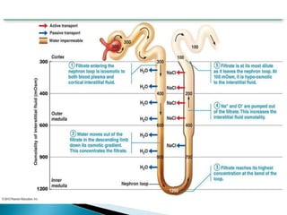
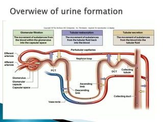
Suprabha P
Assistant prof. Of Zoology
Government Degree College for Women
Begumpet
 It is defined as elimination of metabolic wastes by an
organism at exchange boundaries such as plasma
membrane of unicellular organisms and or excretory
tubules (flame cell, nephridia, malphigean tubules,
nephrons etc) of multicellular organisms.
 Metabolic wastes products are nitrogenous materials
(urea), CO2, pigments (bilirubin), excess of water,
inorganic salts etc.
 It differ from egestion (defecation) and secretion.
Osmoregulation: It is the process of maintaining
constant osmotic conditions in the body. It mainly
involve maintaining constant water and electrolyte
(Na, K and Cl ion) concentration.
Significance of excretion:
 Removal of unwanted metabolic byproducts: Metabolism
of carbohydrates and fats produces CO2and H2O, similarly
Protein metabolism produces nitrogenous wastes in the
cell and tissue which are remove by excretion.
 Removal of toxic wastes: accumulation of excretory
products such as nitrogenous wastes, CO2, pigments
formed by the breakdown of haemoglobin, drugs etc.is
harmful to the body. So, these toxic wastes are removed.
 Osmoregulation of the body: it helps maintaining constant
ionic concentration of body fluid as well as Regulating
water content of the body
 Regulation of body PH
 Thermoregulation: Excretion also helps to maintain a
constant body temperature
 Depending upon the form in which
nitrogenous waste is excreted from the body,
the organisms are grouped as under into
three categories:
 Ammonotelic
 Uricotelic
 Ureotelic
 Those animals which excrete their nitrogenous waste
in the form of ammonia are known as ammonotelic.
 Ammonia is highly soluble in water with which it
forms ammonium hydroxide (NH4OH) which can
damage cells directly by its alkaline caustic action.
 Excretion of ammonia requires large amounts of
water, so there will be more water loss from the
body. That is why such a mode is suitable for aquatic
organisms which have a constant access to water.
 1 gm of ammonia needs about 300 - 500 ml of
water.
 Ammonia is the first metabolic waste product of
protein metabolism and no energy is required to
produce ammonia.
 Examples: All aquatic invertebrates, bony fishes and
aquatic amphibians.
 Those animals which excrete their
nitrogenous waste mainly in the form of uric
acid and urates are known as uricotelic.
 The phenomenon is known as uricotelism.
 Elimination of uric acid requires lesser
amount of water, comparatively less soluble
in water and is less toxic as compared to
ammonia.
 Examples: All terrestrial animals like insects,
reptiles, and birds excrete uric acid as
nitrogenious wastes
Ureotelic Organism
 Those animals that excrete their nitrogenous waste mainly
in the form of urea are known as ureotelic and the
phenomenon is known as ureotelism.
 Urea can be stored in body for considerable periods of
time, and is least toxic. It is eliminated in the form of
urine.
 Examples: Ureotelism is exhibited by semi-terrestrial
animals, e.g. adult amphibians and mammals
 Kidneys are bean-shaped organs, about 11 cm long, 6 cm
wide, 3 cm thick and weigh 150 g.
 They are embedded in, and held in position by, a mass of
adipose tissue.
 Each kidney is enclosed by a thin tough fibrous connective
tissue called renal capsule that protects it from infections
and injuries.
 Around the capsule there is a layer of fat (adipose tissue)
which is further enclosed by another layer of fibrous
membrane known as renal fascia.
 The bean shaped kidney have outer convex surface and
inner concave surface.
 Location: The kidneys lie on the posterior abdominal wall,
one on each side of the vertebral column, behind the
peritoneum and below the diaphragm.
 Position: It is situated at the level of T12-L3. The right
kidney is usually slightly lower than the left, probably
because of the considerable space occupied by the liver.
 The renal artery enters the kidney through the hilum
and then branches progressively to form
the interlobular arteries, and afferent
arterioles, which lead to the glomerularcapillaries.
 The distal ends of the capillaries of each glomerulus
combine to form the efferent arteriole, which leads to
a second capillary network, the peritubular
capillaries, that surrounds the renal tubules
called vasa recta.
 The blood vessels of the venous system progressively
form the interlobular vein, arcuate vein, interlobar
vein, and renal vein, which leaves the kidney beside
the renal artery and ureter.
Longitudinal section of the kidney shows following
parts.
 Capsule: It is an outermost covering composed of
fibrous tissue surrounding the kidney.
 Cortex: It is a reddish-brown layer of tissue
immediately below the capsule and outside the renal
It consists of renal corpuscles and convoluted
tubules.
 Medulla: It is the innermost layer, consisting of
conical areas called the renal pyramids separated by
renal columns.
 There are 8-18 renal pyramids in each kidney.
 The apex of each pyramid is called a renal
papilla, and each papilla projects into a small
depression, called a minor calyx (plural calyces).
 Several minor calyces unite to form a major calyx. In
turn, the major calyces join to form a funnel shaped
structure called renal pelvis that collects urine and
leads to ureter.
Each nephron has two major
portions:
 A Renal corpuscle
(Malpighian body)
 A Renal tubule
 A renal corpuscle consists of a glomerulus surrounded by a
glomerular capsule (Bowman’s capsule).
Glomerulus is a capillary network that arises from an afferent
arteriole and empties into an efferent arteriole.
 The diameter of the efferent arteriole is smaller than that
of the afferent arteriole, which helps maintain a fairly high
blood pressure in the glomerulus.
Bowman’s capsule is double walled cup like structure and it
encloses the glomerulus. The wall of glomerulus and the
Bowman’s capsule consists of a single layer of flattened
epithelial cells.
Glomerular capsule consists of three layers
i) Outer parietal layer consists of squamous epithelium
cells with minute pore of 12nm diameter
called fenestrations.
ii) Middle basement membrane which is selectively
permiable.
iii) The inner visceral layer of large nucleated cell
called podocytes.
Podocytes bears finger like projections known as
podocels. The areas between the two podocels is filtration
slit underlying basement membrane.
 The renal tubule continues from Bowman’s capsule and consists of the
following parts: proximal convolutedtubule (in the renal cortex), loop of
Henle (in the renal medulla), and distal convoluted tubule (in the renal
cortex).
 i) Proximal convoluted tubules (PCT): it is proximal part of renal tubules next
to Bowman’s capsule. It is lined with microvilli. Maximum reabsorption of
water, glucose, amino acids and electrolytes takes place here.
 ii) Loop of Henle: It is U shaped middle portion of renal tubules. It is
composed of ascending and descending loop. Ascending loop is thick walled
and impermeable to water while descending loop is thin walled and
permeable to water. Counter current mechanism is crucial role of loop of
Henle.
 iii) Distal convoluted tubules (DCT): It is the distal part of renal tubules that
leads to collecting ducts. It is similar in structure and function with PCT.
 iv) Collecting tubules: It is not a part of nephron rather it is a part of kidney.
The distal convoluted tubules from several nephrons empty into
a collecting tubule.
 Several collecting tubules then unite to form a papillary duct that empties
urine into a minor calyx and then into major calyx and finally into renal pelvis.

Glomerular filtration
 This takes place through the semipermeable walls of the
glomerular capillaries and Bowman’s capsule.
 The afferent arterioles supplying blood to glomerular
capsule carries useful as well as harmful substances.
 The useful substances are glucose, aminoacids, vitamins,
hormones, electrolytes, ions etc and the harmful
substances are metabolic wastes such as urea, uric acids,
creatinine, ions, etc.
 The diameter of efferent arterioles is narrower than
afferent arterioles. Due to this difference in diameter of
arteries, blood leaving the glomerulus creates the pressure
known as hydrostatic pressure.
 The glomerular hydrostatic pressure forces the blood to
leaves the glomerulus resulting in filtration of blood.
 A capillary hydrostatic pressure of about 55 mmHg builds
up in the glomerulus. However this pressure is opposed by
the osmotic pressure of the blood, provided mainly by
plasma proteins, about 30 mmHg, and by filtrate
hydrostatic pressure of about 15 mmHg in the glomerular
capsule.
 The net filtration pressure is, Therefore: 55-(30 +15) =
10mmHg.
 By the net filtration pressure of 10mmHg, blood is filtered
in the glomerular capsule.
 Water and other small molecules readily pass through the
filtration slits but Blood cells, plasma proteins and other
large molecules are too large to filter through and
therefore remain in the capillaries.
 The filtrate containing large amount of water, glucose,
amino acids, uric acid, urea, electrolytes etc in the
glomerular capsule is known as nephric filtrate or
glomerular filtrate.
 The volume of filtrate formed by both kidneys each minute
is called the glomerular filtration rate (GFR).
 In a healthy adult the GFR is about 125 mL/min, i.e. 180
litres of filtrate are formed each day by the two kidneys
 As the filtrate passes to the renal tubules, useful substances including
some water, electrolytes and organic nutrients such as glucose, amino
acids, vitamins hormones etc are selectively reabsorbed from the filtrate
back into the blood through the renal tubules.
 Reabsorption of some substance is passive, while some substances are
actively transported. Major portion of water is reabsorbed by Osmosis.
 Proximal Convoluted Tubule (PCT): PCT is lined by simple cuboidal brush
border epithelium which increases the surface area for reabsorption.
 Nearly all of the essential nutrients, and 70-80 per cent of electrolytes
and water are reabsorbed by this segment.
 PCT also helps to maintain the pH and ionic balance of the body fluids
by selective secretion of hydrogen ions. Ammonia and potassium ions
into the filtrate and by absorption of HCO3- from it.
 Henle's Loop: Reabsorption in this segment is minimum, However, this
region plays a significant role in the maintenance of high osmolarity of
medullary interstitial fluid. The descending limb of loop of Henle is
permeable to water but almost. impermeable to electrolytes. This
concentrates the filtrate as it moves' down.
 Distal Convoluted Tubule (DCT): Conditional reabsorption of Na+ and
water takes place in this segment. DCT is also capable of reabsorption
of HCO3- and selective secretion of hydrogen and potassium ions and
NH3 to maintain the pH and sodium-potassium balance in blood.
 Collecting Duct: This long duct extends from the cortex of the kidney
to the inner parts of the medulla.
 Large amounts of water could be reabsorbed from this region to
produce a concentrated urine.
 This segment allows passage of small amounts of urea into the
medullary interstitium to keep up the osmolarity.
 It also plays a role in the maintenance of pH and ionic balance of
blood by the selective secretion of H+ and K+ ions.
 Nutrients such as glucose, amino acids, and vitamins are
reabsorbed by active transport.
 Positive charged ions are reabsorbed by active transport while
negative charged ions are reabsorbed most often by passive
transport.
 Water is reabsorbed by osmosis, and small proteins are
reabsorbed by pinocytosis.
 Tubular secretion takes place from the blood in the peritubular
capillaries to the filtrate in the renal tubules and can ensure that
wastes such as creatinine or excess H+ or excess K+ ions are
actively secreted into the filtrate to be excreted.
 Excess K+ ion is secreted in the tubules and in exchange Na+
ion is reabsorbed otherwise it causes a clinical condition called
Hyperkalemia.
 Tubular secretion of hydrogen ions (H+) is very important in
maintaining normal blood pH.
 Substances such as , e.g. drugs including penicillin and aspirin,
may not be entirely filtered out of the blood because of the short
time it remains in the glomerulus.
 Such substances are cleared by secretion from the peritubular
capillaries into the filtrate within the convoluted tubules.
 The tubular filtrate is finally known as urine. Human urine is
usually hypertonic.
Excretory system ppt

Excretory system ppt

  • 1.
    Suprabha P Assistant prof.Of Zoology Government Degree College for Women Begumpet
  • 2.
     It isdefined as elimination of metabolic wastes by an organism at exchange boundaries such as plasma membrane of unicellular organisms and or excretory tubules (flame cell, nephridia, malphigean tubules, nephrons etc) of multicellular organisms.  Metabolic wastes products are nitrogenous materials (urea), CO2, pigments (bilirubin), excess of water, inorganic salts etc.  It differ from egestion (defecation) and secretion. Osmoregulation: It is the process of maintaining constant osmotic conditions in the body. It mainly involve maintaining constant water and electrolyte (Na, K and Cl ion) concentration.
  • 3.
    Significance of excretion: Removal of unwanted metabolic byproducts: Metabolism of carbohydrates and fats produces CO2and H2O, similarly Protein metabolism produces nitrogenous wastes in the cell and tissue which are remove by excretion.  Removal of toxic wastes: accumulation of excretory products such as nitrogenous wastes, CO2, pigments formed by the breakdown of haemoglobin, drugs etc.is harmful to the body. So, these toxic wastes are removed.  Osmoregulation of the body: it helps maintaining constant ionic concentration of body fluid as well as Regulating water content of the body  Regulation of body PH  Thermoregulation: Excretion also helps to maintain a constant body temperature
  • 4.
     Depending uponthe form in which nitrogenous waste is excreted from the body, the organisms are grouped as under into three categories:  Ammonotelic  Uricotelic  Ureotelic
  • 5.
     Those animalswhich excrete their nitrogenous waste in the form of ammonia are known as ammonotelic.  Ammonia is highly soluble in water with which it forms ammonium hydroxide (NH4OH) which can damage cells directly by its alkaline caustic action.  Excretion of ammonia requires large amounts of water, so there will be more water loss from the body. That is why such a mode is suitable for aquatic organisms which have a constant access to water.  1 gm of ammonia needs about 300 - 500 ml of water.  Ammonia is the first metabolic waste product of protein metabolism and no energy is required to produce ammonia.  Examples: All aquatic invertebrates, bony fishes and aquatic amphibians.
  • 6.
     Those animalswhich excrete their nitrogenous waste mainly in the form of uric acid and urates are known as uricotelic.  The phenomenon is known as uricotelism.  Elimination of uric acid requires lesser amount of water, comparatively less soluble in water and is less toxic as compared to ammonia.  Examples: All terrestrial animals like insects, reptiles, and birds excrete uric acid as nitrogenious wastes
  • 7.
    Ureotelic Organism  Thoseanimals that excrete their nitrogenous waste mainly in the form of urea are known as ureotelic and the phenomenon is known as ureotelism.  Urea can be stored in body for considerable periods of time, and is least toxic. It is eliminated in the form of urine.  Examples: Ureotelism is exhibited by semi-terrestrial animals, e.g. adult amphibians and mammals
  • 10.
     Kidneys arebean-shaped organs, about 11 cm long, 6 cm wide, 3 cm thick and weigh 150 g.  They are embedded in, and held in position by, a mass of adipose tissue.  Each kidney is enclosed by a thin tough fibrous connective tissue called renal capsule that protects it from infections and injuries.  Around the capsule there is a layer of fat (adipose tissue) which is further enclosed by another layer of fibrous membrane known as renal fascia.  The bean shaped kidney have outer convex surface and inner concave surface.  Location: The kidneys lie on the posterior abdominal wall, one on each side of the vertebral column, behind the peritoneum and below the diaphragm.  Position: It is situated at the level of T12-L3. The right kidney is usually slightly lower than the left, probably because of the considerable space occupied by the liver.
  • 11.
     The renalartery enters the kidney through the hilum and then branches progressively to form the interlobular arteries, and afferent arterioles, which lead to the glomerularcapillaries.  The distal ends of the capillaries of each glomerulus combine to form the efferent arteriole, which leads to a second capillary network, the peritubular capillaries, that surrounds the renal tubules called vasa recta.  The blood vessels of the venous system progressively form the interlobular vein, arcuate vein, interlobar vein, and renal vein, which leaves the kidney beside the renal artery and ureter.
  • 13.
    Longitudinal section ofthe kidney shows following parts.  Capsule: It is an outermost covering composed of fibrous tissue surrounding the kidney.  Cortex: It is a reddish-brown layer of tissue immediately below the capsule and outside the renal It consists of renal corpuscles and convoluted tubules.  Medulla: It is the innermost layer, consisting of conical areas called the renal pyramids separated by renal columns.  There are 8-18 renal pyramids in each kidney.  The apex of each pyramid is called a renal papilla, and each papilla projects into a small depression, called a minor calyx (plural calyces).  Several minor calyces unite to form a major calyx. In turn, the major calyces join to form a funnel shaped structure called renal pelvis that collects urine and leads to ureter.
  • 14.
    Each nephron hastwo major portions:  A Renal corpuscle (Malpighian body)  A Renal tubule
  • 15.
     A renalcorpuscle consists of a glomerulus surrounded by a glomerular capsule (Bowman’s capsule). Glomerulus is a capillary network that arises from an afferent arteriole and empties into an efferent arteriole.  The diameter of the efferent arteriole is smaller than that of the afferent arteriole, which helps maintain a fairly high blood pressure in the glomerulus. Bowman’s capsule is double walled cup like structure and it encloses the glomerulus. The wall of glomerulus and the Bowman’s capsule consists of a single layer of flattened epithelial cells. Glomerular capsule consists of three layers i) Outer parietal layer consists of squamous epithelium cells with minute pore of 12nm diameter called fenestrations. ii) Middle basement membrane which is selectively permiable. iii) The inner visceral layer of large nucleated cell called podocytes. Podocytes bears finger like projections known as podocels. The areas between the two podocels is filtration slit underlying basement membrane.
  • 16.
     The renaltubule continues from Bowman’s capsule and consists of the following parts: proximal convolutedtubule (in the renal cortex), loop of Henle (in the renal medulla), and distal convoluted tubule (in the renal cortex).  i) Proximal convoluted tubules (PCT): it is proximal part of renal tubules next to Bowman’s capsule. It is lined with microvilli. Maximum reabsorption of water, glucose, amino acids and electrolytes takes place here.  ii) Loop of Henle: It is U shaped middle portion of renal tubules. It is composed of ascending and descending loop. Ascending loop is thick walled and impermeable to water while descending loop is thin walled and permeable to water. Counter current mechanism is crucial role of loop of Henle.  iii) Distal convoluted tubules (DCT): It is the distal part of renal tubules that leads to collecting ducts. It is similar in structure and function with PCT.  iv) Collecting tubules: It is not a part of nephron rather it is a part of kidney. The distal convoluted tubules from several nephrons empty into a collecting tubule.  Several collecting tubules then unite to form a papillary duct that empties urine into a minor calyx and then into major calyx and finally into renal pelvis. 
  • 19.
    Glomerular filtration  Thistakes place through the semipermeable walls of the glomerular capillaries and Bowman’s capsule.  The afferent arterioles supplying blood to glomerular capsule carries useful as well as harmful substances.  The useful substances are glucose, aminoacids, vitamins, hormones, electrolytes, ions etc and the harmful substances are metabolic wastes such as urea, uric acids, creatinine, ions, etc.  The diameter of efferent arterioles is narrower than afferent arterioles. Due to this difference in diameter of arteries, blood leaving the glomerulus creates the pressure known as hydrostatic pressure.  The glomerular hydrostatic pressure forces the blood to leaves the glomerulus resulting in filtration of blood.  A capillary hydrostatic pressure of about 55 mmHg builds up in the glomerulus. However this pressure is opposed by the osmotic pressure of the blood, provided mainly by plasma proteins, about 30 mmHg, and by filtrate hydrostatic pressure of about 15 mmHg in the glomerular capsule.
  • 20.
     The netfiltration pressure is, Therefore: 55-(30 +15) = 10mmHg.  By the net filtration pressure of 10mmHg, blood is filtered in the glomerular capsule.  Water and other small molecules readily pass through the filtration slits but Blood cells, plasma proteins and other large molecules are too large to filter through and therefore remain in the capillaries.  The filtrate containing large amount of water, glucose, amino acids, uric acid, urea, electrolytes etc in the glomerular capsule is known as nephric filtrate or glomerular filtrate.  The volume of filtrate formed by both kidneys each minute is called the glomerular filtration rate (GFR).  In a healthy adult the GFR is about 125 mL/min, i.e. 180 litres of filtrate are formed each day by the two kidneys
  • 21.
     As thefiltrate passes to the renal tubules, useful substances including some water, electrolytes and organic nutrients such as glucose, amino acids, vitamins hormones etc are selectively reabsorbed from the filtrate back into the blood through the renal tubules.  Reabsorption of some substance is passive, while some substances are actively transported. Major portion of water is reabsorbed by Osmosis.  Proximal Convoluted Tubule (PCT): PCT is lined by simple cuboidal brush border epithelium which increases the surface area for reabsorption.  Nearly all of the essential nutrients, and 70-80 per cent of electrolytes and water are reabsorbed by this segment.  PCT also helps to maintain the pH and ionic balance of the body fluids by selective secretion of hydrogen ions. Ammonia and potassium ions into the filtrate and by absorption of HCO3- from it.  Henle's Loop: Reabsorption in this segment is minimum, However, this region plays a significant role in the maintenance of high osmolarity of medullary interstitial fluid. The descending limb of loop of Henle is permeable to water but almost. impermeable to electrolytes. This concentrates the filtrate as it moves' down.
  • 22.
     Distal ConvolutedTubule (DCT): Conditional reabsorption of Na+ and water takes place in this segment. DCT is also capable of reabsorption of HCO3- and selective secretion of hydrogen and potassium ions and NH3 to maintain the pH and sodium-potassium balance in blood.  Collecting Duct: This long duct extends from the cortex of the kidney to the inner parts of the medulla.  Large amounts of water could be reabsorbed from this region to produce a concentrated urine.  This segment allows passage of small amounts of urea into the medullary interstitium to keep up the osmolarity.  It also plays a role in the maintenance of pH and ionic balance of blood by the selective secretion of H+ and K+ ions.  Nutrients such as glucose, amino acids, and vitamins are reabsorbed by active transport.  Positive charged ions are reabsorbed by active transport while negative charged ions are reabsorbed most often by passive transport.  Water is reabsorbed by osmosis, and small proteins are reabsorbed by pinocytosis.
  • 24.
     Tubular secretiontakes place from the blood in the peritubular capillaries to the filtrate in the renal tubules and can ensure that wastes such as creatinine or excess H+ or excess K+ ions are actively secreted into the filtrate to be excreted.  Excess K+ ion is secreted in the tubules and in exchange Na+ ion is reabsorbed otherwise it causes a clinical condition called Hyperkalemia.  Tubular secretion of hydrogen ions (H+) is very important in maintaining normal blood pH.  Substances such as , e.g. drugs including penicillin and aspirin, may not be entirely filtered out of the blood because of the short time it remains in the glomerulus.  Such substances are cleared by secretion from the peritubular capillaries into the filtrate within the convoluted tubules.  The tubular filtrate is finally known as urine. Human urine is usually hypertonic.