QUALITY CONTROL IN
   PATHOLOGY

   QUALITY CONTROL
         IN
          PATHOLOGY
Quality Assurance in Healthcare
• All management systems are now focused
  on getting the job done.
• All promise more efficient and effective
  management
• Some have been effective in making larger
  profits while others have been effective in
  providing a better service to the client
DEFENITION
Quality Control - QC refers to study of those
 errors which are the responsibility of the
 laboratory and the procedures used to
 recognize and minimize them.
This study include all errors arising with in the
 laboratory b/w receipt of specimen and
 dispatch of the report.
Quality assurance:
 It is the sum total of all lab activities that are
 undertaken to ensure generation of accurate
 and reliable results.
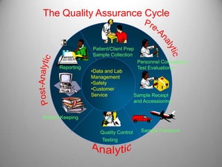
The Quality Assurance Cycle


                  Patient/Client Prep
                  Sample Collection
                                        Personnel Competency
      Reporting                         Test Evaluations
                  •Data and Lab
                  Management
                  •Safety
                  •Customer
                  Service              Sample Receipt
                                       and Accessioning


Record Keeping


                     Quality Control      Sample Transport
                      Testing
The Quality System
  Organizatio   Personnel      Equipment
      n


                   Process
  Purchasing       Control     Information
                (QC & EQA) &
  & Inventory     Specimen
                               Management
                Management


  Documents     Occurrence
                               Assessment
  & Records     Manageme
                    nt


    Process      Customer      Facilities &
  Improvemen      Service        Safety
       t
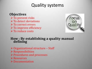
Quality systems
Objectives
 To prevent risks
 To detect deviations
 To correct errors
 To improve efficiency
 To reduce costs

How : By establishing a quality manual
 defining
 Organizational structure – Staff
 Responsibilities
 Procedures and processes
 Resources
 Documentation
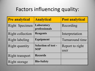
Factors influencing quality:
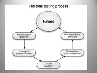
Pre analytical    Analytical            Post analytical
Right Specimen Laboratory               Recording
                  professionals
Right collection Reagents               Interpretation
Right labeling    Equipment             Turnaround time
Right quantity    Selection of test -   Report to right
                  SOP                   user
Right transport   Records

Right storage     Bio-Safety
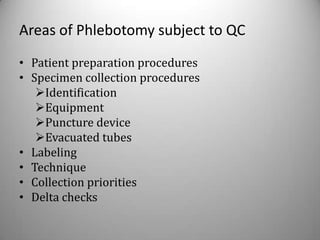
Areas of Phlebotomy subject to QC
• Patient preparation procedures
• Specimen collection procedures
   Identification
   Equipment
   Puncture device
   Evacuated tubes
• Labeling
• Technique
• Collection priorities
• Delta checks
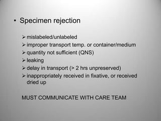
• Specimen rejection

   mislabeled/unlabeled
   improper transport temp. or container/medium
   quantity not sufficient (QNS)
   leaking
   delay in transport (> 2 hrs unpreserved)
   inappropriately received in fixative, or received
    dried up

  MUST COMMUNICATE WITH CARE TEAM
User manual
• Example of QA documentation
• Chart or type form
• Contains information on minimum amount
  of specimens required, special handling
  desired, reference values, TAT etc.
Procedure Manual
•   Standardization purposes
•   Must be updated annually
•   Written in a special format – NCCLS
•   States laboratory policy and procedures that
    apply to each test in the lab
Information found in a Procedure Manual

•   Purpose of the procedure
•   Specimen type and collection method
•   Equipment and supplies required
•   Detailed step-by-step procedure
•   Limitations and variables of the method
•   Corrective actions
•   Method validation
•   Normal values and references
COMPONENTS OF QUALITY
 ASSURANCE

Internal Quality control: IQC
   Nature: Concurrent
   performed by: lab staff
   Objective: Reliable results on a daily basis

External quality assessment: EQA
  Nature: Retrospective to evaluate IQC
  Performed by: Independent agency
  Objective: Ensure inter laboratory
  comparability
IQC
• Based on monitoring the test procedures.
• Measurements on specially prepared
  materials
• Repeated measurements on routine
  specimens.
• Daily statistical analysis of data obtained
• Eliminates differences in random and
  systematic errors between samples and
  standards
EQC
• Evaluation by an outside agency of the
  performance by numerous laboratories of
  specially supplied samples.

• National schemes are known as –
  NEQUAS(National External Quality
  Assessment Scheme).
STANDARDISATION
• Encompass both materials and methods
• Standard material/ reference preparation-
• Used to calibrate an analytical instrument.
• Reference method – Technique that is used in
  association with a reference preparation.
• Working method – Intended to use in
  routine practice.
National Standard and Regulatory Agencies

• World Health Organization(WHO)
• Joint Commission on Accreditation of Healthcare
  Organizations (JCAHO)
• College of American Pathologists (CAP)
• Clinical Laboratory Improvement Amendments of 1988
  (CLIA ’88)
• National Committee for Clinical Laboratory Standards
  (NCCLS)
• National Accrediting Agency for Clinical Laboratory
  Sciences (NAACLS)
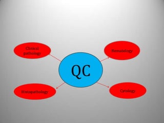
Clinical
                      Hematology
 pathology


                 QC
Histopathology            Cytology
QC IN HEAMATOLOGY
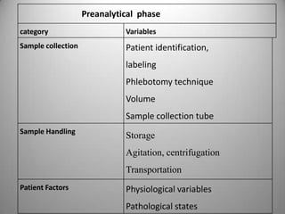
Preanalytical phase
category                      Variables
Sample collection             Patient identification,
                              labeling
                              Phlebotomy technique
                              Volume
                              Sample collection tube
Sample Handling
                              Storage
                              Agitation, centrifugation
                              Transportation
Patient Factors               Physiological variables
                              Pathological states
Types of control materials
• Assayed
   mean calculated by the manufacturer
   must verify in the laboratory
• Unassayed
   less expensive
   must perform data analysis
• “Homemade” or “In-house”
   pooled sera collected in the laboratory
    characterized
   preserved in small quantities for daily use
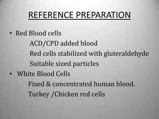
REFERENCE PREPARATION
• Red Blood cells
      ACD/CPD added blood
      Red cells stabilized with gluteraldehyde
      Suitable sized particles
• White Blood Cells
      Fixed & concentrated human blood.
      Turkey /Chicken red cells
• Hemoglobin
     standardized haemolysate
• Platelet
      platelets separated by centrifugation
     (200g for 10') ,fixed by gluteraldehyde.
TEST PROCEDURES
•   Measurement of prepared materials.
•   Repeated measurement/Replicate testing.
•   Duplicate testing.
•   Short hand checking
•   Delta check
•   Daily statistical analysis.
Analysis of Control Materials

• Need data set of at least 20 points, obtained
  over a 30 day period
• Calculate mean, standard deviation,
  coefficient of variation; determine target
  ranges
• Develop Levy-Jennings charts, plot results
Levy-Jennings Chart
 A graphical method for displaying control
 results and evaluating whether a procedure
 is in-control or out-of-control
     Control values are plotted versus time
Gaussian distribution curve
 Cusum chart
ANALYSIS OF EQA DATA


• Deviation index
• Target value & bias
• Youden XY plot - same analysis on two diff.
  control and plot on X & Y axis
QUALITY CONTROL IN
      HISTOPATOHLOGY
Quality assurance in
            HISTOPATHOLOGY
Concept of QC in histopathology lab is
   relatively young
It may be due to,
descriptive nature of report
 lack of objective numerical data
 Individual judgment and bias
 No uniformity of reporting pattern
QUALITY ASSURANCE

This make assessment and implementation of
        QC is more difficult in histopathology
    Even though we can implement QC in
               histopathology
it may be           Pre analytical
                    Analytical
                   Post analytical
A good quality histological section is the
  starting point of an accurate histopathology
  result
According to CAP,
  1. Preanalytical part - all process for
  generation of good section
  2.Analytical part - interpretation of slide
  and accurate diagnosis
  3.Post analytical part - proper dispatching
  of result, storage
Pre analytical
   All process up to submission of slide
majority errors occur in this stage
  Sample accession and identification errors
     Avoided by barcode technology
     Maintain a good referral form with all
     possible details and that should make
     available in sample collection area
Good fixation is very necessary for good
result
Fixation problems are,
 Should be fixed immediately
 Volume of fixative
 Conc. & type of fixative
 Adequate time of fixation
 Space in the container
 It should be cut open for proper internal
   after grossing
 Leakage of fixative
Decalcification problem
   Should remove any traces of calcium salt from
 the tissue
   Always maintain
           proper time
           good decalcifying fluid
           check end point of decalcification
Tissue processing and embedding

 Always maintain good quality reagent
 Periodical changing of processing fluid
 Maintain a appropriate treatment time
 If tissue processor used,
         Ensure complete working
 Use closed type processor
 Temp. of paraffin wax
 Use Tissue Teck system for embedding
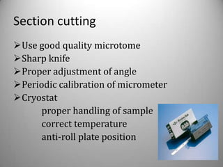
Section cutting
Use good quality microtome
Sharp knife
Proper adjustment of angle
Periodic calibration of micrometer
Cryostat
     proper handling of sample
     correct temperature
     anti-roll plate position
Staining
 Control is used parallel with each batch of
  staining
 Maintain good quality of regent
 Standard operating procedure
 Filter stain regularly
 Special stains are done with suitable controls
 If automatic stainer used, check their working
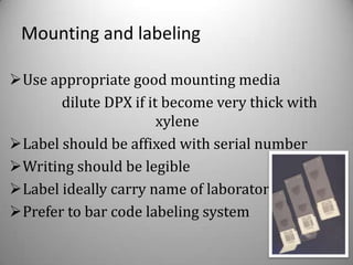
Mounting and labeling

Use appropriate good mounting media
       dilute DPX if it become very thick with
                       xylene
Label should be affixed with serial number
Writing should be legible
Label ideally carry name of laboratory
Prefer to bar code labeling system
Analytical errors
  Assessment of analytical errors are not an
              easy task
Maintained by,
 Intradepartmental consultation ; review selected
  cases by colleague
 Comparison with other reports (cytology, frozen)
 RBRC © by same person – for precision
            © diff. person – for accuracy
 Slide transferring and examination between two
  institution
Post analytical QC
Involves,
 Report generation without any transcription
  error
 Double checking of printed report
  Counter signed by consultant pathologist
 Report dispatch to right person
 Storage of reported material
 Disposal of specimen
 Monitor TAT
EQC in histopathology
*CAP and UK- NQAS – international programmer
In India,

Indian College Of Pathology with collaboration
with Association of Pathologist in North
America(AIPNA) run EQC as a part of NABL
accreditation

Inter-Laboratory Quality Assessment Programe
for Histopathology (ILQA-HP) under ILQA-
Bangalore
EQA programme involves

Scheme divided in to two categories
   asses pre analytical aspect
   asses analytical aspect
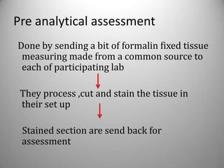
Pre analytical assessment
Done by sending a bit of formalin fixed tissue
 measuring made from a common source to
 each of participating lab

 They process ,cut and stain the tissue in
 their set up

 Stained section are send back for
 assessment
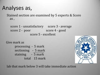
Analyses as,
 Stained section are examined by 5 experts & Score
   as ,

   score 1 - unsatisfactory score 3 - average
   score 2 - poor            score 4 - good
                 score 5 - excellent

 Give mark as
       processing - 5 mark
       sectioning - 5 mark
       staining    - 5 mark
              total 15 mark

 lab that mark below 3 will take immediate action
Analytical aspects,
Section obtained in one common source is
 stained with H&E and distribute to lab along
 with all details

pathologist examine and report was returned
 to nodal centre for assessment
QUALITY CONTROL IN
         CYTOLOGY
Cytopathology QC

Will Require:
  Observation of technical
    procedures
  Review of QI program and
    indicators
  On-site microscopic review
   standardization in reporting
    format
Cytopathology QC
• General Elements of QI

  Technical and procedural (QC)
  Professional/diagnostic activities of
   cytotechnologists and pathologists (QI)
  Quality of the diagnostic report (QC/QI)
Specimen Collection and Receipt

 • Specimens properly identified
 • Instructions available for preferred specimen
   collection/preparation
 • Requisition: complete data requested
   including date, source, physician, LMP,
   pertinent clinical information, etc.
 • Criteria for specimen rejection and
   notification of unacceptable specimens
Cytology Stains

 Stains labeled and dated
 Cytology stains: new requirement for annual
  inventory to ensure proper storage and quality
  (many stains do not expire)
 Papanicolaou stains filtered or replaced
  regularly
 Papanicolaou stain prepared with good reagent
 Regular monitoring of stain characteristics
Instrumentation

• Evidence of active review of results of instrument
  maintenance and function (II)
• Automated instruments (Phase II)
   – Documentation of adherence to manufacturer-
     recommended protocol for implementation
   – Documentation of appropriate technical and
     interpretive training
   – Written procedure to verify diagnostic &
     adequacy performance of screening instrument
Instrumentation

• Automated screening systems
   If tolerance limits exceeded, is there
    documentation of corrective action?
   Documented procedure for handling workload
    during instrument failure
   Documented procedure for handling slides not
    successfully processed
   “Negative” slides subject to 5 year retro review
On-Site Microscopic Review
 • Not meant to be comprehensive rescreen or
   competency review, but a means of facilitating
   evaluation of overall procedures
 • 10 -15 case review recommended including:
        > Unsatisfactory
        > Reactive
        > Positive for all abnormality reported

            “Must have written criteria”
On-Site Microscopic Review
  • Evaluate adequacy, technical quality, labels
  • Determine if significant cells identified
  • Compare with written interpretive report
  • Check requisition for complete information
  • Discrepancies analogous to PAP program
  • Team leader should discuss significant
    discrepancies with laboratory director
  • Record specimen category & discrepancies
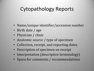
Cytopathology Reports

•   Name/unique identifier/accession number
•   Birth date / age
•   Physician / clinic
•   Anatomic source / type of specimen
•   Collection, receipt, and reporting dates
•   Description of specimen on receipt
•   Interpretation (descriptive terminology)
•   Space for comments / recommendations
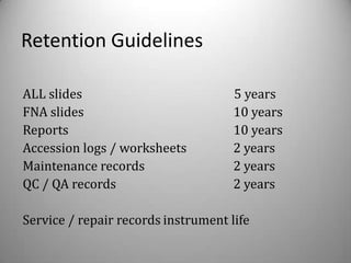
Retention Guidelines

ALL slides                           5 years
FNA slides                           10 years
Reports                              10 years
Accession logs / worksheets          2 years
Maintenance records                  2 years
QC / QA records                      2 years

Service / repair records instrument life
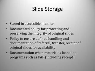
Slide Storage

• Stored in accessible manner
• Documented policy for protecting and
  preserving the integrity of original slides
• Policy to ensure defined handling and
  documentation of referral, transfer, receipt of
  original slides for availability
• Documentation when material is loaned to
  programs such as PAP (including receipt)
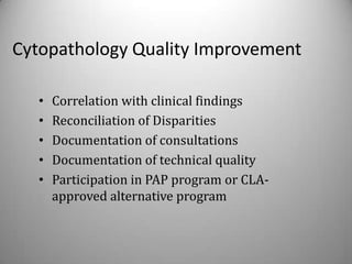
Cytopathology Quality Improvement

   •   Correlation with clinical findings
   •   Reconciliation of Disparities
   •   Documentation of consultations
   •   Documentation of technical quality
   •   Participation in PAP program or CLA-
       approved alternative program
Pap Rescreening
• Laboratory must rescreen a minimum of 10% of each
  cytotechnologist’s initially judged as negative
   Performed by individual qualified to be supervisor
    (3 years experience)
   Must include both high risk and randomly selected
     cases
   Cases not reported until rescreening complete
   Pathologists exempt (but rescreening advised)
EQA IN CYTOLOGY

1. Exchange of slide programme
    A set of gynecological and
   nongynecological
smears are distributed to diff. lab

   rechecking or reassessment of slide
2.Laboratory accreditation and certification
     Indian Academy of cytologist
Accreditation of IAC is based on,

   Workload
   Staff pattern
   Report generation and methodology
   Screening of specimen
   Diagnostic verification
   Follow-up
   Filing of report and slide
   Continuing education
QUALITY CONTROL IN
     CLINICAL PATHOLOGY
QA in CLIP

        > Still in dormant state
        > Require great attention
        > International and national agency
        should come forward
QC in urine analysis
Specimen Collection

     First morning voiding (most concentrated)
     Record collection time
     Type of specimen (e.g. “clean catch”)
     Analyzed within 2 hours of collection
     Free of debris or vaginal secretions
analysis

Performed person should be well trained
Maintain good quality reagents for chemical
 examination with suitable control
Microscopy recheck if needed
Maintain an uniformity in reporting
Microscopic UA

Correlate with cloudiness and other findings
Quality control
  – Consistent volume
  – Centrifugation
  – Well mixed fresh specimen
  – Microscopy (wet mount, use low light)
Accreditation Bodies


• College of Pathologists (CAP) , USA
• Joint Commission on Accreditation of Hospitals
  (JCAHO), USA
• Clinical Pathology Accreditation (CPA), UK Ltd
• National Association of Testing Authorities (NATA),
  Australia
• Department of Standards (DSM) Malaysia –
THANK YOU