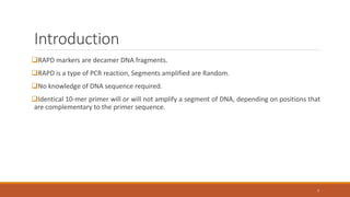
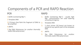
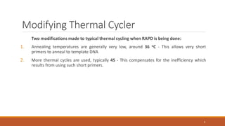
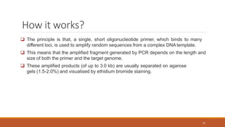
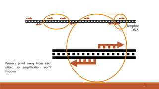
RAPD
(Random Amplification of Polymorphic DNA)
or
(Randomly Amplified Polymorphic DNA)
DR DHAVAL CHAUDHARY
M.V.SC. SCHOLAR
DEPT., OF ANIMAL GENETICS AND BREEDING
Contents
Markers
Classification of Marker
History
Introduction
How it Works?
Interpretation
Procedure
Advantage & Disadvantage
Limitation
Application
Summary
Conclusion
2
Markers
Any genetic trait that can be identified with confidence & relative case and can be
followed in a mapping population is called as marker.
Genetic/ Molecular marker is a specific location on a chromosome that is defined by a
naked eye polymorphism as differences in electrophoretic mobility of specific proteins or
as differences in specific DNA sequence.
3
Classification
4
5
History
Shortly after Kary Mullis invented the Polymerase Chain Reaction (PCR) it was realized that
short primers would bind to several locations in a genome and thus could produce multiple
fragments.
Williams et al. (1990) developed Random Amplified Polymorphic DNA (RAPD) a technique
using very short 10 base primers to generate random fragments from template DNAs.
RAPD fragments can be separated and used as genetic markers or a kind of DNA fingerprint.
6
Introduction
RAPD markers are decamer DNA fragments.
RAPD is a type of PCR reaction, Segments amplified are Random.
No knowledge of DNA sequence required.
Identical 10-mer primer will or will not amplify a segment of DNA, depending on positions that
are complementary to the primer sequence.
7
Components of a PCR and RAPD Reaction
PCR
1.Buffer (containing Mg++)
2.Template DNA
3.2 Primers that flank the fragment of DNA to
be amplified
4.dNTPs
5.Taq DNA Polymerase (or another thermally
stable DNA polymerase)
RAPD
1. Buffer (containing Mg++) - usually high
Mg++ concentrations are used lowering
annealing stringency
2. Template DNA
3. 1 short primer (10 bases) not known to
anneal to any specific part of the template
DNA
4. dNTPs
5. Taq DNA Polymerase (or another
thermally stable DNA polymerase)
8
Modifying Thermal Cycler
Two modifications made to typical thermal cycling when RAPD is being done:
1. Annealing temperatures are generally very low, around 36 oC - This allows very short
primers to anneal to template DNA
2. More thermal cycles are used, typically 45 - This compensates for the inefficiency which
results from using such short primers.
9
How it works?
 The principle is that, a single, short oligonucleotide primer, which binds to many
different loci, is used to amplify random sequences from a complex DNA template.
 This means that the amplified fragment generated by PCR depends on the length and
size of both the primer and the target genome.
 These amplified products (of up to 3.0 kb) are usually separated on agarose
gels (1.5-2.0%) and visualised by ethidium bromide staining.
10
 Nucleotide variation between different sets of template DNAs will result in the presence or
absence of bands because of changes in the priming sites.
 Recently, sequence characterised amplified regions (SCARs) analysis of RAPD polymorphisms
showed that one cause of RAPD polymorphisms is chromosomal rearrangements such as
insertions/deletions.
 In order for PCR to occur:
1)the primers must anneal in a particular orientation
(such that they point towards each other) and,
2)they must anneal within a reasonable distance of one another.
11
12
Template
DNA
 Primer binds to many locations on the template DNA
 Only when primer binding sites are close and oriented in
opposite direction so the primers point toward each other
will amplification take place
13
Template
DNA
Primers point away from each
other, so amplification won’t
happen
14
Template
DNA
Primers point in the same
direction, so amplification
won’t happen
15
Template
DNA
Primers too far apart, so
amplification won’t happen
> 2,000 bases
16
Template
DNA
Primers are just the
right distance apart, so
fragment is amplified
100 - 1,500 bases
17
RAPD Protocol
Reaction mixture
Buffer (10x) 2.5µl
MgCl2 (50mM) 0.75µl
dNTPs (10mM) 0.5µl
Primers(8pmol/µl) 1.0µl
DNA template(30ng/µl) 3.0µl
Distilled water (sterile) 17.25µl
Cycle Conditions
Pre denaturation 940C 4min
Denaturation 940C 1min
Annealing 370C 1min
Extension 720C 2min
Repeat steps 2-4 for 42-45 times
Final extension 720C 5min
Electrophoresis
Electrophoresis conditions
required are :
Matrix:1.5-2% Agarose
Buffer:1x TBE
Voltage:80volts for 30 min,
Visualize under UV light
`
Interpreting RAPD Banding Pattern
18
 Each gel is analysed by scoring the present (1) or absent (0) polymorphic bands in individual
lanes. The scoring can be done based on the banding profiles which is clear and transparent
(Fig.A) otherwise the scoring is very difficult (Fig.B).
A B
Criteria for selecting scoring bands:
1) reproducibility—need to repeat experiments.
2) thickness
3) size and,
4) expected segregation observed in a mapping population.
DNA polymorphism among individuals can be due to:
1) mismatches at the primer site.
2) appearance of a new primer site and,
3) length of the amplified region between primer sites.
The NTSYS-pc software ver. 2.02 is used to estimate genetic similarities with the Jaccard’s coefficient.
19
20
MM 2 3 4 5 6 7 8 9 10
Separated RAPD Fragments 4mM MgCl2
1.2 U Taq
5 pM OPA-16
4mM MgCl2
0.6 U Taq
10 pM OPA-16
2mM MgCl2
1.2 U Taq
10 pM OPA-16
Normal
concentrations are
shown in yellow
text. M = A size
standard
Lowering
Magnesium ion
concentration
results in loss of
the largest
fragment visible
in lanes 2-7
RAPD reactions
were run in groups
of 3 using the same
template and primer,
but varying
Magnesium,
polymerase and
primer
concentrations
Which variable
has the greatest
impact on
fragment
patterns?
Procedure
1. The DNA of a selected species is isolated.
2. An excess of selected decaoligonucleotide added.
3. This mixture is kept in a PCR equipment and is subjected to repeated cycles of DNA
denaturation-renaturation-DNA replication.
4. During this process, the decaoligonucleotide will pair with the homologous sequence present
at different locations in the DNA.
5. DNA replication extend the decaoligonucleotide and copy the sequence continuous with the
sequence with which the selected oligonucleotide has paired.
21
6. The repeated cycles of denaturation - renaturation-DNA replication will amplify this sequence
of DNA.
7. Amplification will takes place only of those regions of the genome that has the sequence
complementary to the decaoligonucleotide at their both ends.
8. After several cycles of amplification the DNA is subjected to gel electrophoresis.
9. The amplified DNA will form a distinct band. it is detected by ethidium bromide staining and
visible fluorescence's under U.V. light
22
23
Isolation of DNA
Keep the tubes in PCR Themocycler
DNA strands separated
Decaoligonucleotide enzyme, primer, Taq DNA
polymerase
Annealing of primer (36°C,2 min)
Primer annealed to template DNA strands
Denature the DNA (94o C, 1 min)
DNA synthesis (72°C, 1.5 min)
24
Complementry strand synethesis
35-45 cycles
Amplified products separated by Gel
electrophoresis
Bands detected by Ethidium bromide statining
25
Advantages
1) No need of prior knowledge of sequence of DNA
2) The level of detectable polymorphisms very high
3) In comparison to RFLP, It is less expensive, faster, requires smaller amount of DNA, requires
less skill
4) Does not involve use of radioisotopes
26
Disadvantages
27
1) Lack of reproducibility
2) RAPD markers are dominant markers.
3)PCR results are very sensitive to amplification conditions and consequently variable between
laboratories and even between assays
Limitation
Nearly all RAPD markers are dominant, i.e. it is not possible to distinguish whether a DNA
segment is amplified from a locus that is heterozygous (1 copy) or homozygous (2 copies).
PCR is an enzymatic reaction, therefore the quality and concentration of template DNA,
concentrations of PCR components, and the PCR cycling conditions may greatly influence the
outcome.
Mismatches between the primer and the template may result in the total absence of PCR
product as well as in a merely decreased amount of the product. Thus, the RAPD results can be
difficult to interpret.
28
Application
Evaluate genetic relationships analysis among native breeds as well between native and exotic
breeds of livestock.
Species differentiation - RAPD markers could differentiate chicken, quail, turkey and other
avian species/strains.
Association with production performance in livestock breeds.
genetic diversity/polymorphism
germplasm characterization
genetic structure of populations & genome mapping
population and evolutionary genetics
animal-plant-microbe interactions
29
Summary
RAPD is a lab technique used to amplify unknown(random) DNA segments.
Then mixture is taken to PCR equipment and the process of DNA denaturation and the
annealing of primer occcurs, then primer extension takes place for 35 to 45 cycles.
DNA hybridizaion occurs at some segment of DNA amplification occurs at a particular site.
DNA is subjected to gel electrophoresis, the amplified DNA will form distinct band detected by
ethidium bromide staining and visible fluorescence’s under U.V. light.
30
RAPD markers exhibit reasonable speed, cost and efficiency compared with other methods.
RAPD can be done in a moderate laboratory. Therefore, despite its reproducibility problem, it
will probably be important until better techniques are developed in terms of cost, time and
labour.
31
Conclusion
THANK YOU!!
32

RAPD

  • 1.
    RAPD (Random Amplification ofPolymorphic DNA) or (Randomly Amplified Polymorphic DNA) DR DHAVAL CHAUDHARY M.V.SC. SCHOLAR DEPT., OF ANIMAL GENETICS AND BREEDING
  • 2.
    Contents Markers Classification of Marker History Introduction Howit Works? Interpretation Procedure Advantage & Disadvantage Limitation Application Summary Conclusion 2
  • 3.
    Markers Any genetic traitthat can be identified with confidence & relative case and can be followed in a mapping population is called as marker. Genetic/ Molecular marker is a specific location on a chromosome that is defined by a naked eye polymorphism as differences in electrophoretic mobility of specific proteins or as differences in specific DNA sequence. 3
  • 4.
  • 5.
  • 6.
    History Shortly after KaryMullis invented the Polymerase Chain Reaction (PCR) it was realized that short primers would bind to several locations in a genome and thus could produce multiple fragments. Williams et al. (1990) developed Random Amplified Polymorphic DNA (RAPD) a technique using very short 10 base primers to generate random fragments from template DNAs. RAPD fragments can be separated and used as genetic markers or a kind of DNA fingerprint. 6
  • 7.
    Introduction RAPD markers aredecamer DNA fragments. RAPD is a type of PCR reaction, Segments amplified are Random. No knowledge of DNA sequence required. Identical 10-mer primer will or will not amplify a segment of DNA, depending on positions that are complementary to the primer sequence. 7
  • 8.
    Components of aPCR and RAPD Reaction PCR 1.Buffer (containing Mg++) 2.Template DNA 3.2 Primers that flank the fragment of DNA to be amplified 4.dNTPs 5.Taq DNA Polymerase (or another thermally stable DNA polymerase) RAPD 1. Buffer (containing Mg++) - usually high Mg++ concentrations are used lowering annealing stringency 2. Template DNA 3. 1 short primer (10 bases) not known to anneal to any specific part of the template DNA 4. dNTPs 5. Taq DNA Polymerase (or another thermally stable DNA polymerase) 8
  • 9.
    Modifying Thermal Cycler Twomodifications made to typical thermal cycling when RAPD is being done: 1. Annealing temperatures are generally very low, around 36 oC - This allows very short primers to anneal to template DNA 2. More thermal cycles are used, typically 45 - This compensates for the inefficiency which results from using such short primers. 9
  • 10.
    How it works? The principle is that, a single, short oligonucleotide primer, which binds to many different loci, is used to amplify random sequences from a complex DNA template.  This means that the amplified fragment generated by PCR depends on the length and size of both the primer and the target genome.  These amplified products (of up to 3.0 kb) are usually separated on agarose gels (1.5-2.0%) and visualised by ethidium bromide staining. 10
  • 11.
     Nucleotide variationbetween different sets of template DNAs will result in the presence or absence of bands because of changes in the priming sites.  Recently, sequence characterised amplified regions (SCARs) analysis of RAPD polymorphisms showed that one cause of RAPD polymorphisms is chromosomal rearrangements such as insertions/deletions.  In order for PCR to occur: 1)the primers must anneal in a particular orientation (such that they point towards each other) and, 2)they must anneal within a reasonable distance of one another. 11
  • 12.
    12 Template DNA  Primer bindsto many locations on the template DNA  Only when primer binding sites are close and oriented in opposite direction so the primers point toward each other will amplification take place
  • 13.
    13 Template DNA Primers point awayfrom each other, so amplification won’t happen
  • 14.
    14 Template DNA Primers point inthe same direction, so amplification won’t happen
  • 15.
    15 Template DNA Primers too farapart, so amplification won’t happen > 2,000 bases
  • 16.
    16 Template DNA Primers are justthe right distance apart, so fragment is amplified 100 - 1,500 bases
  • 17.
    17 RAPD Protocol Reaction mixture Buffer(10x) 2.5µl MgCl2 (50mM) 0.75µl dNTPs (10mM) 0.5µl Primers(8pmol/µl) 1.0µl DNA template(30ng/µl) 3.0µl Distilled water (sterile) 17.25µl Cycle Conditions Pre denaturation 940C 4min Denaturation 940C 1min Annealing 370C 1min Extension 720C 2min Repeat steps 2-4 for 42-45 times Final extension 720C 5min Electrophoresis Electrophoresis conditions required are : Matrix:1.5-2% Agarose Buffer:1x TBE Voltage:80volts for 30 min, Visualize under UV light `
  • 18.
    Interpreting RAPD BandingPattern 18  Each gel is analysed by scoring the present (1) or absent (0) polymorphic bands in individual lanes. The scoring can be done based on the banding profiles which is clear and transparent (Fig.A) otherwise the scoring is very difficult (Fig.B). A B
  • 19.
    Criteria for selectingscoring bands: 1) reproducibility—need to repeat experiments. 2) thickness 3) size and, 4) expected segregation observed in a mapping population. DNA polymorphism among individuals can be due to: 1) mismatches at the primer site. 2) appearance of a new primer site and, 3) length of the amplified region between primer sites. The NTSYS-pc software ver. 2.02 is used to estimate genetic similarities with the Jaccard’s coefficient. 19
  • 20.
    20 MM 2 34 5 6 7 8 9 10 Separated RAPD Fragments 4mM MgCl2 1.2 U Taq 5 pM OPA-16 4mM MgCl2 0.6 U Taq 10 pM OPA-16 2mM MgCl2 1.2 U Taq 10 pM OPA-16 Normal concentrations are shown in yellow text. M = A size standard Lowering Magnesium ion concentration results in loss of the largest fragment visible in lanes 2-7 RAPD reactions were run in groups of 3 using the same template and primer, but varying Magnesium, polymerase and primer concentrations Which variable has the greatest impact on fragment patterns?
  • 21.
    Procedure 1. The DNAof a selected species is isolated. 2. An excess of selected decaoligonucleotide added. 3. This mixture is kept in a PCR equipment and is subjected to repeated cycles of DNA denaturation-renaturation-DNA replication. 4. During this process, the decaoligonucleotide will pair with the homologous sequence present at different locations in the DNA. 5. DNA replication extend the decaoligonucleotide and copy the sequence continuous with the sequence with which the selected oligonucleotide has paired. 21
  • 22.
    6. The repeatedcycles of denaturation - renaturation-DNA replication will amplify this sequence of DNA. 7. Amplification will takes place only of those regions of the genome that has the sequence complementary to the decaoligonucleotide at their both ends. 8. After several cycles of amplification the DNA is subjected to gel electrophoresis. 9. The amplified DNA will form a distinct band. it is detected by ethidium bromide staining and visible fluorescence's under U.V. light 22
  • 23.
    23 Isolation of DNA Keepthe tubes in PCR Themocycler DNA strands separated Decaoligonucleotide enzyme, primer, Taq DNA polymerase Annealing of primer (36°C,2 min) Primer annealed to template DNA strands Denature the DNA (94o C, 1 min) DNA synthesis (72°C, 1.5 min)
  • 24.
    24 Complementry strand synethesis 35-45cycles Amplified products separated by Gel electrophoresis Bands detected by Ethidium bromide statining
  • 25.
  • 26.
    Advantages 1) No needof prior knowledge of sequence of DNA 2) The level of detectable polymorphisms very high 3) In comparison to RFLP, It is less expensive, faster, requires smaller amount of DNA, requires less skill 4) Does not involve use of radioisotopes 26
  • 27.
    Disadvantages 27 1) Lack ofreproducibility 2) RAPD markers are dominant markers. 3)PCR results are very sensitive to amplification conditions and consequently variable between laboratories and even between assays
  • 28.
    Limitation Nearly all RAPDmarkers are dominant, i.e. it is not possible to distinguish whether a DNA segment is amplified from a locus that is heterozygous (1 copy) or homozygous (2 copies). PCR is an enzymatic reaction, therefore the quality and concentration of template DNA, concentrations of PCR components, and the PCR cycling conditions may greatly influence the outcome. Mismatches between the primer and the template may result in the total absence of PCR product as well as in a merely decreased amount of the product. Thus, the RAPD results can be difficult to interpret. 28
  • 29.
    Application Evaluate genetic relationshipsanalysis among native breeds as well between native and exotic breeds of livestock. Species differentiation - RAPD markers could differentiate chicken, quail, turkey and other avian species/strains. Association with production performance in livestock breeds. genetic diversity/polymorphism germplasm characterization genetic structure of populations & genome mapping population and evolutionary genetics animal-plant-microbe interactions 29
  • 30.
    Summary RAPD is alab technique used to amplify unknown(random) DNA segments. Then mixture is taken to PCR equipment and the process of DNA denaturation and the annealing of primer occcurs, then primer extension takes place for 35 to 45 cycles. DNA hybridizaion occurs at some segment of DNA amplification occurs at a particular site. DNA is subjected to gel electrophoresis, the amplified DNA will form distinct band detected by ethidium bromide staining and visible fluorescence’s under U.V. light. 30
  • 31.
    RAPD markers exhibitreasonable speed, cost and efficiency compared with other methods. RAPD can be done in a moderate laboratory. Therefore, despite its reproducibility problem, it will probably be important until better techniques are developed in terms of cost, time and labour. 31 Conclusion
  • 32.