Downloaded 90 times


![References[1-5]
1. Deupree, J.D.; Tutorial in Receptor Binding Techniques;
http://www.unmc.edu/Pharmacology/receptortutorial/; Lurz, Matthew J.; 1998
2. Edwards, R., S. Blincko, and I. Howes, Principles of immunodiagnostic tests
and their development; with specific use of radioisotopes as tracers, in
Immunodiagnostics : a practical approach, The Practical approach series ;
206, R. Edwards, Editor. 1999, Oxford University Press: Oxford ; New York.
p. cm.
3. Fuentes-Arderiu, X.; Glossary of ISO Metrological and Realted Terms and
Definitions Relevant to Clinical Laboratory Sciences;
http://www.westgard.com/isoglossary.htm; Westgard, James O; 1999
4. Kricka, L.J., Principles of Immunochemical Techniques, in Tietz textbook of
clinical chemistry and molecular diagnostics, C.A. Burtis, et al., Editors.
2006, Elsevier Saunders: Philadelphia. p. XXXVI, 2412 s.
5. Linnet, K. and J.C. Boyd, Selection and Analytical Evaluation of Methods -
With Statistical Techniques, in Tietz textbook of clinical chemistry and
molecular diagnostics, C.A. Burtis, et al., Editors. 2006, Elsevier Saunders:
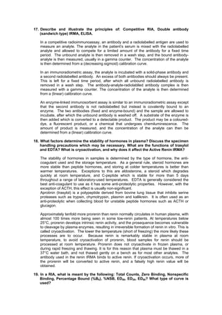
Philadelphia. p. XXXVI, 2412 s.](https://image.slidesharecdn.com/radioimmunoassay-130519164318-phpapp01/85/Radioimmunoassay-13-320.jpg)

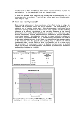
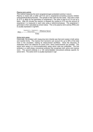
This document provides competency assessment guidelines for staff working in a Radioimmunoassay (RIA) Laboratory. It outlines 11 key areas of training including radiation safety, quality control procedures, assay protocols, equipment operation and maintenance. It also describes the 5 main assays performed - aldosterone, 17-hydroxyprogesterone, human growth hormone, active renin, and 11-deoxycortisol. Staff are expected to demonstrate proficiency in all laboratory standard operating procedures (SOPs) and understanding of RIA principles, techniques, calculations and quality assurance.