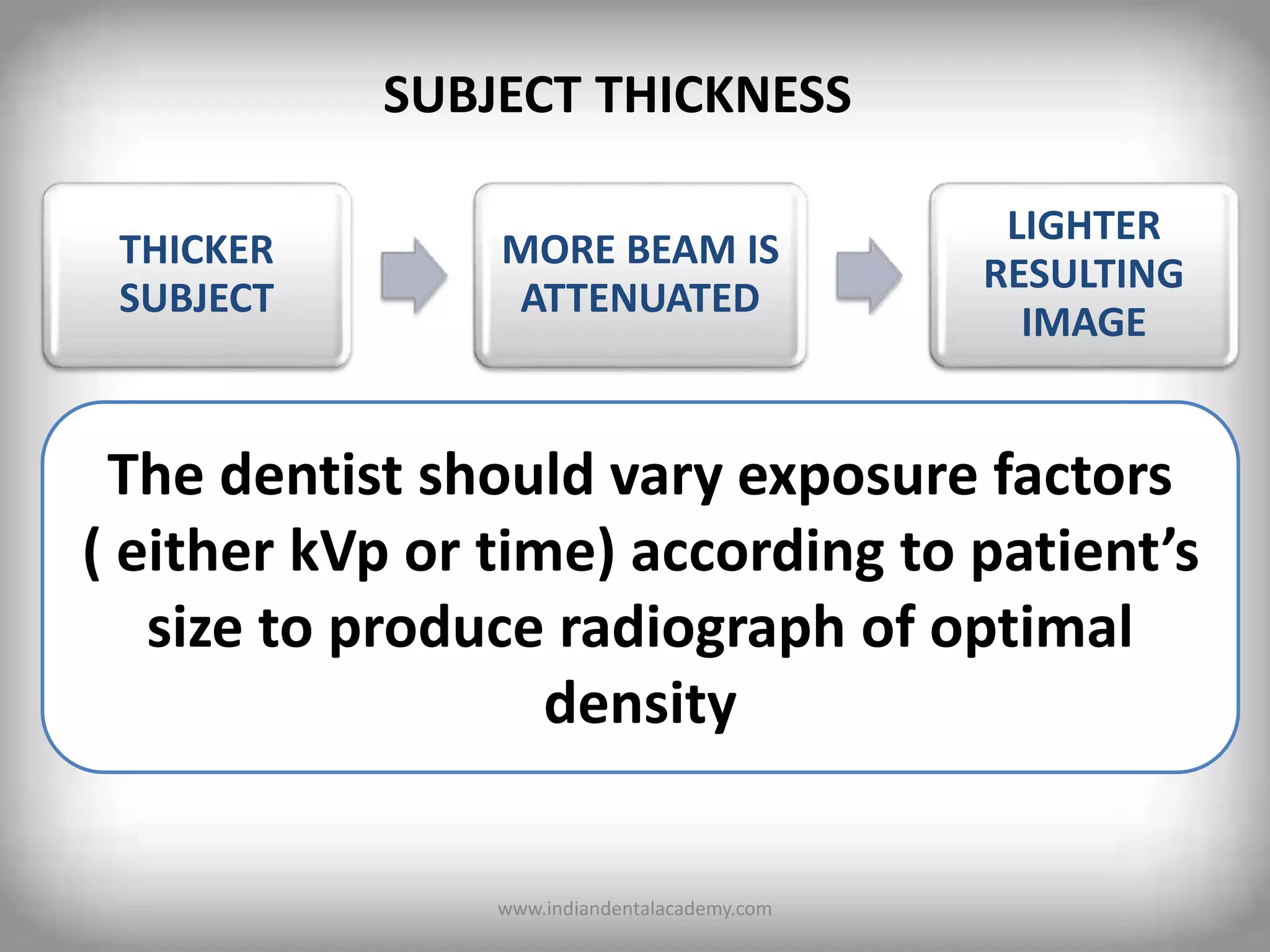
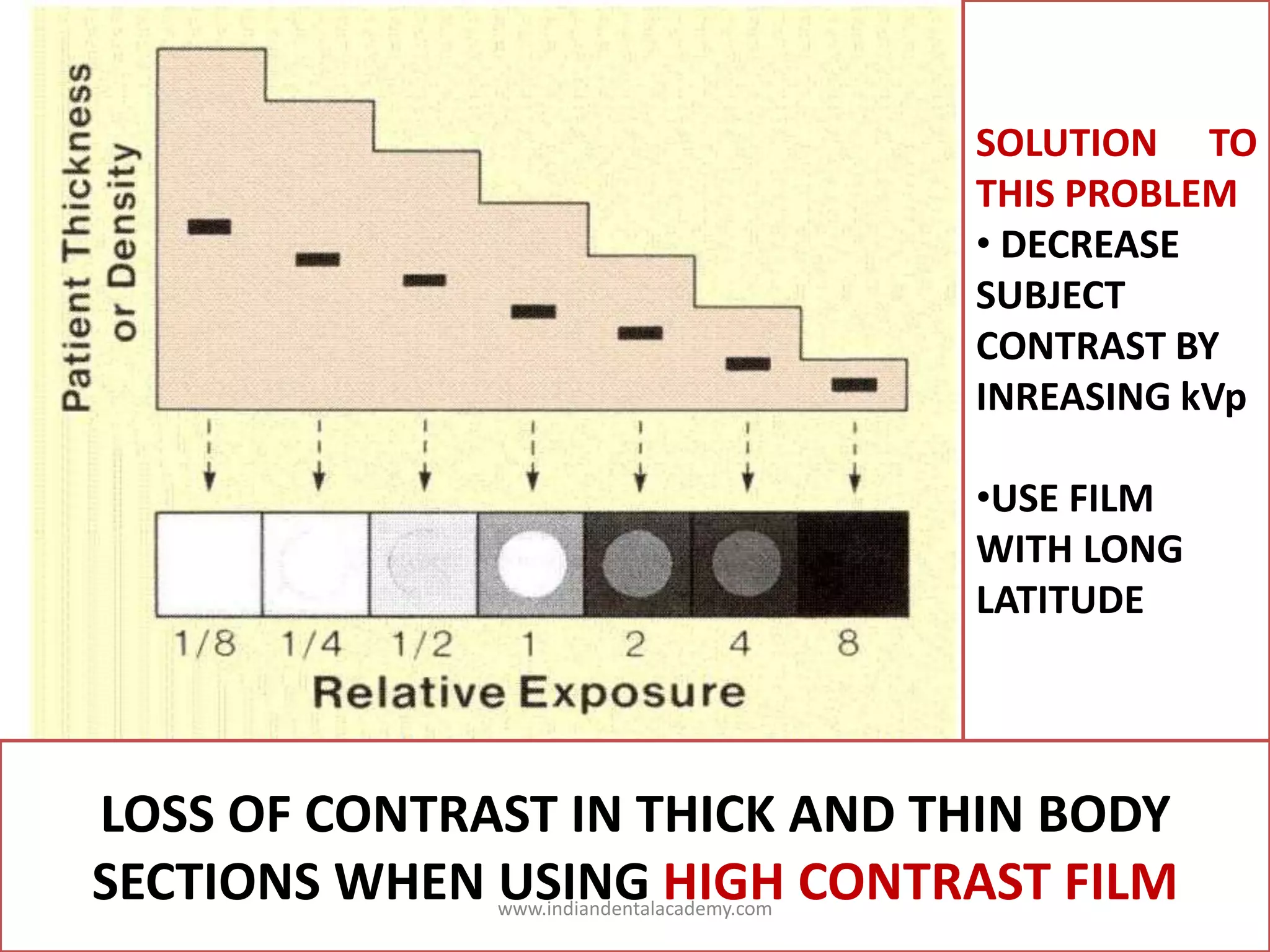
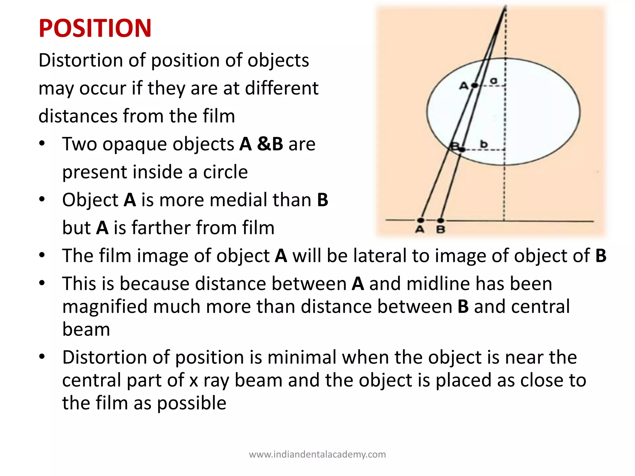
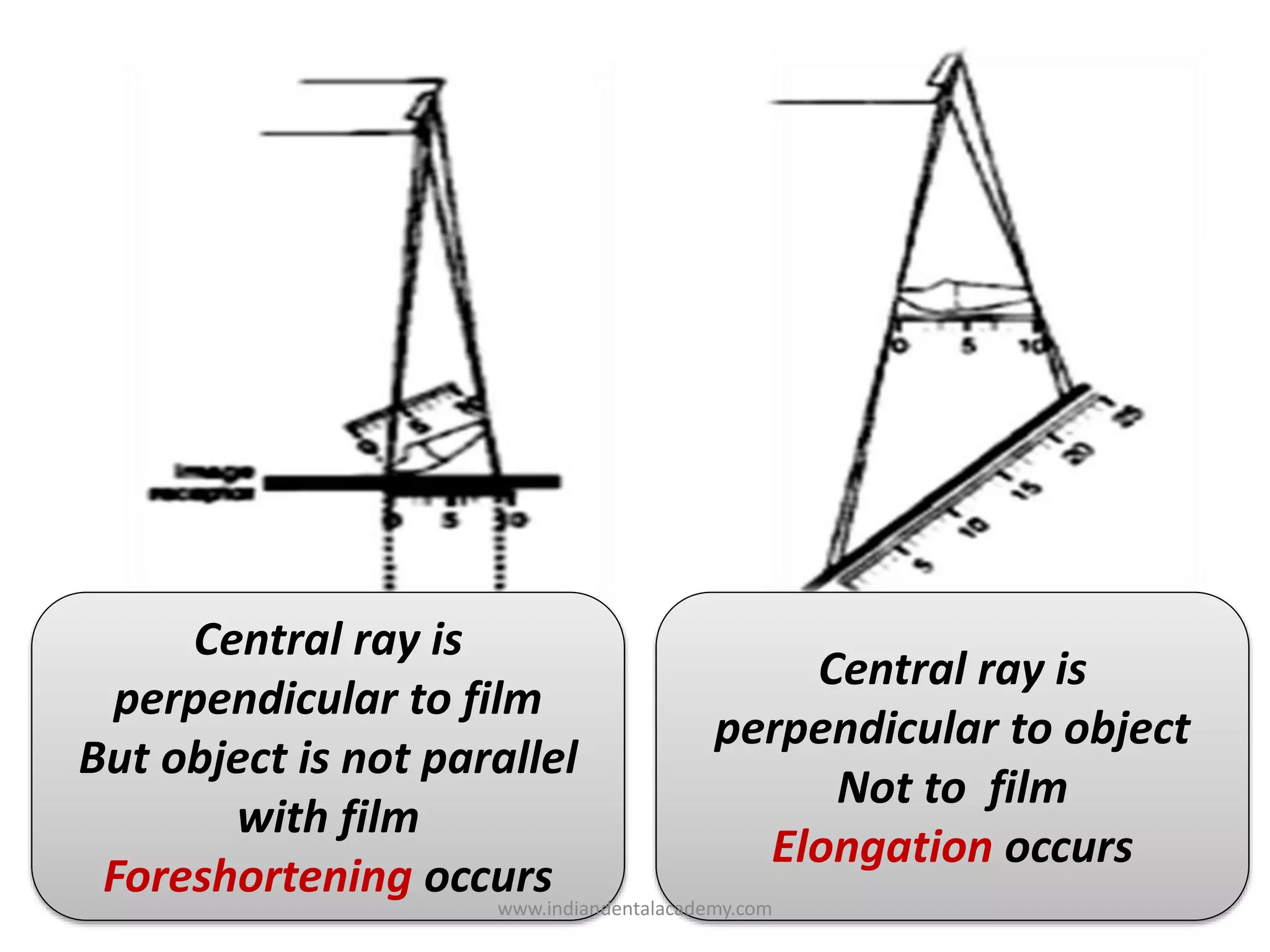
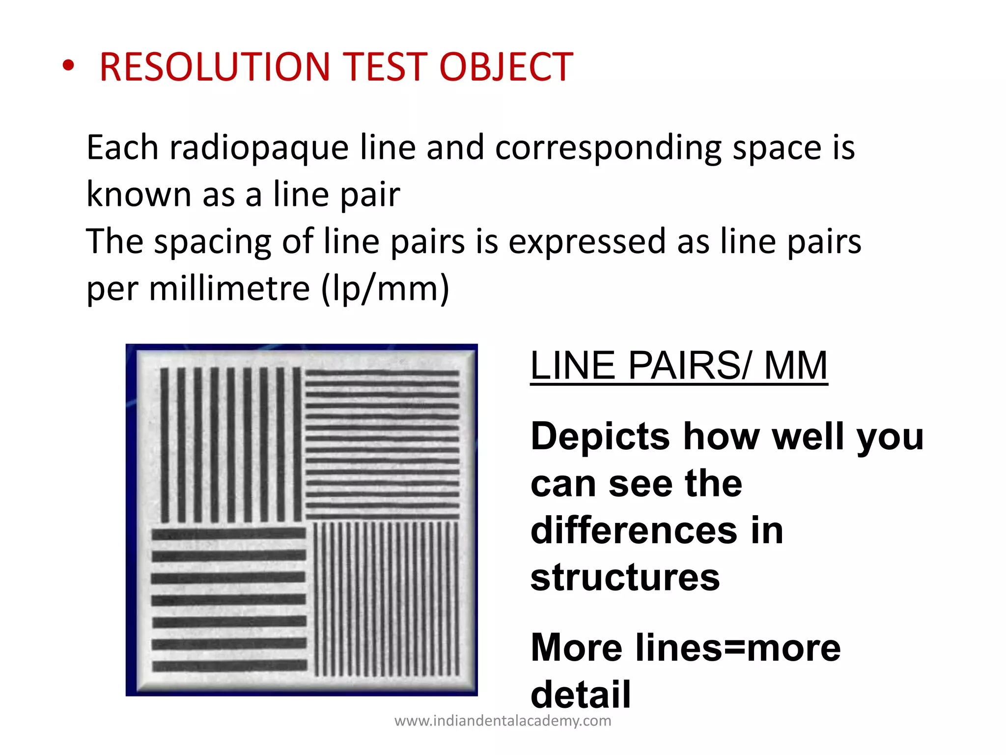
SlideShare a Scribd company logo
RADIOGRAPHIC IMAGE
INDIAN DENTAL
ACADEMY
Leader in continuing
Dental Educationwww.indiandentalacademy.com
INTRODUCTION
Image quality of a radiographic image is described as
“The subjective judgment by clinician of the
overall appearance of a radiograph”
 The ability of a film to record each point in object as
point on film
 But in radiology this point to point reproduction is
never perfect
www.indiandentalacademy.com
5 IMPORTANT COMPONENTS IN IMAGING SYSTEM
Imaging is the Process of Converting Tissue Characteristics into a Visual Imagewww.indiandentalacademy.com
www.indiandentalacademy.com
RADIATION CHARACTERISTICS WHICH
INFLUENCE THE QUALITY OF IMAGE
QUALITY
• VOLTAGE
• kVp
• EXPOSURE TIME
QUANTITY
• AMPERAGE
MILLIAMPERAGE
• MILLIAMPERE -
SECONDS
INTENSITY
• QUALITY
• QUANTITY
• DISTANCE
• INVERSE SQUARE
LAW
• HALF VALUE LAYER
X-RAY BEAM
www.indiandentalacademy.com
X-RAY BEAM QUALITY
• Mean Energy & Penetration Ability of X ray beam
• Wave length determines its energy & penetrating power
• Quality, wavelength, energy are controlled by
TUBE VOLTAGE
www.indiandentalacademy.com
Inside the dental x ray tubehead
• TUBE VOLTAGE :Potential difference between
Cathode & Anode that gives K.E. to electrons &
makes electrons move from cathode to anode
www.indiandentalacademy.com
KILOVOLTAGE PEAK kVp
• Maximum or peak voltage of alternating current
• In an AC polarity of line current alternate so
polarity of x ray tube alternates at
same frequency
Filament -ve
Target +ve
Flow of electrons
Filament +ve
Target -ve
No electron flow
Inverse voltage
reverse biaswww.indiandentalacademy.com
INCREASE IN
VOLTAGE
INCREASES
SPEED OF
ELECTRONS
ELECTRONS
STRIKE TARGET
WITH GREATER
FORCE AND
ENERGY
PENETRATING
BEAM WITH A
SHORTER
WAVELENGTH
Voltage determines speed of electrons
www.indiandentalacademy.com
1kV=1000V
DENTAL RADIOGRAPHY
65 TO 100kV
85 TO 100 kV SHORTER
WAVELENGTH MORE ENERGY
OVER PENETRATION
65 TO 75kV LONGER
WAVELENGTH LESS ENERGY
DONOT ALLOW AEQUATE
PENETRATION
www.indiandentalacademy.com
EXPOSURE TIME
• Time during which x rays are produced
• It is measured in impulses as x rays are created in a
series of bursts or pulses rather than in a continuous
stream
• 1 impulse occurs in 1/60 of second ( 60impulses in 1s)
www.indiandentalacademy.com
X-RAY BEAM QUANTITY
 It is the number of x rays produced in
dental x ray unit
 AMPERAGE determines amount of electrons passing
through the cathode filament
 AMPERE - unit used to measure no of electrons or
current flowing through the cathode filament
www.indiandentalacademy.com
 1milli ampere = 1/1000 of ampere
 A change in mA does not change K.E of electrons
 So only quantity is changed
 In dental radiography use of 7 to 15mA is required
www.indiandentalacademy.com
MILLIAMPERE- SECONDS
• Both mA and s have a direct influence on no of
electrons produced by cathode filament
• 1 mAs = 6.25 x 10¹⁵ electrons per second
• When milliamperage is increased the exposure time
must be decreased and vice versa, if the density of
exposed radiograph is to remain same
Milliamperes X Exposure Time = mAs
www.indiandentalacademy.com
X RAY BEAM INTENSITY
• INTENSITY is defined as product of quality (energy
of each photon) quantity (no of x ray photons) per
unit area per unit time of exposure
• Higher kvp , mA, exposure time,
Intensity of x ray beam
INTENSITY = NO OF PHOTONS X ENERGY OF EACH PHOTON
AREA X EXPOSURE RATE
www.indiandentalacademy.com
DISTANCE
• The distance between
source of radiation and film
has marked effect on intensity
• The distances to be considered are
SOD , OID, SID
• As x rays travel from source their
intensity decreases
• The relation between distance and intensity
is explained by inverse square law.
s
I
o
www.indiandentalacademy.com
INVERSE SQUARE LAW
• The intensity of radiation is inversely proportional to
square of distance from source of radiation
x
xD = 1 ft
Area = x2
Intensity = 144
units/sq.inch
D = 2 ft
Area = 4x2
Intensity = 36
units/sq.inch
D = 3 ft
Area = 3x2
Intensity = 16
units/sq.inch
2x
2x
I1/I 2= D2
2 / D 1
2
www.indiandentalacademy.com
HALF VALUE LAYER
• To reduce intensity of x ray beam , aluminum filters are
placed in path of beam
• These are used to remove low energy, less penetrating,
long wavelength x rays
• Al filters increase mean penetrating
capacity of x rays while reducing
their intensity
• The thickness of material which
reduce intensity by half is
called half value layer.
www.indiandentalacademy.com
Ag HALIDE
CRYSTALS
X-rays LIGHT
Grains of
metallic
silver
This gives film its
dark appearance
The Overall degree of darkening of an exposed
film is called its RADIOGRAPHIC DENSITY
www.indiandentalacademy.com
X-ray tube
Object
Plot of incident x-ray
beam intensity
Plot of transmitted x-ray
beam intensity
www.indiandentalacademy.com
Consider the light transmitted through an area of a
film
Incident light
intensity (Ii )
Transmitted light
intensity (It)
Transmittance (Transmission ratio)= It / Ii
Opacity = 1 / Transmittance = Ii / It
Density = Log10 Opacity = Log10 Ii / It
www.indiandentalacademy.com
Range of densities on a film
Ii It Transmit
tance
Opacity Density
100
0.01
0.1
1.0
10.0
100
0.0001
0.001
0.01
0.1
1
10000
1000
100
10
1
4
3
2
1
0
4 3 2 1 0
www.indiandentalacademy.com
1
2
3
4
1 2 3 4
Log relative exposure
Density
Shoulder
Toe
CHARACTERISTIC CURVE /HURTER AND DRIFFIELD CURVE
GF
DMax
Straight line
portion
SHOULDER AND TOE
VERY HIGH AND VERY LOW VARIATION
IN EXPOSURE MAKE VERY SMALL
CHANGES IN DENSITY
GREATEST DIAGNOSTIC
VALUE WHEN STRUCTURES
OF INTEREST ARE IMAGED
ON RELATIVELY STRAIGHT
PORTION OF GRAPH
BETWEEN 0.6 AND 3.0
GROSS FOG OR BASE PLUS FOG MINIMAL DENSITY SHOWN WHEN
AN UNEXPOSED FILM IS PROCESSED {0.07+0.05= 0.12(0.2to 0.3)}www.indiandentalacademy.com
when other exposure factors
remain constant
EFFECT ON DENSITY
INCREASED KVp INCREASED DENSITY
DECREASED KVp DECREASED DENSITY
INCREASED mA INCREASED DENSITY
DECREASED mA DECREASED DENSITY
INCREASED TIME INCREASED DENSITY
DECREASED TIME DECREASED DENSITY
EXPOSURE FACTORS
www.indiandentalacademy.com
SUBJECT THICKNESS
THICKER
SUBJECT
MORE BEAM IS
ATTENUATED
LIGHTER
RESULTING
IMAGE
The dentist should vary exposure factors
( either kVp or time) according to patient’s
size to produce radiograph of optimal
density
www.indiandentalacademy.com
SUBJECT DENSITY
 Greater the density of a structure in subject, greater
is attenuation of beam
 Dense objects ( which are strong absorbers) cause the
image to be light ( radiopaque)
 Low density objects are weak absorbers, allow most
photons to pass through and cast a dark area on film
( radiolucent)
Order Of Decreasing Density
Enamel >Dentin > Cementum>bone>muscle>fat>airwww.indiandentalacademy.com
INCREASED DENSITY
If developer solution is too hot
Prolonged developing
DECREASED DENSITY
If developer solution is cold
Developing time is too short
Underactive/exhausted
solutions
www.indiandentalacademy.com
CONTRAST
• It is defined as difference in densities between light
and dark regions on a radiograph
AN IMAGE WHICH
SHOWS BOTH DARK
AND LIGHT AREAS
HAS
HIGH CONTRAST/
SHORT GRAY SCALE
AN IMAGE WHICH
SHOWS ONLY LIGHT
GRAY AND DARK
GRAY ZONES HAS
LOW CONTRAST/
LONG GRAY SCALE
www.indiandentalacademy.com
A.SUBJECT CONTRAST
 Range of characteristics of subject that influences
radiographic contrast
 It is the difference in x ray intensity transmitted
through one part of subject as compared to other
 This difference in intensity of beam caused by object
is subject contrast
MUSCLE BONE
www.indiandentalacademy.com
1.THICKNESS DIFFERENCE I
 No .of x rays through thin part
are greater than thick part IS
IS - Intensity of x ray beam IL
transmitted through thin part
IL - Intensity of x ray beam through
thick part
SUBJECT CONTRAST = IS/IL
IS > IL
www.indiandentalacademy.com
2. DENSITY DIFFERENCE
 The difference in density between body tissues is
one of the most important factor causing subject
contrast
 The greater density of tissue (mass/ volume)
greater is the ability to attenuation
 For e.g: consider same thickness of ice floating in
water
 Water attenuates more of beam than ice
www.indiandentalacademy.com
3. ATOMIC NUMBER
 Attenuation of x ray beam by photoelectric effect
makes most important contribution to subject contrast
 Photoelectric effect is increased in substances with high
atomic number
 Bone (ca # 20, P # 15) attenuates more x rays than
muscle(H #1, N#7)
 Muscle and fat due to their little difference in atomic
number show little difference in attenuation
www.indiandentalacademy.com
Contrast media:
 The use of contrast materials with high atomic
number( 53 # I , 56 # Ba) gives high subject contrast
 Photoelectric absorptions of x rays in barium and
iodine are proportionally much greater than in bone
and tissue
www.indiandentalacademy.com
 Low kVp produce high subject contrast, provided
the kVp is high enough to penetrate part being
examined adequately
 If the kVp is too low almost all xrays are attenuated
in pt and never reach film
Increased
kVp
More
penetration
in to tissues
Interact
by
compton
effect
Increased
scattered
radiation
Image
noise and
decreased
contrast
4.RADIATION QUALITY
www.indiandentalacademy.com
 In general , low kVp gives high subject contrast (short scale
contrast)
 High kVp gives low subject contrast(long scale contrast)
SHORT
SCALE
EVERY THING
IS BLACK/
WHITE
ON FILM
WITH FEWER
SHADES OF
GRAY IN
BETWEEN
LONG
SCALE
LONG SCALE
OF SHADES OF
GRAY IN
BETWEEN
LIGHTEST AND
DARKEST
PORTIONS OF
IMAGE
www.indiandentalacademy.com
GRIDS
• The grid which is placed between subject and film ,
preferentially removes the scattered radiation and
spares primary photons
• This reduces nonimaging
exposures
• Increase subject contrast
www.indiandentalacademy.com
B. FILM CONTRAST
• It describes capacity of radiographic film to display
differences in subject contast
• A high contrast film reveals areas of small
difference in subject contrast more clearly than
does a low contrast film
Greater the slope
of curve greater
is the contrast
So film A Shows
greater change in
optical density
than B
Film B with
shallow slope
has wide
latitude
So B records
greater range of
exposures
within useful
density rangewww.indiandentalacademy.com
SOLUTION TO
THIS PROBLEM
• DECREASE
SUBJECT
CONTRAST BY
INREASING kVp
•USE FILM
WITH LONG
LATITUDE
LOSS OF CONTRAST IN THICK AND THIN BODY
SECTIONS WHEN USING HIGH CONTRAST FILMwww.indiandentalacademy.com
C. SCATTER AND FOG
• Reduces contrast
• Scattered radiation that reaches film/ film-screen
combination produces unwanted density
SCATTERED RADIATION OCCURS DUE TO SO TO MINIMISE
INCREASED THICKNESS OF PART
INCREASED FIELD SIZE COLLIMATE BEAM TO SIZE OF FILM
USE GRIDS IN EXTRAORAL
RADIOGRAPHY
INCREASED ENERGY OF BEAM USE LOW KVPwww.indiandentalacademy.com
FOG
 Defined as those silver halide crystals that are
developed even though they are not exposed by light/
x rays
 If accidental exposure occurs – exposure fog
Although origin of both is different , both cause
unwanted film density which lowers contrast
Fog is increased by
 High speed film, improper film storage
 Excessive time/temp of developer
 Contaminated/exhausted developerwww.indiandentalacademy.com
FILM
CHARACTERISTICS
RADIOGRAPHIC
SPEED
RADIOGRAPHIC
NOISE
RADIOGRAPHIC
MOTTLE
ARTIFACT
LATITUDE
www.indiandentalacademy.com
SPEED
• refers to “amount of radiation required to produce
an image of standard density”
• how efficiently the light-sensitive agents in a film
emulsion react to energy (e.g., x-rays or light)
• Expressed as reciprocal of exposure ((in roentgens)
required to produce an optical density of 1 above
gross fog
• Controlled largely by size of silver halide grains and
their silver contentwww.indiandentalacademy.com
• Fast film requires relatively less amount of radiation
than a slow film
• The film speeds most commonly used in dental
imaging are D(ultra speed),E(Ekta speed,F(insight )
• D-speed is the slowest film of the three, and F-
speed is the fastest.
• Insight film is preferred as it requires only half of
exposure ultraspeed and offers comparable contrast
and resolution
www.indiandentalacademy.com
FLAT CRYSTALS INTERCEPT MORE LIGHT BUT THE TOTAL AMOUNT OF
SILVER DOES NOT INCREASE.
INCREASE IN SPEED WITH LESS NOTICEABLE GRAIN.
AS A RESULT, EKTASPEED PLUS FILM DELIVERS EXCELLENT IMAGE
QUALITY WHILE AT THE SAME TIME REDUCED RADIATION
Conventional
silver-halide
crystals are
pebble shaped
KODAK
T-GRAIN
Emulsion
crystals
Are flat
INSIGHT
ULTRA
SPEED
www.indiandentalacademy.com
• Film speed can be increased slightly by processing
at high temp, this is achieved at expense of
increased film fog and graininess
• Adding radiosensitive dyes to film emulsion
• Processing in depleted solutions lowers speed
www.indiandentalacademy.com
LATITUDE
• Film latitude is a measure of range of exposures
that can be recorded as distinguishable densities on
a film
• A film optimized to record wide latitude can record
a subject with a wide range of subject contrast
Curve with Long straight
line portion and shallow
slope has wide latitude .
wide variations in amount
of radiation exiting the
subject can be recorded
www.indiandentalacademy.com
• Wide latitude films are used when both osseous
structures of skull and soft tissues of facial region must
be recorded
• Wide latitude films have lower contrast
• To some extent operator can modify latitude of an
image
• High kVp produce image with wide latitude and low
contrast
• Wide latitude film is recommended for imaging
structures with a wide range of subject densities
www.indiandentalacademy.com
RADIOGRAPHIC NOISE
• It is the appearance of uneven density of a
uniformly exposed radiographic film
• Seen on small areas of film as localised variations in
density
• Primarily due to Radiographic Mottle And Artifacts
• Artifacts are defects caused by errors in film
handling, such as fingerprints, bends, errors in film
processing such as splashing of developer or fixer
on a film or marks or scratches from wrong handlingwww.indiandentalacademy.com
• Mottle may be seen as film graininess which is
caused by visibility of silver grains in film emulsion
especially when magnification is used to examine
an image
• Two important causes are screen structure mottle,
quantum mottle
• Screen structure mottle is graininess caused by
screen phosphors
• Most evident when fast screens with large crystals
are used
www.indiandentalacademy.com
• X-ray interactions are random events.
• Therefore, “uniform” x-ray exposure does not produce
uniform film darkening.
• Regional variability in the film darkening is called
“QUANTUM MOTTLE.”
• Caused by fluctuation in number of photons per unit of
beam cross-sectional area absorbed by intensifying
screen
• Quantum mottle decreases as x-ray exposure is
increased.
www.indiandentalacademy.com
• Consequently, “fast” screen-film systems will have
more quantum mottle than “slower” systems
• Longer exposure required by slower systems tend to
average out beam pattern and there by reduce
quantum mottle
Quantum mottle
masks low contrast
objects
Quantum mottle is
worse for small
objects
www.indiandentalacademy.com
MAGNIFICATION
• When an object is placed in path of
beam it will cast a shadow on film that
will show some degree of
enlargement.
• If the object is round and flat like a coin, its
magnified image will be round but larger than coin
• amount of magnification (M)
SIZE OF IMAGE
SIZE OF OBJECT
M =
www.indiandentalacademy.com
To achieve minimal
magnification
1.Keep focus film
distance as large as
possible(SID)
2.Keep film as close to
object as possible(OID)
www.indiandentalacademy.com
INCREASING SOURCE OBJECT DISTANCE
RESULTS IN IMAGE WITH INCREASED
SHARPNESS AND LESS MAGNIFICATION
THIS IS DUE TO DECREASED
DIVERGENCE
www.indiandentalacademy.com
Target
16” Target
8”
Target
16”
INCREASE SOURCE-OBJECT DISTANCE, DECREASE
MAGNIFICATION
DECREASE OBJECT-FILM DISTANCE, DECREASE MAGNIFICATION
www.indiandentalacademy.com
DECREASING OBJECT AND IMAGE RECEPTOR
DISTANCE INCREASES SHARPNESS AND LESS
MAGNIFICATION
www.indiandentalacademy.com
DISTORTION
• It is misrepresentation of true size and shape of object being
radiographed
• Distortion results from unequal
magnification of different parts
of same object
Amount of distortion depends on
• Thickness
• Position
• shape
www.indiandentalacademy.com
THICKNESS
• Thick objects are more distorted as different parts
are at different distances from x ray film,
each part will be magnified by
different amount
• This will cause Shape of the
image of most thick objects
to be distorted
• Only The part of the thick object
that is parallel to the film will be
undistorted
www.indiandentalacademy.com
POSITION
Distortion of position of objects
may occur if they are at different
distances from the film
• Two opaque objects A &B are
present inside a circle
• Object A is more medial than B
but A is farther from film
• The film image of object A will be lateral to image of object of B
• This is because distance between A and midline has been
magnified much more than distance between B and central
beam
• Distortion of position is minimal when the object is near the
central part of x ray beam and the object is placed as close to
the film as possible
www.indiandentalacademy.com
• Size distortion
• Occurs due to improper orientation of film and
object. To minimize
1. Position the film parallel to long axis of object
2. Orient central ray perpendicular to object and film
BUT AS OID
INCREASES THERE IS
SOME AMOUNT OF
MAGNIFICATION ,
LOSS OF DEFINITION
BY INCREASING
UNSHARPNESS
PARALLELING
TECHNIQUE BEST
INCORPORATES
IMAGING
PRINCIPLES
www.indiandentalacademy.com
Central ray should be perpendicular to long
axes of tooth and film .if it is not, then
distortion occurs
www.indiandentalacademy.com
Central ray is
perpendicular to film
But object is not parallel
with film
Foreshortening occurs
Central ray is
perpendicular to object
Not to film
Elongation occurs
www.indiandentalacademy.com
• SHAPE DISTORTION
• The sphere that lies in center of beam
will exhibit round (undistorted)
magnified image
• The image of laterally placed sphere
will be an ellipse because x ray beam
sees diameter of sphere that is
not parallel to film
www.indiandentalacademy.com
SHARPNESS
• SHARPNESS is a ability of x ray film or
film screen system to define an edge precisely
• E.g DEJ, a thin trabecular pattern
• An unsharp edge can be easily seen if contrast is
high but a sharp edge may be poorly visible if
contrast low.
www.indiandentalacademy.com
SHARPNESS
A B
Distance (mm)
Density
D1
D2
0 5 10 15 20
The boundary between two areas A & B appears very sharp
www.indiandentalacademy.com
Unsharpness
A
Distance (mm)
Density
D1
D2
0 .2 .4 .6 .8
The boundary between two areas A & B appears unsharp
B
The steeper the slope the more sharp the image appears. The shallower the slope
the more blurred the image
www.indiandentalacademy.com
MOTION UNSHARPNESS
• Caused by motion of examined
object/film during exposure
• Depends only on velocity(v) of motion(mm/sec)and
exposure time(t) (seconds)
• Restricting movement of pt and
• reducing exposure time helps to
resolve problem
www.indiandentalacademy.com
ABSORPTION UNSHARPNESS
• Due to gradual change in x-ray absorption across an
object’s edge or boundary
• Thickness of absorber presented to beam changes
• Effect is poorly defined margin of solid objects
X-Ray
Tube
X-Ray
Tube
X-Ray
Tube
www.indiandentalacademy.com
PENUMBRA
Pene = almost ; umbra = shadow
• It is the region of partial illumination
that surrounds umbra / complete image
• UMBRA is the region of complete image
www.indiandentalacademy.com
• Focal spot is not a point. it has finite dimensions ,
usually ranging from 0.3 -2mmsquare
• The focal spot acts as if it were composed of many
point sources of x rays with each point source
forming its own image of an object
• The edges of each of these images will not be in
exactly the same spot on film
www.indiandentalacademy.com
• Fig . Shows edge of an object formed by
two x ray sources A &B which
represents opposite ends of
focal spot
• If A & B are only sources of x-rays
there would be two
overlapping images recorded
on the film
• But A and B are only opposite
ends of focal spot, a continuous line
of x ray sources
• Object edge is imaged many times
between edge B and edge A
www.indiandentalacademy.com
• The image formed by line(focal spot)
of x rays is not sharp at the edge and
varies in density
• This zone of unsharpness is called
GEOMETRIC UNSHARPNESS,
PENUMBRA, EDGE GRADIENT
• Which represent area at which margins caused by
many point sources of x rays in the focal spot overlap
www.indiandentalacademy.com
• The resulting zone of unsharpness cause loss of image
clarity by reducing sharpness and resolution
• 3 methods to minimize loss of clarity and improve
quality are
1.use as small as an effective focal spot
2.increase the distance between focal spot and object by
using long open ended cylinder(increase SID)
3.minimize the distance between object and film
( decreasedOID)
www.indiandentalacademy.com
Larger the focal spot greater the loss
of image sharpness
www.indiandentalacademy.com
SMALL ANGLE has
greater wearing
effect on target
INCREASED
IMAGE
SHARPNESS AND
RESOLUTION
but small
effective focal
spot
LOSS OF IMAGE
CLARITY
Decreased heat
generation,
prolonged tube
life
LARGE ANGLE
distributes electron
beam over large
surface
www.indiandentalacademy.com
RESOLUTION
• The resolution of a system is its ability to
demonstrate closely spaced structures in the
subject as separate entities in the image
E.g. The trabeculae pattern of bone
• The resolution of an image refers to its ability
to show small structures separately
• The smaller the structures visible the higher
the resolution of the image/system
www.indiandentalacademy.com
MEASUREMENT OF RESOLUTION
• The resolution is expressed in terms of the smallest
spacing /highest spatial frequency of high and low
dense structures visible in the image
• This is done subjectively by radiographing a test
object consist of a grid of closely spaced lines,
alternately radiopaque and radiolucent.
www.indiandentalacademy.com
• RESOLUTION TEST OBJECT
Each radiopaque line and corresponding space is
known as a line pair
The spacing of line pairs is expressed as line pairs
per millimetre (lp/mm)
LINE PAIRS/ MM
Depicts how well you
can see the
differences in
structures
More lines=more
detailwww.indiandentalacademy.com
COMPARISON OF RESOLUTION
• The resolution of different imaging systems may be
compared by reference to the Modulation Transfer
Function (MTF) of each system
Modulation = frequency of change (of structure ,
etc.)
• MTF is a method of assessing the success with which
modulations of structure (detail) in an object are
transferred into modulations of density or luminance
in the image
www.indiandentalacademy.com
Modulation in the image
• MTF = ------------------------------------
Modulation in the object
–MTF = 1 means that image reproduces
exactly the variations in the object.
–It is a characteristic of imaging systems
that, as the detail in the object becomes
finer, the ability of the system to record
that detail becomes progressively reduced.
www.indiandentalacademy.com
TO PRODUCE RADIOGRAPHS OF OPTIMAL
DIAGNOSTIC QUALITY
• Radiographs should record complete areas of
interest on the image
• In case of IOPA, full length of roots and 2mm of
periapical bone must be visible
• If evidence of pathology is present area of entire
lesion plus some surrounding normal bone should
show on one radiograph
www.indiandentalacademy.com
• Radiographs should have least possible amount of
distortion
• Most distortion is caused by improper angulation of
x ray beam rather than by curvature of structures
being examined or inappropriate positioning of
receptor
• Close attention to proper positioning of receptor
and x ray tube results in diagnostically useful
images
www.indiandentalacademy.com
Simple quality control procedures assure that
high-quality patient radiograph
PATIENT POSITIONING
• Film should be close to object to reduce OID
• Center the beam to area of interest
• Get area of interest parallel to beam /film
• Restrain motion of patient and communicate
with patient
• Use short exposure times
www.indiandentalacademy.com
IMAGE RECEPTOR
• Use correct screen-film
combination
• Low contrast films have wider latitude and
large margin of error in producing acceptable
radiograph
• Use highest speed system that will provide
adequate contrast and density
www.indiandentalacademy.com
PROCESSING
• Although exposure factors are crucial in
influencing density, contrast faulty processing
can adversely affect diagnostic quality of
properly exposed radiographs
• Even the best exposure conditions can be
compromised with poor processing technique
www.indiandentalacademy.com
Method 1: Sensitometry and Densitometry
• A sensitometer is used to expose a film, followed by
standard processing of the film
• processed film will have a defined pattern of optical
densities that are measured with a densitometer
• The densitometer measurements are compared to the
densities of films exposed in ideal conditions
• A change in densitometer values indicates a problem with
either the development time, temp or the developer
solutions
• Advantages
• Accuracy and Speed
• Disadvantage
• Expense of additional equipment
www.indiandentalacademy.com
Method 2: Stepwedge
• An aluminum stepwedge , with each step of the aluminum at
1 millimeter thick and 3 to 4 mm wide, with at least six steps
• A film is exposed through the stepwedge ,with the same
machine settings, film placement and stepwedge placement
used for each daily exposure
• The processed film is compared visually with a reference film
• A change in density of one or more steps indicates a problem
with either the development time, temperature or the
developer solutions
Advantage
• Cost effectiveness
Disadvantage
• Less precision
www.indiandentalacademy.com
• Method 3: Reference Film
• A film exposed and processed under ideal conditions is
attached to the corner of a view box as a reference film
• Subsequent films are compared with the reference film
• Advantage
• Cost effectiveness
• Disadvantage
• Least sensitivity
www.indiandentalacademy.com
INTENSIFYING SCREENS AND EXTRAORAL
CASETTES
• Visual inspection of cassette integrity
• Examination of intensifying screen for scratches
• Development of an unexposed film that has been in
the cassette exposed to normal lighting for one
hour or more
www.indiandentalacademy.com
DARK ROOM INTEGRITY
• While in a darkroom with the safelight on, place a
metal object (such as a coin) on unwrapped film for
a period that is equivalent to the time required for
a typical darkroom procedure
• Develop the film
• Detection of the object indicates a problem with
the safelight or light leaks in the darkroom
www.indiandentalacademy.com
X RAY MACHINE
• Should be inspected by qualified expert
• The following parameters should be checked
• X ray out put
• Collimation
• Beam energy, timer, mA
• Tube head stability
• Focal spot size
www.indiandentalacademy.com
CONCLUSION
• Quality of image is influenced at every stage
• We must identify the mistakes and try to improve them.
• But we must realize that in improving one aspect we may
weaken another.
• There is a complex interrelationship between many of the
factors concerned, including all of the image characteristics
discussed .
• Only when we have studied all the ramifications of image
production we will be able to understand fully how quality
may be optimized.
www.indiandentalacademy.com
REFERENCES
1. Stuart C White , Michael J Pharoah ,
Textbook Of Oral Radiology Principles And
Interpretation
2. Christensens Physics Of Diagnostic Radiology
www.indiandentalacademy.com
Quality leads! ! It is the difference between
being average or being number one
www.indiandentalacademy.com

More Related Content

What's hot

The control of scattered radiation
The control of scattered radiationThe control of scattered radiation
The control of scattered radiation
Nitish Rajput
 
Radiographic artifacts
Radiographic artifactsRadiographic artifacts
Radiographic artifactsRad Tech
 
Magnification(macro and micro radiography), distortion
Magnification(macro and micro radiography), distortionMagnification(macro and micro radiography), distortion
Magnification(macro and micro radiography), distortion
parthajyotidas11
 
Distortion
DistortionDistortion
Distortionmr_koky
 
Flim screen physics
Flim screen physicsFlim screen physics
Flim screen physics
Muhammad Wasim Awan
 
Radiographic factor
Radiographic factorRadiographic factor
Radiographic factor
Dr,saket Jain
 
Image quality, digital technology and radiation protection
Image quality, digital technology and radiation protectionImage quality, digital technology and radiation protection
Image quality, digital technology and radiation protectionRad Tech
 
Quality of radiograph by dr ashok
Quality of radiograph by dr  ashokQuality of radiograph by dr  ashok
Quality of radiograph by dr ashok
Ashok Sharma
 
Radiographic image4
Radiographic image4Radiographic image4
Radiographic image4mr_koky
 
CR and DR ppt
CR and DR pptCR and DR ppt
CR and DR ppt
shonimaindiaultrasound
 
Grids
GridsGrids
Grids
aslam bs
 
Radiographic artifacts
Radiographic artifactsRadiographic artifacts
Radiographic artifacts
Ajith Y
 
Radiographic film
Radiographic filmRadiographic film
Radiographic filmRad Tech
 
CT artifact
CT artifact CT artifact
CT artifact
Ganesan Yogananthem
 
Radiographic Intensifying Screen
Radiographic Intensifying ScreenRadiographic Intensifying Screen
Radiographic Intensifying Screen
Tarique Ajij
 
Interactions of X-ray & matter & Attenuation - Dr. Sayak Datta
Interactions of X-ray & matter & Attenuation - Dr. Sayak DattaInteractions of X-ray & matter & Attenuation - Dr. Sayak Datta
Interactions of X-ray & matter & Attenuation - Dr. Sayak Datta
SayakDatta
 
Radiographic Grid.pptx
Radiographic Grid.pptxRadiographic Grid.pptx
Radiographic Grid.pptx
Dr. Dheeraj Kumar
 
factor affecting quality of radiograph
factor affecting quality of radiographfactor affecting quality of radiograph
factor affecting quality of radiograph
Islamfaiq9
 
Radiographic exposure and image quality
Radiographic exposure and image qualityRadiographic exposure and image quality
Radiographic exposure and image qualityRad Tech
 
Geometric Properties Distortion
Geometric Properties DistortionGeometric Properties Distortion
Geometric Properties Distortion
lambertrad2014
 

What's hot (20)

The control of scattered radiation
The control of scattered radiationThe control of scattered radiation
The control of scattered radiation
 
Radiographic artifacts
Radiographic artifactsRadiographic artifacts
Radiographic artifacts
 
Magnification(macro and micro radiography), distortion
Magnification(macro and micro radiography), distortionMagnification(macro and micro radiography), distortion
Magnification(macro and micro radiography), distortion
 
Distortion
DistortionDistortion
Distortion
 
Flim screen physics
Flim screen physicsFlim screen physics
Flim screen physics
 
Radiographic factor
Radiographic factorRadiographic factor
Radiographic factor
 
Image quality, digital technology and radiation protection
Image quality, digital technology and radiation protectionImage quality, digital technology and radiation protection
Image quality, digital technology and radiation protection
 
Quality of radiograph by dr ashok
Quality of radiograph by dr  ashokQuality of radiograph by dr  ashok
Quality of radiograph by dr ashok
 
Radiographic image4
Radiographic image4Radiographic image4
Radiographic image4
 
CR and DR ppt
CR and DR pptCR and DR ppt
CR and DR ppt
 
Grids
GridsGrids
Grids
 
Radiographic artifacts
Radiographic artifactsRadiographic artifacts
Radiographic artifacts
 
Radiographic film
Radiographic filmRadiographic film
Radiographic film
 
CT artifact
CT artifact CT artifact
CT artifact
 
Radiographic Intensifying Screen
Radiographic Intensifying ScreenRadiographic Intensifying Screen
Radiographic Intensifying Screen
 
Interactions of X-ray & matter & Attenuation - Dr. Sayak Datta
Interactions of X-ray & matter & Attenuation - Dr. Sayak DattaInteractions of X-ray & matter & Attenuation - Dr. Sayak Datta
Interactions of X-ray & matter & Attenuation - Dr. Sayak Datta
 
Radiographic Grid.pptx
Radiographic Grid.pptxRadiographic Grid.pptx
Radiographic Grid.pptx
 
factor affecting quality of radiograph
factor affecting quality of radiographfactor affecting quality of radiograph
factor affecting quality of radiograph
 
Radiographic exposure and image quality
Radiographic exposure and image qualityRadiographic exposure and image quality
Radiographic exposure and image quality
 
Geometric Properties Distortion
Geometric Properties DistortionGeometric Properties Distortion
Geometric Properties Distortion
 

Similar to Radiographic image quality /prosthodontic courses

Image characteristics
Image characteristicsImage characteristics
Image characteristics
Bhargavi Sood
 
medical equipment lec 4 x-ray
medical equipment lec 4 x-raymedical equipment lec 4 x-ray
medical equipment lec 4 x-ray
cairo university
 
Unit 1 ppt notes /BM8702 /RADIOLOGICAL EQUIPMENTS
Unit 1 ppt notes /BM8702 /RADIOLOGICAL EQUIPMENTSUnit 1 ppt notes /BM8702 /RADIOLOGICAL EQUIPMENTS
Unit 1 ppt notes /BM8702 /RADIOLOGICAL EQUIPMENTS
KPR INSTITUE OF ENGINEERING AND TECHNOLOGY
 
Radiology.pptx
Radiology.pptxRadiology.pptx
Radiology.pptx
khaziathar777
 
Exposure factors2
Exposure factors2Exposure factors2
Exposure factors2mr_koky
 
Chapter 3 Radiation Characteristics
Chapter 3 Radiation CharacteristicsChapter 3 Radiation Characteristics
Chapter 3 Radiation Characteristics
KatieHenkel1
 
X ray attenuation & law of exponential attenuation
X ray attenuation & law of exponential attenuation X ray attenuation & law of exponential attenuation
X ray attenuation & law of exponential attenuation
ravi christian
 
Rdaiography.pptx
Rdaiography.pptxRdaiography.pptx
Rdaiography.pptx
MohammadImran65702
 
ATTENUATION AND FILTERS.pptx
ATTENUATION AND FILTERS.pptxATTENUATION AND FILTERS.pptx
ATTENUATION AND FILTERS.pptx
dypradio
 
Factors affecting x-ray beam quality and quantity
Factors affecting x-ray beam quality and quantityFactors affecting x-ray beam quality and quantity
Factors affecting x-ray beam quality and quantity
ravi christian
 
Medical Equipment presentation 4
Medical Equipment presentation 4Medical Equipment presentation 4
Medical Equipment presentation 4
cairo university
 
X ray attenuation
X ray attenuationX ray attenuation
X ray attenuation
aslam bs
 
Factors controlling the beam of x-ray
Factors controlling the beam of x-rayFactors controlling the beam of x-ray
Factors controlling the beam of x-ray
Anu Jose
 
Medical imaging
Medical imagingMedical imaging
Medical imaging
Mazni Hanisah
 
factors_affecting_xray_production.pdf
factors_affecting_xray_production.pdffactors_affecting_xray_production.pdf
factors_affecting_xray_production.pdf
Jisni Cheruvathoor
 
Contrast
ContrastContrast
Contrastmr_koky
 
2. x ray emission
2. x ray emission2. x ray emission
2. x ray emission
AbuIshaq
 
IMAGE CHARACTERSTICS AND PROJECTION GEOMETRY
 IMAGE CHARACTERSTICS AND PROJECTION GEOMETRY IMAGE CHARACTERSTICS AND PROJECTION GEOMETRY
IMAGE CHARACTERSTICS AND PROJECTION GEOMETRY
dr. amit bansal
 

Similar to Radiographic image quality /prosthodontic courses (20)

Image characteristics
Image characteristicsImage characteristics
Image characteristics
 
medical equipment lec 4 x-ray
medical equipment lec 4 x-raymedical equipment lec 4 x-ray
medical equipment lec 4 x-ray
 
Unit 1 ppt notes /BM8702 /RADIOLOGICAL EQUIPMENTS
Unit 1 ppt notes /BM8702 /RADIOLOGICAL EQUIPMENTSUnit 1 ppt notes /BM8702 /RADIOLOGICAL EQUIPMENTS
Unit 1 ppt notes /BM8702 /RADIOLOGICAL EQUIPMENTS
 
Radiology.pptx
Radiology.pptxRadiology.pptx
Radiology.pptx
 
Exposure factors2
Exposure factors2Exposure factors2
Exposure factors2
 
Chapter 3 Radiation Characteristics
Chapter 3 Radiation CharacteristicsChapter 3 Radiation Characteristics
Chapter 3 Radiation Characteristics
 
X ray attenuation & law of exponential attenuation
X ray attenuation & law of exponential attenuation X ray attenuation & law of exponential attenuation
X ray attenuation & law of exponential attenuation
 
Rdaiography.pptx
Rdaiography.pptxRdaiography.pptx
Rdaiography.pptx
 
ATTENUATION AND FILTERS.pptx
ATTENUATION AND FILTERS.pptxATTENUATION AND FILTERS.pptx
ATTENUATION AND FILTERS.pptx
 
Factors affecting x-ray beam quality and quantity
Factors affecting x-ray beam quality and quantityFactors affecting x-ray beam quality and quantity
Factors affecting x-ray beam quality and quantity
 
Medical Equipment presentation 4
Medical Equipment presentation 4Medical Equipment presentation 4
Medical Equipment presentation 4
 
X ray attenuation
X ray attenuationX ray attenuation
X ray attenuation
 
Density
DensityDensity
Density
 
Factors controlling the beam of x-ray
Factors controlling the beam of x-rayFactors controlling the beam of x-ray
Factors controlling the beam of x-ray
 
Medical imaging
Medical imagingMedical imaging
Medical imaging
 
factors_affecting_xray_production.pdf
factors_affecting_xray_production.pdffactors_affecting_xray_production.pdf
factors_affecting_xray_production.pdf
 
X ray physics
X ray physicsX ray physics
X ray physics
 
Contrast
ContrastContrast
Contrast
 
2. x ray emission
2. x ray emission2. x ray emission
2. x ray emission
 
IMAGE CHARACTERSTICS AND PROJECTION GEOMETRY
 IMAGE CHARACTERSTICS AND PROJECTION GEOMETRY IMAGE CHARACTERSTICS AND PROJECTION GEOMETRY
IMAGE CHARACTERSTICS AND PROJECTION GEOMETRY
 

More from Indian dental academy

Indian Dentist - relocate to united kingdom
Indian Dentist - relocate to united kingdomIndian Dentist - relocate to united kingdom
Indian Dentist - relocate to united kingdom
Indian dental academy
 
1ST, 2ND AND 3RD ORDER BENDS IN STANDARD EDGEWISE APPLIANCE SYSTEM /Fixed ort...
1ST, 2ND AND 3RD ORDER BENDS IN STANDARD EDGEWISE APPLIANCE SYSTEM /Fixed ort...1ST, 2ND AND 3RD ORDER BENDS IN STANDARD EDGEWISE APPLIANCE SYSTEM /Fixed ort...
1ST, 2ND AND 3RD ORDER BENDS IN STANDARD EDGEWISE APPLIANCE SYSTEM /Fixed ort...
Indian dental academy
 
Invisalign -invisible aligners course in india
Invisalign -invisible aligners course in india Invisalign -invisible aligners course in india
Invisalign -invisible aligners course in india
Indian dental academy
 
Invisible aligners for your orthodontics pratice
Invisible aligners for your orthodontics praticeInvisible aligners for your orthodontics pratice
Invisible aligners for your orthodontics pratice
Indian dental academy
 
online fixed orthodontics course
online fixed orthodontics courseonline fixed orthodontics course
online fixed orthodontics course
Indian dental academy
 
online orthodontics course
online orthodontics courseonline orthodontics course
online orthodontics course
Indian dental academy
 
Development of muscles of mastication / dental implant courses
Development of muscles of mastication / dental implant coursesDevelopment of muscles of mastication / dental implant courses
Development of muscles of mastication / dental implant courses
Indian dental academy
 
Corticosteriods uses in dentistry/ oral surgery courses  
Corticosteriods uses in dentistry/ oral surgery courses  Corticosteriods uses in dentistry/ oral surgery courses  
Corticosteriods uses in dentistry/ oral surgery courses  
Indian dental academy
 
Cytotoxicity of silicone materials used in maxillofacial prosthesis / dental ...
Cytotoxicity of silicone materials used in maxillofacial prosthesis / dental ...Cytotoxicity of silicone materials used in maxillofacial prosthesis / dental ...
Cytotoxicity of silicone materials used in maxillofacial prosthesis / dental ...
Indian dental academy
 
Diagnosis and treatment planning in completely endntulous arches/dental courses
Diagnosis and treatment planning in completely endntulous arches/dental coursesDiagnosis and treatment planning in completely endntulous arches/dental courses
Diagnosis and treatment planning in completely endntulous arches/dental courses
Indian dental academy
 
Properties of Denture base materials /rotary endodontic courses
Properties of Denture base materials /rotary endodontic coursesProperties of Denture base materials /rotary endodontic courses
Properties of Denture base materials /rotary endodontic courses
Indian dental academy
 
Use of modified tooth forms in complete denture occlusion / dental implant...
Use of modified  tooth forms  in  complete denture occlusion / dental implant...Use of modified  tooth forms  in  complete denture occlusion / dental implant...
Use of modified tooth forms in complete denture occlusion / dental implant...
Indian dental academy
 
Dental luting cements / oral surgery courses  
Dental   luting cements / oral surgery courses  Dental   luting cements / oral surgery courses  
Dental luting cements / oral surgery courses  
Indian dental academy
 
Dental casting alloys/ oral surgery courses  
Dental casting alloys/ oral surgery courses  Dental casting alloys/ oral surgery courses  
Dental casting alloys/ oral surgery courses  
Indian dental academy
 
Dental casting investment materials/endodontic courses
Dental casting investment materials/endodontic coursesDental casting investment materials/endodontic courses
Dental casting investment materials/endodontic courses
Indian dental academy
 
Dental casting waxes/ oral surgery courses  
Dental casting waxes/ oral surgery courses  Dental casting waxes/ oral surgery courses  
Dental casting waxes/ oral surgery courses  
Indian dental academy
 
Dental ceramics/prosthodontic courses
Dental ceramics/prosthodontic coursesDental ceramics/prosthodontic courses
Dental ceramics/prosthodontic courses
Indian dental academy
 
Dental implant/ oral surgery courses  
Dental implant/ oral surgery courses  Dental implant/ oral surgery courses  
Dental implant/ oral surgery courses  
Indian dental academy
 
Dental perspective/cosmetic dentistry courses
Dental perspective/cosmetic dentistry coursesDental perspective/cosmetic dentistry courses
Dental perspective/cosmetic dentistry courses
Indian dental academy
 
Dental tissues and their replacements/ oral surgery courses  
Dental tissues and their replacements/ oral surgery courses  Dental tissues and their replacements/ oral surgery courses  
Dental tissues and their replacements/ oral surgery courses  
Indian dental academy
 

More from Indian dental academy (20)

Indian Dentist - relocate to united kingdom
Indian Dentist - relocate to united kingdomIndian Dentist - relocate to united kingdom
Indian Dentist - relocate to united kingdom
 
1ST, 2ND AND 3RD ORDER BENDS IN STANDARD EDGEWISE APPLIANCE SYSTEM /Fixed ort...
1ST, 2ND AND 3RD ORDER BENDS IN STANDARD EDGEWISE APPLIANCE SYSTEM /Fixed ort...1ST, 2ND AND 3RD ORDER BENDS IN STANDARD EDGEWISE APPLIANCE SYSTEM /Fixed ort...
1ST, 2ND AND 3RD ORDER BENDS IN STANDARD EDGEWISE APPLIANCE SYSTEM /Fixed ort...
 
Invisalign -invisible aligners course in india
Invisalign -invisible aligners course in india Invisalign -invisible aligners course in india
Invisalign -invisible aligners course in india
 
Invisible aligners for your orthodontics pratice
Invisible aligners for your orthodontics praticeInvisible aligners for your orthodontics pratice
Invisible aligners for your orthodontics pratice
 
online fixed orthodontics course
online fixed orthodontics courseonline fixed orthodontics course
online fixed orthodontics course
 
online orthodontics course
online orthodontics courseonline orthodontics course
online orthodontics course
 
Development of muscles of mastication / dental implant courses
Development of muscles of mastication / dental implant coursesDevelopment of muscles of mastication / dental implant courses
Development of muscles of mastication / dental implant courses
 
Corticosteriods uses in dentistry/ oral surgery courses  
Corticosteriods uses in dentistry/ oral surgery courses  Corticosteriods uses in dentistry/ oral surgery courses  
Corticosteriods uses in dentistry/ oral surgery courses  
 
Cytotoxicity of silicone materials used in maxillofacial prosthesis / dental ...
Cytotoxicity of silicone materials used in maxillofacial prosthesis / dental ...Cytotoxicity of silicone materials used in maxillofacial prosthesis / dental ...
Cytotoxicity of silicone materials used in maxillofacial prosthesis / dental ...
 
Diagnosis and treatment planning in completely endntulous arches/dental courses
Diagnosis and treatment planning in completely endntulous arches/dental coursesDiagnosis and treatment planning in completely endntulous arches/dental courses
Diagnosis and treatment planning in completely endntulous arches/dental courses
 
Properties of Denture base materials /rotary endodontic courses
Properties of Denture base materials /rotary endodontic coursesProperties of Denture base materials /rotary endodontic courses
Properties of Denture base materials /rotary endodontic courses
 
Use of modified tooth forms in complete denture occlusion / dental implant...
Use of modified  tooth forms  in  complete denture occlusion / dental implant...Use of modified  tooth forms  in  complete denture occlusion / dental implant...
Use of modified tooth forms in complete denture occlusion / dental implant...
 
Dental luting cements / oral surgery courses  
Dental   luting cements / oral surgery courses  Dental   luting cements / oral surgery courses  
Dental luting cements / oral surgery courses  
 
Dental casting alloys/ oral surgery courses  
Dental casting alloys/ oral surgery courses  Dental casting alloys/ oral surgery courses  
Dental casting alloys/ oral surgery courses  
 
Dental casting investment materials/endodontic courses
Dental casting investment materials/endodontic coursesDental casting investment materials/endodontic courses
Dental casting investment materials/endodontic courses
 
Dental casting waxes/ oral surgery courses  
Dental casting waxes/ oral surgery courses  Dental casting waxes/ oral surgery courses  
Dental casting waxes/ oral surgery courses  
 
Dental ceramics/prosthodontic courses
Dental ceramics/prosthodontic coursesDental ceramics/prosthodontic courses
Dental ceramics/prosthodontic courses
 
Dental implant/ oral surgery courses  
Dental implant/ oral surgery courses  Dental implant/ oral surgery courses  
Dental implant/ oral surgery courses  
 
Dental perspective/cosmetic dentistry courses
Dental perspective/cosmetic dentistry coursesDental perspective/cosmetic dentistry courses
Dental perspective/cosmetic dentistry courses
 
Dental tissues and their replacements/ oral surgery courses  
Dental tissues and their replacements/ oral surgery courses  Dental tissues and their replacements/ oral surgery courses  
Dental tissues and their replacements/ oral surgery courses  
 

Recently uploaded

A Strategic Approach: GenAI in Education
A Strategic Approach: GenAI in EducationA Strategic Approach: GenAI in Education
A Strategic Approach: GenAI in Education
Peter Windle
 
Operation Blue Star - Saka Neela Tara
Operation Blue Star   -  Saka Neela TaraOperation Blue Star   -  Saka Neela Tara
Operation Blue Star - Saka Neela Tara
Balvir Singh
 
2024.06.01 Introducing a competency framework for languag learning materials ...
2024.06.01 Introducing a competency framework for languag learning materials ...2024.06.01 Introducing a competency framework for languag learning materials ...
2024.06.01 Introducing a competency framework for languag learning materials ...
Sandy Millin
 
How libraries can support authors with open access requirements for UKRI fund...
How libraries can support authors with open access requirements for UKRI fund...How libraries can support authors with open access requirements for UKRI fund...
How libraries can support authors with open access requirements for UKRI fund...
Jisc
 
TESDA TM1 REVIEWER FOR NATIONAL ASSESSMENT WRITTEN AND ORAL QUESTIONS WITH A...
TESDA TM1 REVIEWER  FOR NATIONAL ASSESSMENT WRITTEN AND ORAL QUESTIONS WITH A...TESDA TM1 REVIEWER  FOR NATIONAL ASSESSMENT WRITTEN AND ORAL QUESTIONS WITH A...
TESDA TM1 REVIEWER FOR NATIONAL ASSESSMENT WRITTEN AND ORAL QUESTIONS WITH A...
EugeneSaldivar
 
Overview on Edible Vaccine: Pros & Cons with Mechanism
Overview on Edible Vaccine: Pros & Cons with MechanismOverview on Edible Vaccine: Pros & Cons with Mechanism
Overview on Edible Vaccine: Pros & Cons with Mechanism
DeeptiGupta154
 
CLASS 11 CBSE B.St Project AIDS TO TRADE - INSURANCE
CLASS 11 CBSE B.St Project AIDS TO TRADE - INSURANCECLASS 11 CBSE B.St Project AIDS TO TRADE - INSURANCE
CLASS 11 CBSE B.St Project AIDS TO TRADE - INSURANCE
BhavyaRajput3
 
Model Attribute Check Company Auto Property
Model Attribute  Check Company Auto PropertyModel Attribute  Check Company Auto Property
Model Attribute Check Company Auto Property
Celine George
 
Honest Reviews of Tim Han LMA Course Program.pptx
Honest Reviews of Tim Han LMA Course Program.pptxHonest Reviews of Tim Han LMA Course Program.pptx
Honest Reviews of Tim Han LMA Course Program.pptx
timhan337
 
Adversarial Attention Modeling for Multi-dimensional Emotion Regression.pdf
Adversarial Attention Modeling for Multi-dimensional Emotion Regression.pdfAdversarial Attention Modeling for Multi-dimensional Emotion Regression.pdf
Adversarial Attention Modeling for Multi-dimensional Emotion Regression.pdf
Po-Chuan Chen
 
Mule 4.6 & Java 17 Upgrade | MuleSoft Mysore Meetup #46
Mule 4.6 & Java 17 Upgrade | MuleSoft Mysore Meetup #46Mule 4.6 & Java 17 Upgrade | MuleSoft Mysore Meetup #46
Mule 4.6 & Java 17 Upgrade | MuleSoft Mysore Meetup #46
MysoreMuleSoftMeetup
 
Lapbook sobre os Regimes Totalitários.pdf
Lapbook sobre os Regimes Totalitários.pdfLapbook sobre os Regimes Totalitários.pdf
Lapbook sobre os Regimes Totalitários.pdf
Jean Carlos Nunes Paixão
 
Embracing GenAI - A Strategic Imperative
Embracing GenAI - A Strategic ImperativeEmbracing GenAI - A Strategic Imperative
Embracing GenAI - A Strategic Imperative
Peter Windle
 
Welcome to TechSoup New Member Orientation and Q&A (May 2024).pdf
Welcome to TechSoup   New Member Orientation and Q&A (May 2024).pdfWelcome to TechSoup   New Member Orientation and Q&A (May 2024).pdf
Welcome to TechSoup New Member Orientation and Q&A (May 2024).pdf
TechSoup
 
1.4 modern child centered education - mahatma gandhi-2.pptx
1.4 modern child centered education - mahatma gandhi-2.pptx1.4 modern child centered education - mahatma gandhi-2.pptx
1.4 modern child centered education - mahatma gandhi-2.pptx
JosvitaDsouza2
 
Supporting (UKRI) OA monographs at Salford.pptx
Supporting (UKRI) OA monographs at Salford.pptxSupporting (UKRI) OA monographs at Salford.pptx
Supporting (UKRI) OA monographs at Salford.pptx
Jisc
 
The Accursed House by Émile Gaboriau.pptx
The Accursed House by Émile Gaboriau.pptxThe Accursed House by Émile Gaboriau.pptx
The Accursed House by Émile Gaboriau.pptx
DhatriParmar
 
Biological Screening of Herbal Drugs in detailed.
Biological Screening of Herbal Drugs in detailed.Biological Screening of Herbal Drugs in detailed.
Biological Screening of Herbal Drugs in detailed.
Ashokrao Mane college of Pharmacy Peth-Vadgaon
 
Unit 2- Research Aptitude (UGC NET Paper I).pdf
Unit 2- Research Aptitude (UGC NET Paper I).pdfUnit 2- Research Aptitude (UGC NET Paper I).pdf
Unit 2- Research Aptitude (UGC NET Paper I).pdf
Thiyagu K
 
The geography of Taylor Swift - some ideas
The geography of Taylor Swift - some ideasThe geography of Taylor Swift - some ideas
The geography of Taylor Swift - some ideas
GeoBlogs
 

Recently uploaded (20)

A Strategic Approach: GenAI in Education
A Strategic Approach: GenAI in EducationA Strategic Approach: GenAI in Education
A Strategic Approach: GenAI in Education
 
Operation Blue Star - Saka Neela Tara
Operation Blue Star   -  Saka Neela TaraOperation Blue Star   -  Saka Neela Tara
Operation Blue Star - Saka Neela Tara
 
2024.06.01 Introducing a competency framework for languag learning materials ...
2024.06.01 Introducing a competency framework for languag learning materials ...2024.06.01 Introducing a competency framework for languag learning materials ...
2024.06.01 Introducing a competency framework for languag learning materials ...
 
How libraries can support authors with open access requirements for UKRI fund...
How libraries can support authors with open access requirements for UKRI fund...How libraries can support authors with open access requirements for UKRI fund...
How libraries can support authors with open access requirements for UKRI fund...
 
TESDA TM1 REVIEWER FOR NATIONAL ASSESSMENT WRITTEN AND ORAL QUESTIONS WITH A...
TESDA TM1 REVIEWER  FOR NATIONAL ASSESSMENT WRITTEN AND ORAL QUESTIONS WITH A...TESDA TM1 REVIEWER  FOR NATIONAL ASSESSMENT WRITTEN AND ORAL QUESTIONS WITH A...
TESDA TM1 REVIEWER FOR NATIONAL ASSESSMENT WRITTEN AND ORAL QUESTIONS WITH A...
 
Overview on Edible Vaccine: Pros & Cons with Mechanism
Overview on Edible Vaccine: Pros & Cons with MechanismOverview on Edible Vaccine: Pros & Cons with Mechanism
Overview on Edible Vaccine: Pros & Cons with Mechanism
 
CLASS 11 CBSE B.St Project AIDS TO TRADE - INSURANCE
CLASS 11 CBSE B.St Project AIDS TO TRADE - INSURANCECLASS 11 CBSE B.St Project AIDS TO TRADE - INSURANCE
CLASS 11 CBSE B.St Project AIDS TO TRADE - INSURANCE
 
Model Attribute Check Company Auto Property
Model Attribute  Check Company Auto PropertyModel Attribute  Check Company Auto Property
Model Attribute Check Company Auto Property
 
Honest Reviews of Tim Han LMA Course Program.pptx
Honest Reviews of Tim Han LMA Course Program.pptxHonest Reviews of Tim Han LMA Course Program.pptx
Honest Reviews of Tim Han LMA Course Program.pptx
 
Adversarial Attention Modeling for Multi-dimensional Emotion Regression.pdf
Adversarial Attention Modeling for Multi-dimensional Emotion Regression.pdfAdversarial Attention Modeling for Multi-dimensional Emotion Regression.pdf
Adversarial Attention Modeling for Multi-dimensional Emotion Regression.pdf
 
Mule 4.6 & Java 17 Upgrade | MuleSoft Mysore Meetup #46
Mule 4.6 & Java 17 Upgrade | MuleSoft Mysore Meetup #46Mule 4.6 & Java 17 Upgrade | MuleSoft Mysore Meetup #46
Mule 4.6 & Java 17 Upgrade | MuleSoft Mysore Meetup #46
 
Lapbook sobre os Regimes Totalitários.pdf
Lapbook sobre os Regimes Totalitários.pdfLapbook sobre os Regimes Totalitários.pdf
Lapbook sobre os Regimes Totalitários.pdf
 
Embracing GenAI - A Strategic Imperative
Embracing GenAI - A Strategic ImperativeEmbracing GenAI - A Strategic Imperative
Embracing GenAI - A Strategic Imperative
 
Welcome to TechSoup New Member Orientation and Q&A (May 2024).pdf
Welcome to TechSoup   New Member Orientation and Q&A (May 2024).pdfWelcome to TechSoup   New Member Orientation and Q&A (May 2024).pdf
Welcome to TechSoup New Member Orientation and Q&A (May 2024).pdf
 
1.4 modern child centered education - mahatma gandhi-2.pptx
1.4 modern child centered education - mahatma gandhi-2.pptx1.4 modern child centered education - mahatma gandhi-2.pptx
1.4 modern child centered education - mahatma gandhi-2.pptx
 
Supporting (UKRI) OA monographs at Salford.pptx
Supporting (UKRI) OA monographs at Salford.pptxSupporting (UKRI) OA monographs at Salford.pptx
Supporting (UKRI) OA monographs at Salford.pptx
 
The Accursed House by Émile Gaboriau.pptx
The Accursed House by Émile Gaboriau.pptxThe Accursed House by Émile Gaboriau.pptx
The Accursed House by Émile Gaboriau.pptx
 
Biological Screening of Herbal Drugs in detailed.
Biological Screening of Herbal Drugs in detailed.Biological Screening of Herbal Drugs in detailed.
Biological Screening of Herbal Drugs in detailed.
 
Unit 2- Research Aptitude (UGC NET Paper I).pdf
Unit 2- Research Aptitude (UGC NET Paper I).pdfUnit 2- Research Aptitude (UGC NET Paper I).pdf
Unit 2- Research Aptitude (UGC NET Paper I).pdf
 
The geography of Taylor Swift - some ideas
The geography of Taylor Swift - some ideasThe geography of Taylor Swift - some ideas
The geography of Taylor Swift - some ideas
 

Radiographic image quality /prosthodontic courses

  • 1. RADIOGRAPHIC IMAGE INDIAN DENTAL ACADEMY Leader in continuing Dental Educationwww.indiandentalacademy.com
  • 2. INTRODUCTION Image quality of a radiographic image is described as “The subjective judgment by clinician of the overall appearance of a radiograph”  The ability of a film to record each point in object as point on film  But in radiology this point to point reproduction is never perfect www.indiandentalacademy.com
  • 3. 5 IMPORTANT COMPONENTS IN IMAGING SYSTEM Imaging is the Process of Converting Tissue Characteristics into a Visual Imagewww.indiandentalacademy.com
  • 5. RADIATION CHARACTERISTICS WHICH INFLUENCE THE QUALITY OF IMAGE QUALITY • VOLTAGE • kVp • EXPOSURE TIME QUANTITY • AMPERAGE MILLIAMPERAGE • MILLIAMPERE - SECONDS INTENSITY • QUALITY • QUANTITY • DISTANCE • INVERSE SQUARE LAW • HALF VALUE LAYER X-RAY BEAM www.indiandentalacademy.com
  • 6. X-RAY BEAM QUALITY • Mean Energy & Penetration Ability of X ray beam • Wave length determines its energy & penetrating power • Quality, wavelength, energy are controlled by TUBE VOLTAGE www.indiandentalacademy.com
  • 7. Inside the dental x ray tubehead • TUBE VOLTAGE :Potential difference between Cathode & Anode that gives K.E. to electrons & makes electrons move from cathode to anode www.indiandentalacademy.com
  • 8. KILOVOLTAGE PEAK kVp • Maximum or peak voltage of alternating current • In an AC polarity of line current alternate so polarity of x ray tube alternates at same frequency Filament -ve Target +ve Flow of electrons Filament +ve Target -ve No electron flow Inverse voltage reverse biaswww.indiandentalacademy.com
  • 9. INCREASE IN VOLTAGE INCREASES SPEED OF ELECTRONS ELECTRONS STRIKE TARGET WITH GREATER FORCE AND ENERGY PENETRATING BEAM WITH A SHORTER WAVELENGTH Voltage determines speed of electrons www.indiandentalacademy.com
  • 10. 1kV=1000V DENTAL RADIOGRAPHY 65 TO 100kV 85 TO 100 kV SHORTER WAVELENGTH MORE ENERGY OVER PENETRATION 65 TO 75kV LONGER WAVELENGTH LESS ENERGY DONOT ALLOW AEQUATE PENETRATION www.indiandentalacademy.com
  • 11. EXPOSURE TIME • Time during which x rays are produced • It is measured in impulses as x rays are created in a series of bursts or pulses rather than in a continuous stream • 1 impulse occurs in 1/60 of second ( 60impulses in 1s) www.indiandentalacademy.com
  • 12. X-RAY BEAM QUANTITY  It is the number of x rays produced in dental x ray unit  AMPERAGE determines amount of electrons passing through the cathode filament  AMPERE - unit used to measure no of electrons or current flowing through the cathode filament www.indiandentalacademy.com
  • 13.  1milli ampere = 1/1000 of ampere  A change in mA does not change K.E of electrons  So only quantity is changed  In dental radiography use of 7 to 15mA is required www.indiandentalacademy.com
  • 14. MILLIAMPERE- SECONDS • Both mA and s have a direct influence on no of electrons produced by cathode filament • 1 mAs = 6.25 x 10¹⁵ electrons per second • When milliamperage is increased the exposure time must be decreased and vice versa, if the density of exposed radiograph is to remain same Milliamperes X Exposure Time = mAs www.indiandentalacademy.com
  • 15. X RAY BEAM INTENSITY • INTENSITY is defined as product of quality (energy of each photon) quantity (no of x ray photons) per unit area per unit time of exposure • Higher kvp , mA, exposure time, Intensity of x ray beam INTENSITY = NO OF PHOTONS X ENERGY OF EACH PHOTON AREA X EXPOSURE RATE www.indiandentalacademy.com
  • 16. DISTANCE • The distance between source of radiation and film has marked effect on intensity • The distances to be considered are SOD , OID, SID • As x rays travel from source their intensity decreases • The relation between distance and intensity is explained by inverse square law. s I o www.indiandentalacademy.com
  • 17. INVERSE SQUARE LAW • The intensity of radiation is inversely proportional to square of distance from source of radiation x xD = 1 ft Area = x2 Intensity = 144 units/sq.inch D = 2 ft Area = 4x2 Intensity = 36 units/sq.inch D = 3 ft Area = 3x2 Intensity = 16 units/sq.inch 2x 2x I1/I 2= D2 2 / D 1 2 www.indiandentalacademy.com
  • 18. HALF VALUE LAYER • To reduce intensity of x ray beam , aluminum filters are placed in path of beam • These are used to remove low energy, less penetrating, long wavelength x rays • Al filters increase mean penetrating capacity of x rays while reducing their intensity • The thickness of material which reduce intensity by half is called half value layer. www.indiandentalacademy.com
  • 20. Ag HALIDE CRYSTALS X-rays LIGHT Grains of metallic silver This gives film its dark appearance The Overall degree of darkening of an exposed film is called its RADIOGRAPHIC DENSITY www.indiandentalacademy.com
  • 21. X-ray tube Object Plot of incident x-ray beam intensity Plot of transmitted x-ray beam intensity www.indiandentalacademy.com
  • 22. Consider the light transmitted through an area of a film Incident light intensity (Ii ) Transmitted light intensity (It) Transmittance (Transmission ratio)= It / Ii Opacity = 1 / Transmittance = Ii / It Density = Log10 Opacity = Log10 Ii / It www.indiandentalacademy.com
  • 23. Range of densities on a film Ii It Transmit tance Opacity Density 100 0.01 0.1 1.0 10.0 100 0.0001 0.001 0.01 0.1 1 10000 1000 100 10 1 4 3 2 1 0 4 3 2 1 0 www.indiandentalacademy.com
  • 24. 1 2 3 4 1 2 3 4 Log relative exposure Density Shoulder Toe CHARACTERISTIC CURVE /HURTER AND DRIFFIELD CURVE GF DMax Straight line portion SHOULDER AND TOE VERY HIGH AND VERY LOW VARIATION IN EXPOSURE MAKE VERY SMALL CHANGES IN DENSITY GREATEST DIAGNOSTIC VALUE WHEN STRUCTURES OF INTEREST ARE IMAGED ON RELATIVELY STRAIGHT PORTION OF GRAPH BETWEEN 0.6 AND 3.0 GROSS FOG OR BASE PLUS FOG MINIMAL DENSITY SHOWN WHEN AN UNEXPOSED FILM IS PROCESSED {0.07+0.05= 0.12(0.2to 0.3)}www.indiandentalacademy.com
  • 25. when other exposure factors remain constant EFFECT ON DENSITY INCREASED KVp INCREASED DENSITY DECREASED KVp DECREASED DENSITY INCREASED mA INCREASED DENSITY DECREASED mA DECREASED DENSITY INCREASED TIME INCREASED DENSITY DECREASED TIME DECREASED DENSITY EXPOSURE FACTORS www.indiandentalacademy.com
  • 26. SUBJECT THICKNESS THICKER SUBJECT MORE BEAM IS ATTENUATED LIGHTER RESULTING IMAGE The dentist should vary exposure factors ( either kVp or time) according to patient’s size to produce radiograph of optimal density www.indiandentalacademy.com
  • 27. SUBJECT DENSITY  Greater the density of a structure in subject, greater is attenuation of beam  Dense objects ( which are strong absorbers) cause the image to be light ( radiopaque)  Low density objects are weak absorbers, allow most photons to pass through and cast a dark area on film ( radiolucent) Order Of Decreasing Density Enamel >Dentin > Cementum>bone>muscle>fat>airwww.indiandentalacademy.com
  • 28. INCREASED DENSITY If developer solution is too hot Prolonged developing DECREASED DENSITY If developer solution is cold Developing time is too short Underactive/exhausted solutions www.indiandentalacademy.com
  • 29. CONTRAST • It is defined as difference in densities between light and dark regions on a radiograph AN IMAGE WHICH SHOWS BOTH DARK AND LIGHT AREAS HAS HIGH CONTRAST/ SHORT GRAY SCALE AN IMAGE WHICH SHOWS ONLY LIGHT GRAY AND DARK GRAY ZONES HAS LOW CONTRAST/ LONG GRAY SCALE www.indiandentalacademy.com
  • 31. A.SUBJECT CONTRAST  Range of characteristics of subject that influences radiographic contrast  It is the difference in x ray intensity transmitted through one part of subject as compared to other  This difference in intensity of beam caused by object is subject contrast MUSCLE BONE www.indiandentalacademy.com
  • 32. 1.THICKNESS DIFFERENCE I  No .of x rays through thin part are greater than thick part IS IS - Intensity of x ray beam IL transmitted through thin part IL - Intensity of x ray beam through thick part SUBJECT CONTRAST = IS/IL IS > IL www.indiandentalacademy.com
  • 33. 2. DENSITY DIFFERENCE  The difference in density between body tissues is one of the most important factor causing subject contrast  The greater density of tissue (mass/ volume) greater is the ability to attenuation  For e.g: consider same thickness of ice floating in water  Water attenuates more of beam than ice www.indiandentalacademy.com
  • 34. 3. ATOMIC NUMBER  Attenuation of x ray beam by photoelectric effect makes most important contribution to subject contrast  Photoelectric effect is increased in substances with high atomic number  Bone (ca # 20, P # 15) attenuates more x rays than muscle(H #1, N#7)  Muscle and fat due to their little difference in atomic number show little difference in attenuation www.indiandentalacademy.com
  • 35. Contrast media:  The use of contrast materials with high atomic number( 53 # I , 56 # Ba) gives high subject contrast  Photoelectric absorptions of x rays in barium and iodine are proportionally much greater than in bone and tissue www.indiandentalacademy.com
  • 36.  Low kVp produce high subject contrast, provided the kVp is high enough to penetrate part being examined adequately  If the kVp is too low almost all xrays are attenuated in pt and never reach film Increased kVp More penetration in to tissues Interact by compton effect Increased scattered radiation Image noise and decreased contrast 4.RADIATION QUALITY www.indiandentalacademy.com
  • 37.  In general , low kVp gives high subject contrast (short scale contrast)  High kVp gives low subject contrast(long scale contrast) SHORT SCALE EVERY THING IS BLACK/ WHITE ON FILM WITH FEWER SHADES OF GRAY IN BETWEEN LONG SCALE LONG SCALE OF SHADES OF GRAY IN BETWEEN LIGHTEST AND DARKEST PORTIONS OF IMAGE www.indiandentalacademy.com
  • 38. GRIDS • The grid which is placed between subject and film , preferentially removes the scattered radiation and spares primary photons • This reduces nonimaging exposures • Increase subject contrast www.indiandentalacademy.com
  • 39. B. FILM CONTRAST • It describes capacity of radiographic film to display differences in subject contast • A high contrast film reveals areas of small difference in subject contrast more clearly than does a low contrast film Greater the slope of curve greater is the contrast So film A Shows greater change in optical density than B Film B with shallow slope has wide latitude So B records greater range of exposures within useful density rangewww.indiandentalacademy.com
  • 40. SOLUTION TO THIS PROBLEM • DECREASE SUBJECT CONTRAST BY INREASING kVp •USE FILM WITH LONG LATITUDE LOSS OF CONTRAST IN THICK AND THIN BODY SECTIONS WHEN USING HIGH CONTRAST FILMwww.indiandentalacademy.com
  • 41. C. SCATTER AND FOG • Reduces contrast • Scattered radiation that reaches film/ film-screen combination produces unwanted density SCATTERED RADIATION OCCURS DUE TO SO TO MINIMISE INCREASED THICKNESS OF PART INCREASED FIELD SIZE COLLIMATE BEAM TO SIZE OF FILM USE GRIDS IN EXTRAORAL RADIOGRAPHY INCREASED ENERGY OF BEAM USE LOW KVPwww.indiandentalacademy.com
  • 42. FOG  Defined as those silver halide crystals that are developed even though they are not exposed by light/ x rays  If accidental exposure occurs – exposure fog Although origin of both is different , both cause unwanted film density which lowers contrast Fog is increased by  High speed film, improper film storage  Excessive time/temp of developer  Contaminated/exhausted developerwww.indiandentalacademy.com
  • 44. SPEED • refers to “amount of radiation required to produce an image of standard density” • how efficiently the light-sensitive agents in a film emulsion react to energy (e.g., x-rays or light) • Expressed as reciprocal of exposure ((in roentgens) required to produce an optical density of 1 above gross fog • Controlled largely by size of silver halide grains and their silver contentwww.indiandentalacademy.com
  • 45. • Fast film requires relatively less amount of radiation than a slow film • The film speeds most commonly used in dental imaging are D(ultra speed),E(Ekta speed,F(insight ) • D-speed is the slowest film of the three, and F- speed is the fastest. • Insight film is preferred as it requires only half of exposure ultraspeed and offers comparable contrast and resolution www.indiandentalacademy.com
  • 46. FLAT CRYSTALS INTERCEPT MORE LIGHT BUT THE TOTAL AMOUNT OF SILVER DOES NOT INCREASE. INCREASE IN SPEED WITH LESS NOTICEABLE GRAIN. AS A RESULT, EKTASPEED PLUS FILM DELIVERS EXCELLENT IMAGE QUALITY WHILE AT THE SAME TIME REDUCED RADIATION Conventional silver-halide crystals are pebble shaped KODAK T-GRAIN Emulsion crystals Are flat INSIGHT ULTRA SPEED www.indiandentalacademy.com
  • 47. • Film speed can be increased slightly by processing at high temp, this is achieved at expense of increased film fog and graininess • Adding radiosensitive dyes to film emulsion • Processing in depleted solutions lowers speed www.indiandentalacademy.com
  • 48. LATITUDE • Film latitude is a measure of range of exposures that can be recorded as distinguishable densities on a film • A film optimized to record wide latitude can record a subject with a wide range of subject contrast Curve with Long straight line portion and shallow slope has wide latitude . wide variations in amount of radiation exiting the subject can be recorded www.indiandentalacademy.com
  • 49. • Wide latitude films are used when both osseous structures of skull and soft tissues of facial region must be recorded • Wide latitude films have lower contrast • To some extent operator can modify latitude of an image • High kVp produce image with wide latitude and low contrast • Wide latitude film is recommended for imaging structures with a wide range of subject densities www.indiandentalacademy.com
  • 50. RADIOGRAPHIC NOISE • It is the appearance of uneven density of a uniformly exposed radiographic film • Seen on small areas of film as localised variations in density • Primarily due to Radiographic Mottle And Artifacts • Artifacts are defects caused by errors in film handling, such as fingerprints, bends, errors in film processing such as splashing of developer or fixer on a film or marks or scratches from wrong handlingwww.indiandentalacademy.com
  • 51. • Mottle may be seen as film graininess which is caused by visibility of silver grains in film emulsion especially when magnification is used to examine an image • Two important causes are screen structure mottle, quantum mottle • Screen structure mottle is graininess caused by screen phosphors • Most evident when fast screens with large crystals are used www.indiandentalacademy.com
  • 52. • X-ray interactions are random events. • Therefore, “uniform” x-ray exposure does not produce uniform film darkening. • Regional variability in the film darkening is called “QUANTUM MOTTLE.” • Caused by fluctuation in number of photons per unit of beam cross-sectional area absorbed by intensifying screen • Quantum mottle decreases as x-ray exposure is increased. www.indiandentalacademy.com
  • 53. • Consequently, “fast” screen-film systems will have more quantum mottle than “slower” systems • Longer exposure required by slower systems tend to average out beam pattern and there by reduce quantum mottle Quantum mottle masks low contrast objects Quantum mottle is worse for small objects www.indiandentalacademy.com
  • 55. MAGNIFICATION • When an object is placed in path of beam it will cast a shadow on film that will show some degree of enlargement. • If the object is round and flat like a coin, its magnified image will be round but larger than coin • amount of magnification (M) SIZE OF IMAGE SIZE OF OBJECT M = www.indiandentalacademy.com
  • 56. To achieve minimal magnification 1.Keep focus film distance as large as possible(SID) 2.Keep film as close to object as possible(OID) www.indiandentalacademy.com
  • 57. INCREASING SOURCE OBJECT DISTANCE RESULTS IN IMAGE WITH INCREASED SHARPNESS AND LESS MAGNIFICATION THIS IS DUE TO DECREASED DIVERGENCE www.indiandentalacademy.com
  • 58. Target 16” Target 8” Target 16” INCREASE SOURCE-OBJECT DISTANCE, DECREASE MAGNIFICATION DECREASE OBJECT-FILM DISTANCE, DECREASE MAGNIFICATION www.indiandentalacademy.com
  • 59. DECREASING OBJECT AND IMAGE RECEPTOR DISTANCE INCREASES SHARPNESS AND LESS MAGNIFICATION www.indiandentalacademy.com
  • 60. DISTORTION • It is misrepresentation of true size and shape of object being radiographed • Distortion results from unequal magnification of different parts of same object Amount of distortion depends on • Thickness • Position • shape www.indiandentalacademy.com
  • 61. THICKNESS • Thick objects are more distorted as different parts are at different distances from x ray film, each part will be magnified by different amount • This will cause Shape of the image of most thick objects to be distorted • Only The part of the thick object that is parallel to the film will be undistorted www.indiandentalacademy.com
  • 62. POSITION Distortion of position of objects may occur if they are at different distances from the film • Two opaque objects A &B are present inside a circle • Object A is more medial than B but A is farther from film • The film image of object A will be lateral to image of object of B • This is because distance between A and midline has been magnified much more than distance between B and central beam • Distortion of position is minimal when the object is near the central part of x ray beam and the object is placed as close to the film as possible www.indiandentalacademy.com
  • 63. • Size distortion • Occurs due to improper orientation of film and object. To minimize 1. Position the film parallel to long axis of object 2. Orient central ray perpendicular to object and film BUT AS OID INCREASES THERE IS SOME AMOUNT OF MAGNIFICATION , LOSS OF DEFINITION BY INCREASING UNSHARPNESS PARALLELING TECHNIQUE BEST INCORPORATES IMAGING PRINCIPLES www.indiandentalacademy.com
  • 64. Central ray should be perpendicular to long axes of tooth and film .if it is not, then distortion occurs www.indiandentalacademy.com
  • 65. Central ray is perpendicular to film But object is not parallel with film Foreshortening occurs Central ray is perpendicular to object Not to film Elongation occurs www.indiandentalacademy.com
  • 66. • SHAPE DISTORTION • The sphere that lies in center of beam will exhibit round (undistorted) magnified image • The image of laterally placed sphere will be an ellipse because x ray beam sees diameter of sphere that is not parallel to film www.indiandentalacademy.com
  • 67. SHARPNESS • SHARPNESS is a ability of x ray film or film screen system to define an edge precisely • E.g DEJ, a thin trabecular pattern • An unsharp edge can be easily seen if contrast is high but a sharp edge may be poorly visible if contrast low. www.indiandentalacademy.com
  • 68. SHARPNESS A B Distance (mm) Density D1 D2 0 5 10 15 20 The boundary between two areas A & B appears very sharp www.indiandentalacademy.com
  • 69. Unsharpness A Distance (mm) Density D1 D2 0 .2 .4 .6 .8 The boundary between two areas A & B appears unsharp B The steeper the slope the more sharp the image appears. The shallower the slope the more blurred the image www.indiandentalacademy.com
  • 70. MOTION UNSHARPNESS • Caused by motion of examined object/film during exposure • Depends only on velocity(v) of motion(mm/sec)and exposure time(t) (seconds) • Restricting movement of pt and • reducing exposure time helps to resolve problem www.indiandentalacademy.com
  • 71. ABSORPTION UNSHARPNESS • Due to gradual change in x-ray absorption across an object’s edge or boundary • Thickness of absorber presented to beam changes • Effect is poorly defined margin of solid objects X-Ray Tube X-Ray Tube X-Ray Tube www.indiandentalacademy.com
  • 72. PENUMBRA Pene = almost ; umbra = shadow • It is the region of partial illumination that surrounds umbra / complete image • UMBRA is the region of complete image www.indiandentalacademy.com
  • 73. • Focal spot is not a point. it has finite dimensions , usually ranging from 0.3 -2mmsquare • The focal spot acts as if it were composed of many point sources of x rays with each point source forming its own image of an object • The edges of each of these images will not be in exactly the same spot on film www.indiandentalacademy.com
  • 74. • Fig . Shows edge of an object formed by two x ray sources A &B which represents opposite ends of focal spot • If A & B are only sources of x-rays there would be two overlapping images recorded on the film • But A and B are only opposite ends of focal spot, a continuous line of x ray sources • Object edge is imaged many times between edge B and edge A www.indiandentalacademy.com
  • 75. • The image formed by line(focal spot) of x rays is not sharp at the edge and varies in density • This zone of unsharpness is called GEOMETRIC UNSHARPNESS, PENUMBRA, EDGE GRADIENT • Which represent area at which margins caused by many point sources of x rays in the focal spot overlap www.indiandentalacademy.com
  • 76. • The resulting zone of unsharpness cause loss of image clarity by reducing sharpness and resolution • 3 methods to minimize loss of clarity and improve quality are 1.use as small as an effective focal spot 2.increase the distance between focal spot and object by using long open ended cylinder(increase SID) 3.minimize the distance between object and film ( decreasedOID) www.indiandentalacademy.com
  • 77. Larger the focal spot greater the loss of image sharpness www.indiandentalacademy.com
  • 78. SMALL ANGLE has greater wearing effect on target INCREASED IMAGE SHARPNESS AND RESOLUTION but small effective focal spot LOSS OF IMAGE CLARITY Decreased heat generation, prolonged tube life LARGE ANGLE distributes electron beam over large surface www.indiandentalacademy.com
  • 79. RESOLUTION • The resolution of a system is its ability to demonstrate closely spaced structures in the subject as separate entities in the image E.g. The trabeculae pattern of bone • The resolution of an image refers to its ability to show small structures separately • The smaller the structures visible the higher the resolution of the image/system www.indiandentalacademy.com
  • 80. MEASUREMENT OF RESOLUTION • The resolution is expressed in terms of the smallest spacing /highest spatial frequency of high and low dense structures visible in the image • This is done subjectively by radiographing a test object consist of a grid of closely spaced lines, alternately radiopaque and radiolucent. www.indiandentalacademy.com
  • 81. • RESOLUTION TEST OBJECT Each radiopaque line and corresponding space is known as a line pair The spacing of line pairs is expressed as line pairs per millimetre (lp/mm) LINE PAIRS/ MM Depicts how well you can see the differences in structures More lines=more detailwww.indiandentalacademy.com
  • 82. COMPARISON OF RESOLUTION • The resolution of different imaging systems may be compared by reference to the Modulation Transfer Function (MTF) of each system Modulation = frequency of change (of structure , etc.) • MTF is a method of assessing the success with which modulations of structure (detail) in an object are transferred into modulations of density or luminance in the image www.indiandentalacademy.com
  • 83. Modulation in the image • MTF = ------------------------------------ Modulation in the object –MTF = 1 means that image reproduces exactly the variations in the object. –It is a characteristic of imaging systems that, as the detail in the object becomes finer, the ability of the system to record that detail becomes progressively reduced. www.indiandentalacademy.com
  • 84. TO PRODUCE RADIOGRAPHS OF OPTIMAL DIAGNOSTIC QUALITY • Radiographs should record complete areas of interest on the image • In case of IOPA, full length of roots and 2mm of periapical bone must be visible • If evidence of pathology is present area of entire lesion plus some surrounding normal bone should show on one radiograph www.indiandentalacademy.com
  • 85. • Radiographs should have least possible amount of distortion • Most distortion is caused by improper angulation of x ray beam rather than by curvature of structures being examined or inappropriate positioning of receptor • Close attention to proper positioning of receptor and x ray tube results in diagnostically useful images www.indiandentalacademy.com
  • 86. Simple quality control procedures assure that high-quality patient radiograph PATIENT POSITIONING • Film should be close to object to reduce OID • Center the beam to area of interest • Get area of interest parallel to beam /film • Restrain motion of patient and communicate with patient • Use short exposure times www.indiandentalacademy.com
  • 87. IMAGE RECEPTOR • Use correct screen-film combination • Low contrast films have wider latitude and large margin of error in producing acceptable radiograph • Use highest speed system that will provide adequate contrast and density www.indiandentalacademy.com
  • 88. PROCESSING • Although exposure factors are crucial in influencing density, contrast faulty processing can adversely affect diagnostic quality of properly exposed radiographs • Even the best exposure conditions can be compromised with poor processing technique www.indiandentalacademy.com
  • 89. Method 1: Sensitometry and Densitometry • A sensitometer is used to expose a film, followed by standard processing of the film • processed film will have a defined pattern of optical densities that are measured with a densitometer • The densitometer measurements are compared to the densities of films exposed in ideal conditions • A change in densitometer values indicates a problem with either the development time, temp or the developer solutions • Advantages • Accuracy and Speed • Disadvantage • Expense of additional equipment www.indiandentalacademy.com
  • 90. Method 2: Stepwedge • An aluminum stepwedge , with each step of the aluminum at 1 millimeter thick and 3 to 4 mm wide, with at least six steps • A film is exposed through the stepwedge ,with the same machine settings, film placement and stepwedge placement used for each daily exposure • The processed film is compared visually with a reference film • A change in density of one or more steps indicates a problem with either the development time, temperature or the developer solutions Advantage • Cost effectiveness Disadvantage • Less precision www.indiandentalacademy.com
  • 91. • Method 3: Reference Film • A film exposed and processed under ideal conditions is attached to the corner of a view box as a reference film • Subsequent films are compared with the reference film • Advantage • Cost effectiveness • Disadvantage • Least sensitivity www.indiandentalacademy.com
  • 92. INTENSIFYING SCREENS AND EXTRAORAL CASETTES • Visual inspection of cassette integrity • Examination of intensifying screen for scratches • Development of an unexposed film that has been in the cassette exposed to normal lighting for one hour or more www.indiandentalacademy.com
  • 93. DARK ROOM INTEGRITY • While in a darkroom with the safelight on, place a metal object (such as a coin) on unwrapped film for a period that is equivalent to the time required for a typical darkroom procedure • Develop the film • Detection of the object indicates a problem with the safelight or light leaks in the darkroom www.indiandentalacademy.com
  • 94. X RAY MACHINE • Should be inspected by qualified expert • The following parameters should be checked • X ray out put • Collimation • Beam energy, timer, mA • Tube head stability • Focal spot size www.indiandentalacademy.com
  • 95. CONCLUSION • Quality of image is influenced at every stage • We must identify the mistakes and try to improve them. • But we must realize that in improving one aspect we may weaken another. • There is a complex interrelationship between many of the factors concerned, including all of the image characteristics discussed . • Only when we have studied all the ramifications of image production we will be able to understand fully how quality may be optimized. www.indiandentalacademy.com
  • 96. REFERENCES 1. Stuart C White , Michael J Pharoah , Textbook Of Oral Radiology Principles And Interpretation 2. Christensens Physics Of Diagnostic Radiology www.indiandentalacademy.com
  • 97. Quality leads! ! It is the difference between being average or being number one www.indiandentalacademy.com