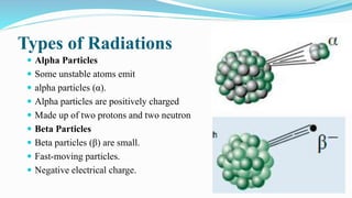
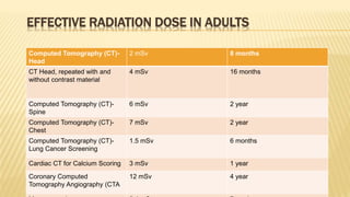
Radiations
 X-rays are forms of radiant energy, like light or radio waves.
 Unlike light, x-rays can penetrate the body.
 Which allows a technologist to produce pictures of internal structures.
 X-ray examinations play an important role in diagnosis.
 There are actually two kinds of radiation.
 One is more energetic than the other.
 It has so much energy it can knock electrons out of atoms this process
known as ionization.
Radiations Effect
 Ionizing radiation can affect the atoms in living things.
 It poses a health risk by damaging tissue and DNA.
 less energetic, types of nonionizing radiation (including radio waves,
microwaves—and visible light).
Types of Radiations
 Alpha Particles
 Some unstable atoms emit
 alpha particles (α).
 Alpha particles are positively charged
 Made up of two protons and two neutron
 Beta Particles
 Beta particles (β) are small.
 Fast-moving particles.
 Negative electrical charge.
Types of Radiations
 Gamma Rays
 Gamma rays (γ) are weightless packets of energy
 Gamma rays are pure energy.
 Gamma rays are similar to visible light.
 But have much higher energy.
Penetrating Powers of Alpha Particles, Beta Particles, Gamma Rays and
X-Rays
Measuring radiation dosage
 The scientific unit of measurement for radiation dose is the millisieverts (mSv).
 Other radiation dose measurement units include rad, rem, roentgen, Sievert, and
gray.
 The term effective dose is used when referring to the radiation risk averaged over
the entire body.
 The effective dose accounts for the relative sensitivities of the different tissues
exposed.
More importantly.
 it allows for quantification of risk and comparison to more familiar sources of
exposure that range from natural background radiation to radiographic medical
procedures.
Naturally-occurring "background" radiation exposure
Radon:
 It is a colorless, odorless, tasteless radioactive gas that comes from the
decay of radium, which is present in nearly all rocks and soils. Most of our
exposure to naturally occurring radiation is from indoor radon.
 Since radon gas emits alpha particles, inhaling it can cause cancer.
 Radon causes an estimated 20,000 lung cancer deaths each year.
Radiation from the Ground and from Space
 Some exposure to natural radiation comes from other elements in Earth’s
crust, such as thorium and potassium.
 Another natural source is cosmic (space) radiation
Man-Made Radiation Radiation
 Most medical exposure comes from the use of standard x-rays and CT scans.
 Drugs with radioactive material attached, known as radiopharmaceuticals.
 Radiation therapy use radiation to treat patients.
Exposure to Ionizing Radiation
RADIATION DOSE FROM CT EXAMINATIONS
 It varies from patient to patient.
 Depending on size of the body part examined.
 The type of procedure
 The type of CT equipment and its operation.
 The actual dose from a procedure could be two or three times
larger or smaller than the estimates.
EFFECTIVE RADIATION DOSE IN ADULTS
For this procedure * An adult’s approximate
effective radiation dose is
Comparable to natural
background radiation for:
Ct Abdomen and Pelvis 10 mSv 3 year
Ct Abdomen and Pelvis,
repeated with and without
contrast materia
20 mSv 7 year
Intravenous Pyelogram (IVP) 3 mSv 1 year
Radiography (X-ray)-Spine 1.5 mSv 6 month
Ct Colonography 6 mSv 2 year
EFFECTIVE RADIATION DOSE IN ADULTS
Computed Tomography (CT)-
Head
2 mSv 8 months
CT Head, repeated with and
without contrast material
4 mSv 16 months
Computed Tomography (CT)-
Spine
6 mSv 2 year
Computed Tomography (CT)-
Chest
7 mSv 2 year
Computed Tomography (CT)-
Lung Cancer Screening
1.5 mSv 6 months
Cardiac CT for Calcium Scoring 3 mSv 1 year
Coronary Computed
Tomography Angiography (CTA
12 mSv 4 year
RADIATION RISKS FROM CT
 As in many aspects of medicine, there are both benefits and
risks associated with the use of CT. The main risks are those
associated with
 Test results that demonstrate a benign or incidental finding,
leading to unneeded, possibly invasive, follow-up tests that
may present additional risks and
 The increased possibility of cancer induction from x-ray
radiation exposure
RADIATION RISKS
 Radiation can damage living tissue by changing cell
structure and damaging DNA.
 The amount of damage depends upon the type of radiation,
its energy the total amount of radiation absorbed.
 The most important risk from exposure to radiation is cancer.
 The higher the radiation dose, the greater the chance of
developing cancer.
 The chance of developing cancer, not the seriousness of the
cancer, increases as the radiation dose increases.
RADIATION RISKS
 Cancers caused by radiation do not appear until years after the
radiation exposure.
 Some people are more likely to develop cancer from radiation
exposure than others.
 Damage to genetic material in reproductive cells can cause genetic
mutations,
THE RISKS OF CT SCANS FOR CHILDREN
 Radiation exposure from CT scans affects adults and
children differently.
 Children are considerably more sensitive to radiation than
adults because of their growing bodies and the rapid pace at
which the cells in their bodies divide
 Individuals who have had multiple CT scans before the age
of 15 were found to have an increased risk of
developing leukemia brain tumors and other cancers in the
decade following their first scan.
CT room shielding
 Computed tomography rooms typically have high workloads and
high kilovoltage technique settings.
As a result, at least 1/16-Inch lead shielding or equivalent is required
for the walls, doors, floors, ceilings, and operator's barrier.
The concrete equivalence of 1/16-inch thick lead would be about 4 to
6 inches of standard-density concrete (147 pounds per cubic foot).
The Radiation Protection Advisor for the hospital is responsible for
advising the actual shielding required and this will depend on a
multitude of factors.
LEAD PLASTERBOARD
1.5 LEAD LINED DOOR
Single door set
Viewing window
Lead plasterboard
• Forms a complete shielded envelope to the room. The shielding
usually extends to the full structural height of the room.
1.5 lead lined door
• A 1.5 leaf door set is usually used for patient entrance. This allows the
passage of the patient trolley.
Single door sets
• A single door set is required as passage from the scanner room to the
control room.
Viewing window
• Shielded frame and lead glass window enables the operator to see
procedures from the control room.
Radiation protection
What is being done to reduce the level of radiation
exposure from CT
• In response to concerns about the increased risk of cancer associated with CT and
other imaging procedures that use ionizing radiation several organizations and
government agencies have developed guidelines and recommendations regarding
the appropriate use of these procedures.
• In 2010, the U.S. Food and Drug Administration (FDA) launched the Initiative
to Reduce Unnecessary Radiation Exposure from Medical Imaging.
• This initiative focuses on the safe use of medical imaging devices, informed
decision-making about when to use specific imaging procedures, and increasing
patients’ awareness of their radiation exposure
• The NIH Clinical Center requires that radiation dose exposures from
CT and other imaging procedures be included in the electronic medical
records of patients treated at the center.
Radiation hazards in CT scan

Radiation hazards in CT scan

  • 1.
    Radiations  X-rays areforms of radiant energy, like light or radio waves.  Unlike light, x-rays can penetrate the body.  Which allows a technologist to produce pictures of internal structures.  X-ray examinations play an important role in diagnosis.  There are actually two kinds of radiation.  One is more energetic than the other.  It has so much energy it can knock electrons out of atoms this process known as ionization.
  • 2.
    Radiations Effect  Ionizingradiation can affect the atoms in living things.  It poses a health risk by damaging tissue and DNA.  less energetic, types of nonionizing radiation (including radio waves, microwaves—and visible light).
  • 3.
    Types of Radiations Alpha Particles  Some unstable atoms emit  alpha particles (α).  Alpha particles are positively charged  Made up of two protons and two neutron  Beta Particles  Beta particles (β) are small.  Fast-moving particles.  Negative electrical charge.
  • 4.
    Types of Radiations Gamma Rays  Gamma rays (γ) are weightless packets of energy  Gamma rays are pure energy.  Gamma rays are similar to visible light.  But have much higher energy.
  • 5.
    Penetrating Powers ofAlpha Particles, Beta Particles, Gamma Rays and X-Rays
  • 6.
    Measuring radiation dosage The scientific unit of measurement for radiation dose is the millisieverts (mSv).  Other radiation dose measurement units include rad, rem, roentgen, Sievert, and gray.  The term effective dose is used when referring to the radiation risk averaged over the entire body.  The effective dose accounts for the relative sensitivities of the different tissues exposed. More importantly.  it allows for quantification of risk and comparison to more familiar sources of exposure that range from natural background radiation to radiographic medical procedures.
  • 7.
    Naturally-occurring "background" radiationexposure Radon:  It is a colorless, odorless, tasteless radioactive gas that comes from the decay of radium, which is present in nearly all rocks and soils. Most of our exposure to naturally occurring radiation is from indoor radon.  Since radon gas emits alpha particles, inhaling it can cause cancer.  Radon causes an estimated 20,000 lung cancer deaths each year.
  • 8.
    Radiation from theGround and from Space  Some exposure to natural radiation comes from other elements in Earth’s crust, such as thorium and potassium.  Another natural source is cosmic (space) radiation
  • 9.
    Man-Made Radiation Radiation Most medical exposure comes from the use of standard x-rays and CT scans.  Drugs with radioactive material attached, known as radiopharmaceuticals.  Radiation therapy use radiation to treat patients.
  • 10.
  • 11.
    RADIATION DOSE FROMCT EXAMINATIONS  It varies from patient to patient.  Depending on size of the body part examined.  The type of procedure  The type of CT equipment and its operation.  The actual dose from a procedure could be two or three times larger or smaller than the estimates.
  • 12.
    EFFECTIVE RADIATION DOSEIN ADULTS For this procedure * An adult’s approximate effective radiation dose is Comparable to natural background radiation for: Ct Abdomen and Pelvis 10 mSv 3 year Ct Abdomen and Pelvis, repeated with and without contrast materia 20 mSv 7 year Intravenous Pyelogram (IVP) 3 mSv 1 year Radiography (X-ray)-Spine 1.5 mSv 6 month Ct Colonography 6 mSv 2 year
  • 13.
    EFFECTIVE RADIATION DOSEIN ADULTS Computed Tomography (CT)- Head 2 mSv 8 months CT Head, repeated with and without contrast material 4 mSv 16 months Computed Tomography (CT)- Spine 6 mSv 2 year Computed Tomography (CT)- Chest 7 mSv 2 year Computed Tomography (CT)- Lung Cancer Screening 1.5 mSv 6 months Cardiac CT for Calcium Scoring 3 mSv 1 year Coronary Computed Tomography Angiography (CTA 12 mSv 4 year
  • 14.
    RADIATION RISKS FROMCT  As in many aspects of medicine, there are both benefits and risks associated with the use of CT. The main risks are those associated with  Test results that demonstrate a benign or incidental finding, leading to unneeded, possibly invasive, follow-up tests that may present additional risks and  The increased possibility of cancer induction from x-ray radiation exposure
  • 15.
    RADIATION RISKS  Radiationcan damage living tissue by changing cell structure and damaging DNA.  The amount of damage depends upon the type of radiation, its energy the total amount of radiation absorbed.  The most important risk from exposure to radiation is cancer.  The higher the radiation dose, the greater the chance of developing cancer.  The chance of developing cancer, not the seriousness of the cancer, increases as the radiation dose increases.
  • 16.
    RADIATION RISKS  Cancerscaused by radiation do not appear until years after the radiation exposure.  Some people are more likely to develop cancer from radiation exposure than others.  Damage to genetic material in reproductive cells can cause genetic mutations,
  • 17.
    THE RISKS OFCT SCANS FOR CHILDREN  Radiation exposure from CT scans affects adults and children differently.  Children are considerably more sensitive to radiation than adults because of their growing bodies and the rapid pace at which the cells in their bodies divide  Individuals who have had multiple CT scans before the age of 15 were found to have an increased risk of developing leukemia brain tumors and other cancers in the decade following their first scan.
  • 18.
    CT room shielding Computed tomography rooms typically have high workloads and high kilovoltage technique settings. As a result, at least 1/16-Inch lead shielding or equivalent is required for the walls, doors, floors, ceilings, and operator's barrier. The concrete equivalence of 1/16-inch thick lead would be about 4 to 6 inches of standard-density concrete (147 pounds per cubic foot).
  • 20.
    The Radiation ProtectionAdvisor for the hospital is responsible for advising the actual shielding required and this will depend on a multitude of factors. LEAD PLASTERBOARD 1.5 LEAD LINED DOOR Single door set Viewing window
  • 21.
    Lead plasterboard • Formsa complete shielded envelope to the room. The shielding usually extends to the full structural height of the room.
  • 22.
    1.5 lead lineddoor • A 1.5 leaf door set is usually used for patient entrance. This allows the passage of the patient trolley.
  • 23.
    Single door sets •A single door set is required as passage from the scanner room to the control room.
  • 24.
    Viewing window • Shieldedframe and lead glass window enables the operator to see procedures from the control room.
  • 25.
  • 26.
    What is beingdone to reduce the level of radiation exposure from CT • In response to concerns about the increased risk of cancer associated with CT and other imaging procedures that use ionizing radiation several organizations and government agencies have developed guidelines and recommendations regarding the appropriate use of these procedures. • In 2010, the U.S. Food and Drug Administration (FDA) launched the Initiative to Reduce Unnecessary Radiation Exposure from Medical Imaging. • This initiative focuses on the safe use of medical imaging devices, informed decision-making about when to use specific imaging procedures, and increasing patients’ awareness of their radiation exposure
  • 27.
    • The NIHClinical Center requires that radiation dose exposures from CT and other imaging procedures be included in the electronic medical records of patients treated at the center.