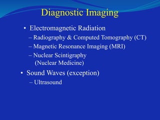
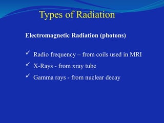
Types of Radiation
ElectromagneticRadiation (photons)
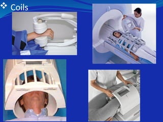
Radio frequency – from coils used in MRI
X-Rays - from xray tube
Gamma rays - from nuclear decay
9.
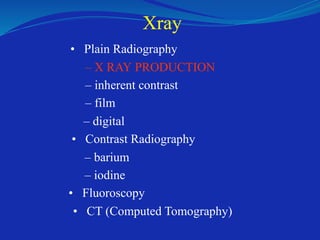
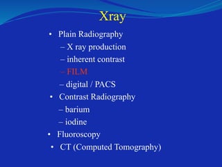
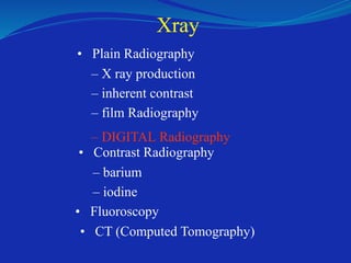
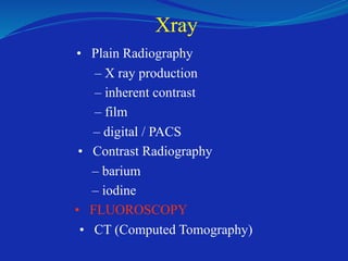
Xray
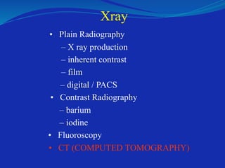
• Plain Radiography
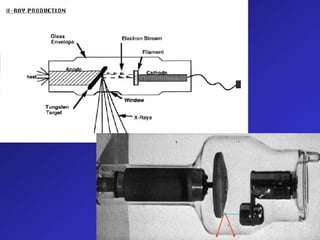
–X RAY PRODUCTION
– inherent contrast
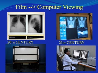
– film
– digital
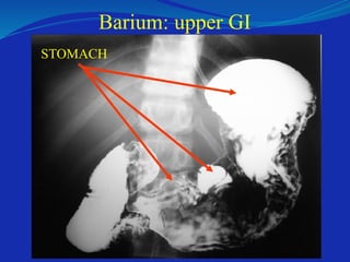
• Contrast Radiography
– barium
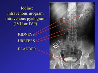
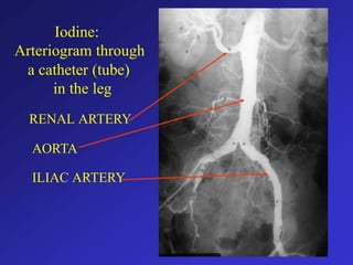
– iodine
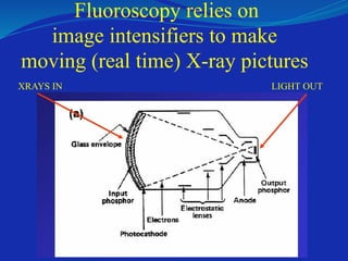
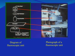
• Fluoroscopy
• CT (Computed Tomography)
11.
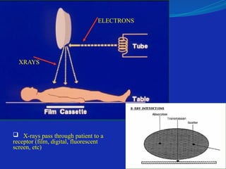
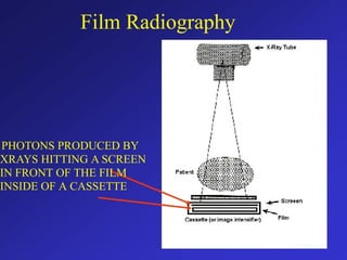
X-rays passthrough patient to a
receptor (film, digital, fluorescent
screen, etc)
ELECTRONS
XRAYS
12.
January 1896 -First x-ray made in
public
Routine x-ray current technology
Wilhelm Conrad Roentgen
1845-1923
13.
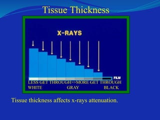
A diagnosticimage is composed of
differences in contrast between tissues!
Tissue thickness affectsx-rays attenuation.
WHITE GRAY BLACK
Tissue Thickness
LESS GET THROUGH>>MORE GET THROUGH
16.
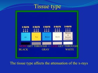
The tissue typeaffects the attenuation of the x-rays
BLACK GRAY WHITE
Tissue type
MORE GET THROUGH >> LESS GET THROUGH
17.
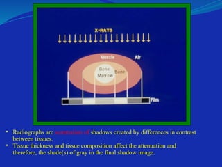
• Radiographs aresummation of shadows created by differences in contrast
between tissues.
• Tissue thickness and tissue composition affect the attenuation and
therefore, the shade(s) of gray in the final shadow image.
18.
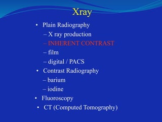
Xray
• Plain Radiography
–X ray production
– INHERENT CONTRAST
– film
– digital / PACS
• Contrast Radiography
– barium
– iodine
• Fluoroscopy
• CT (Computed Tomography)
19.
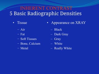
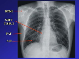
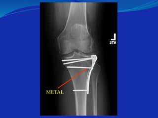
INHERENT CONTRAST
5 BasicRadiographic Densities
• Tissue
–
–
–
–
–
Air
Fat
Soft Tissues
Bone, Calcium
Metal
• Appearance on XRAY
–
–
–
–
–
Black
Dark Gray
Gray
White
Really White
20.
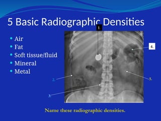
5 Basic RadiographicDensities
Air
Fat
Soft tissue/fluid
Mineral
Metal
1.
2.
3.
4.
5.
Name these radiographic densities.
Xray
• Plain Radiography
–X ray production
– inherent contrast
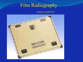
– film Radiography
– DIGITAL Radiography
• Contrast Radiography
– barium
– iodine
• Fluoroscopy
• CT (Computed Tomography)
27.
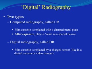
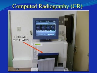
‘Digital’ Radiography
• Twotypes
– Computed radiography, called CR
• Film cassette is replaced with a charged metal plate
• After exposure, plate is ‘read’ in a special device
– Digital radiography, called DR
• Film cassette is replaced by a charged sensor (like in a
digital camera or video camera)
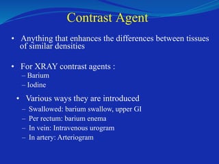
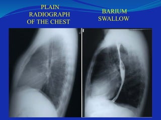
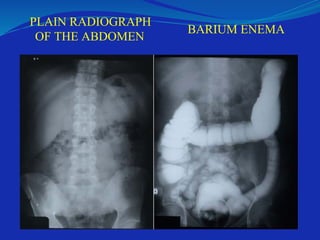
Contrast Agent
• Anythingthat enhances the differences between tissues
of similar densities
• For XRAY contrast agents :
– Barium
– Iodine
• Various ways they are introduced
–
–
–
–
Swallowed: barium swallow, upper GI
Per rectum: barium enema
In vein: Intravenous urogram
In artery: Arteriogram
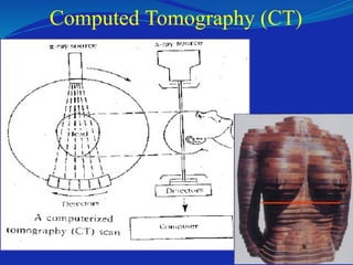
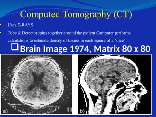
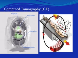
• Uses X-RAYS
•Tube & Detector spins together around the patient Computer performs
calculations to estimate density of tissues in each square of a ‘slice’
Computed Tomography (CT)
Brain Image 1974, Matrix 80 x 80
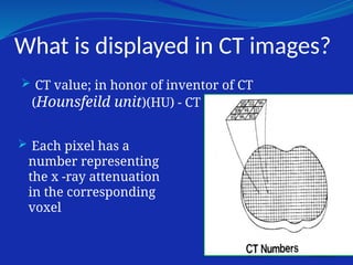
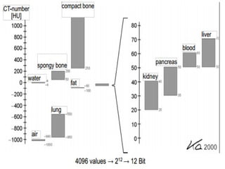
What is displayedin CT images?
CT value; in honor of inventor of CT
(Hounsfeild unit)(HU) - CT numbers.
Each pixel has a
number representing
the x -ray attenuation
in the corresponding
voxel
44.
Cont…
Water andwater equivalent tissue (0HU)
Air corresponds (-1000HU).
Tissues denser than water are given positive CT
numbers.
The Hounsfield scale has no upper limit.but; for
medical application (-1024HU to +3071HU)
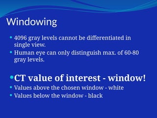
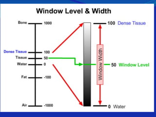
Windowing
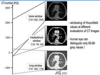
4096 graylevels cannot be differentiated in
single view.
Human eye can only distinguish max. of 60-80
gray levels.
CT value of interest - window!
Values above the chosen window - white
Values below the window - black
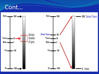
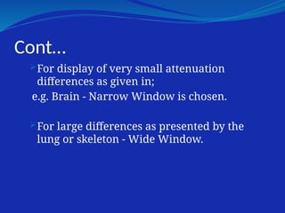
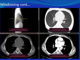
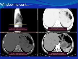
Cont…
For display ofvery small attenuation
differences as given in;
e.g. Brain - Narrow Window is chosen.
For large differences as presented by the
lung or skeleton - Wide Window.
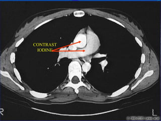
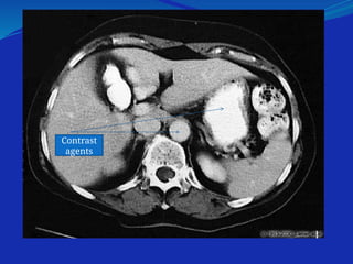
Contrast for CT
•Iodine injected into an arm vein
• Iodine or Barium diluted in water given orally
for abdomen scans
• There are some risks
– Allergic reaction
– Kidney damage
• Enhances the blood vessels and organs
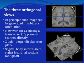
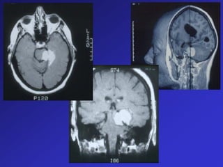
The three orthogonal
views
In principle slice image can
be generated in arbitrary
orientation.
However; for CT mostly a
transverse (x/y plane) is
scanned directly
Z-axis - perpendicular scan
plane
Sagittal body sections (left /
right) & coronal sections
(ant /post).
57.
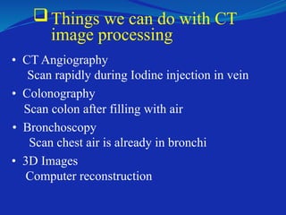
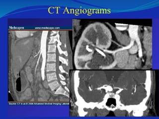
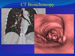
Things we cando with CT
image processing
• CT Angiography
Scan rapidly during Iodine injection in vein
• Colonography
Scan colon after filling with air
• Bronchoscopy
Scan chest air is already in bronchi
• 3D Images
Computer reconstruction
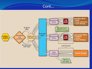
Human Radiation Response
effect of x-rays is the result of interactions at the
atomic level
atomic interactions take the form of ionization
or excitation of orbital electrons and result in
the deposition of energy in tissue
Cont…
The abnormalmolecule in time
- function improperly
- cease to function or death of cell
At each stage in the sequence, it is possible to
repair radiation damage and recover.
65.
Cont…
radiation responseoccurs within minutes or
days after exposure, it is classified as an early
effect of radiation.
On the other hand, if the human injury is not
observed for months or years, it is called a late
effect of radiation.
66.
Cont…
Most humanresponses have been observed to
occur after exposure to rather large radiation
doses.
However, we are cautious and assume that
even small doses are harmful.
67.
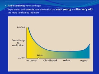
• Radio-sensitivity varieswith age.
Experiments with animals have shown that the very young and the very old
are more sensitive to radiation.
68.
EARLY EFFECTS OFRADIATION ON
HUMANS
1. Acute radiation syndrome
a.Hematologic syndrome
b.Gastrointestinal syndrome
c.Central nervous system syndrome
2. Local tissue damage
d.Skin
e.Gonads
3. Hematologic depression
69.
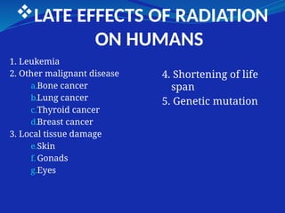
LATE EFFECTS OFRADIATION
ON HUMANS
1. Leukemia
2. Other malignant disease
a.Bone cancer
b.Lung cancer
c.Thyroid cancer
d.Breast cancer
3. Local tissue damage
e.Skin
f. Gonads
g.Eyes
4. Shortening of life
span
5. Genetic mutation
70.
Effects of FetalIrradiation
1.Prenatal death
2.Neonatal death
3.Congenital malformation
4.Childhood malignancy
5.Diminished growth and
development
71.
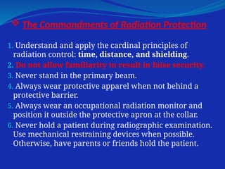
The Commandmentsof Radiation Protection
1. Understand and apply the cardinal principles of
radiation control: time, distance, and shielding.
2. Do not allow familiarity to result in false security.
3. Never stand in the primary beam.
4. Always wear protective apparel when not behind a
protective barrier.
5. Always wear an occupational radiation monitor and
position it outside the protective apron at the collar.
6. Never hold a patient during radiographic examination.
Use mechanical restraining devices when possible.
Otherwise, have parents or friends hold the patient.
72.
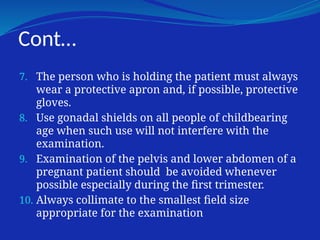
Cont…
7. The personwho is holding the patient must always
wear a protective apron and, if possible, protective
gloves.
8. Use gonadal shields on all people of childbearing
age when such use will not interfere with the
examination.
9. Examination of the pelvis and lower abdomen of a
pregnant patient should be avoided whenever
possible especially during the first trimester.
10. Always collimate to the smallest field size
appropriate for the examination
Magnetic Resonance Imaging
•Starts with a really strong magnet
– Supercooled with Liquid Helium / Nitrogen
• Transmit radio wave pulses into patient
• Listen for return radio waves caused by
interaction with protons (water) in the
patient’s body
• Process the frequency and phase of the
returned signals by computer
76.
Magnetic Resonance Imaging
Starts with a really strong magnet
– Supercooled with Liquid Helium / Nitrogen
Transmit radio wave pulses into patient
Listen for return radio waves caused by interaction
with protons (water) in the patient’s body
Process the frequency and phase of the returned
signals
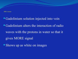
MRI Contrast
Gadoliniumsolution injected into vein
Gadolinium alters the interaction of radio
waves with the protons in water so that it
gives MORE signal
Shows up as white on images
82.
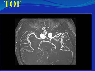
Magnetic Resonance Angiography
May be done after injecting contrast (Gadolinium) or
without(TOF)
Flowing blood changes the way that radio waves
interact with the water in blood
– May give more signal (IN TOF)
– May give less signal (AS FLOW VOID)
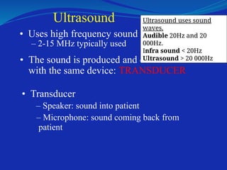
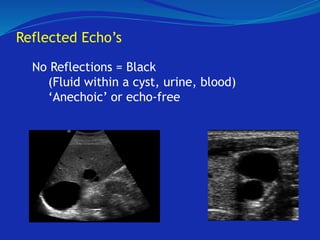
Ultrasound
• Uses highfrequency sound
– 2-15 MHz typically used
• The sound is produced and detected
with the same device: TRANSDUCER
• Transducer
– Speaker: sound into patient
– Microphone: sound coming back from
patient
Ultrasound uses sound
waves.
Audible 20Hz and 20
000Hz.
Infra sound < 20Hz
Ultrasound > 20 000Hz
86.
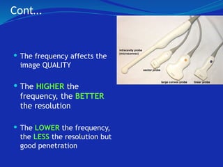
Cont…
The frequencyaffects the
image QUALITY
The HIGHER the
frequency, the BETTER
the resolution
The LOWER the frequency,
the LESS the resolution but
good penetration
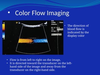
• Color FlowImaging
• The direction of
blood flow is
indicated by the
display color
• Flow is from left to right on the image,
• It is directed toward the transducer on the left-
hand side of the image and away from the
transducer on the right-hand side.
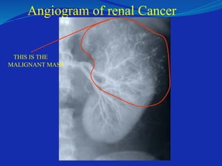
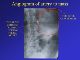
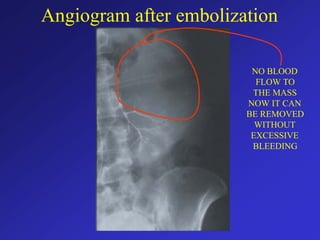
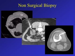
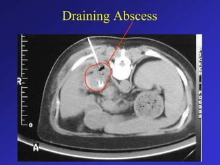
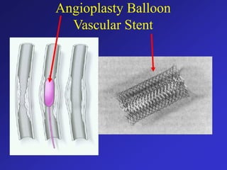
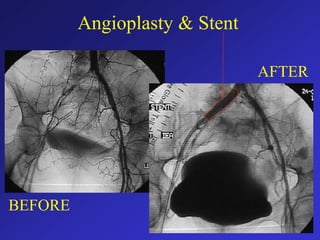
Interventional Radiology
• Needlesfor biopsy or fluid removal
• Catheters to make angiograms
• Catheters with balloons to open blood vessels
• Stents to hold blood vessels open
• Coils and material to block blood vessels
• Catheters to drain abscesses
• Tubes for feeding
Etc…
93.
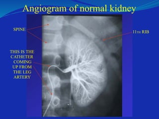
Angiogram of normalkidney
SPINE
THIS IS THE
CATHETER
COMING
UP FROM
THE LEG
ARTERY
11TH RIB


![ONFH[AVN HIP] -TRIPLE REGIME -A NOVAL SURGICAL CONCEPT .pptx](https://cdn.slidesharecdn.com/ss_thumbnails/onfhavnhip2026koaconcalicutdrgokuldevdrmashraf-260210064517-213ec005-thumbnail.jpg?width=640&height=640&fit=bounds)


![CTEV [ clubfoot] DR ARUN LAL ,DR MOHAMED ASHRAF travancore medical college k...](https://cdn.slidesharecdn.com/ss_thumbnails/ctevclubfootdrarunlaldrmohamedashraftravancoremedicalcollegekollamkeralaindia-260208063247-18fc466c-thumbnail.jpg?width=640&height=640&fit=bounds)

