
Recommended
More Related Content
What's hot
What's hot (20)
Similar to medical radiation safety awareness
Similar to medical radiation safety awareness (20)
More from Mmedsc Hahm
More from Mmedsc Hahm (20)
Recently uploaded
Recently uploaded (20)
medical radiation safety awareness
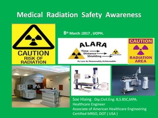
- 1. Medical Radiation Safety Awareness Soe Hlaing . Dip.Civil.Eng: B,S.BSC,MPA. Healthcare Engineer Associate of American Healthcare Engineering Certified MRSO, DOT ( USA ) 8th March :2017 , UOPH.
- 2. We live in Sea of Radiation COSMIC p+ , ē , α2+ TERRESTRIAL ( Th 90,U , Ra, K 40 ) AIR Inhaled Radon,Thoron FOOD and WATER INTERNAL α2+ β Γ rays
- 3. Sources of Radiation (620 mrem /year) Natural(50%) • Cosmic • Terrestrial • Radon • Internal (food and water) Man-Made(50%) • Medical • Industrial • Occupational • Consumer products 360 mrem/yr NCRP Report 1994 620 mrem/yr NCRP Report 2009
- 4. Man-Made Radiation Consumer Products 2% Fiesta ware , uranium glass Industrials < 0.1 % Occupational < 0.1 % Medical 48%
- 5. Radon Gas
- 6. Orders of magnitude (radiation) Source: http://en.wikipedia.or g/wiki/Orders_of_ma gnitude_(radiation) Updated: 2017-02- 02T12:26Z
- 7. Radiation Risk Perceptions • Why are we interested in radiation risk? • Perceptions drive our beliefs, decisions and reactions to radiation. • Fear of Radiation is Common • Understanding these fear is helpful for understanding response to radiation • Without understanding fears take over - we are most afraid of what we know the least about?
- 8. Two Types of Radiation Views Common Views • Mysterious • ( cannot detect by our five senses) • Sinister ( Unknown Danger) • Deadly ( Causes Cancer ) Scientists’ views • Parts of Natural World easily measurable • Risks are well known since 1896 90 min- 21milli sec • Major Benefit in Medicine treatment for Cancer
- 9. The LINEAR NO-THRESHOLD MODEL (LNT) • The model used in radiation protection to estimate the long-term, biological damage caused by ionizing radiation • It assumed that the damage is directly proportional (“linear”) to the dose of radiation, at all dose levels • Radiation is always considered harmful with no safety threshold
- 10. Threshold and Hormesis Model • Threshold: very small exposures are harmless • Radiation Hormesis Model: radiation at very small doses can be beneficial • Not enough data clearly in favor of any theory. No definitive answer. However, LNT model is used worldwide for radiation protection regulations
- 11. Competing Theories on Hazard
- 12. Radiation Protection Purposes • it is assumed that any dose above zero can increase the risk of radiation-induced cancer (i.e., that there is no threshold) • Epidemiologic studies have found that the estimated lifetime risk of dying from cancer is greater by about 0.004% per mSv (0.04% per rem) of radiation dose to the whole body (NRC, 1990) • Ref: An Evaluation of Radiation Exposure Guidance for Military Operations: Interim Report (1997). J. Christopher Johnson and Susan Thaul, Editors. National Academy of Sciences. ISBN 0-309-05895-3.
- 13. What are the effects of radiation? • Large doses of radiation from some procedures may cause temporary skin burns. However, a greater concern is that radiation may cause cancer. There is no conclusive evidence that radiation causes cancer, but large population studies have shown a slight increase in cancer even from small amounts of radiation. • Ref: RadiologyInfo.org
- 14. External Radiation Protection • Time , Distance , Shielding • Neutron absorbed by low density materials • Beta stopped by plastic or wood • Photons shielded by lead or other dense materials • Bremasstrahlung with Beta emitters?
- 15. Internal Radiation Protection • Routes of Entry • Prevention of Intakes • Detecting and Quantifying Intakes • Medical Intervention • Regulatory Limits ALIs and DACs • Notification Requirements (in or external irradiation) • Abbr: ALIs- Annual limit Intake • DACs- Derived Air Concentrations
- 16. Radiation Doses • Different from medicine dose. Intensity and length of exposure • Different types of units and Measurement. • Absorbed dose is used to assess the potential for biochemical changes in specific tissues. Tell us energy deposit in a small volume of tissue. Unit- milligray (mGy) • Equivalent dose is used to assess how much biological damage is expected from the absorbed dose. (Different types of radiation have different damaging properties.) Addresses the impact that the type of radiation has on that tissue. Unit- milliSievert (mSv) • Effective dose is used to assess the potential for long-term effects that might occur in the future
- 17. • Effective dose is a calculated value, measured in mSv, that takes three factors into account: 1 .the absorbed dose to all organs of the body, 2.the relative harm level of the radiation, and 3.the sensitivities of each organ to radiation. eg: the head is less sensitive than < the chest. • Effective dose relates to the overall long-term risk to a person from a procedure and is useful for comparing risks from different procedures. Effective Dose
- 18. Effective Dose • Effective dose is not intended to apply to a specific patient. • The actual risk depending on the size of the patient and the type of procedure. • Example of CT of the abdomen, • Typical absorbed dose: 20 mGy • Typical equivalent dose: 20 mSv • Typical effective dose: 15 mSv
- 22. Dose Limits for Occupational Exposure of any Workers • Following limits be not exceeded, a. An effective dose of 20 mSv/yr averaged over 5 consecutive yrs: b. An effective dose of 50 mSv in any single year ( 16-18 6mSv) c. An equivalent dose to lens of eyes of 150 mSv in a yr (50 ) d. An equivalent dose to the extremities (hand and feet) or skin of 500 mSv in a year ( for16-18 aged 150 mSv) Ref: IAEA SAFETY GUIDE No-G-1.1 page19 .para.3.1
- 23. Recommended Doses • The value of 50 mSv in a year that you quote is a number recommended by the Health Physics Society (HPS) in * position paper in 2010* Radiation doses that exceed a minimum (threshold) level can cause undesirable effects such as depression of the blood cell-forming process (threshold dose = 500 mSv, 50 rem) or cataracts (threshold dose = 5,000 mSv, 500 rem)*
- 24. IAEA .BSS Intervention Situation • Type 1. Emergency Exposure Situation * Accident and other temporary exposure • Type 2. Chronic Exposure Situation * Natural Exposure – such as radon to work place and building * Radioactive Residues from past events radioactive contamination Ref: BSS (Ref. 2. para.3.1)
- 26. ALARA and Inverse Square Law • ALARA • As Low As Reasonable Achievable • inverse-square law • specified physical quantity or intensity is inversely proportional to the square of the distance from the source of that physical quantity.
- 27. Nature of Rays and Shielding
- 29. X Ray and Fluoroscopy Safety • Sources of exposure 1. Primary Beam - very intense exposure in beam - Small beam diameter 2. Scattered radiation - low intensity - large area of exposure - from housing leakage - from any target material
- 31. Sources of Exposure • X-Rays leakage • Sky shine- Scattered • Inadequate shielding • Not following safety procedures -by passing interlocks etc: • Not using or heeding radiation instruments • Not using PPE, lead apron , etc:
- 32. Principles of Radiation Protection • Minimize Time around x-ray machines when in operation • Minimize Distance between you and the x-ray source • Utilize Shielding whenever possible -lead aprons, lead-lined gloves, collar / thyroid shields, shield booths
- 33. Sources of Exposure of Staff in PET • *Radiation Sources - Syringes containing PET doses - Calibration and transmission sources - Patients and their specimens • *Staff exposures - Staff not in room during CT potion - PET technologist is twice that of non-PET tech in Nuclear medicine dept: “Carey-Clinical PET, AAPM meeting July 2001”
- 34. ALARA in PET-CT Time - Minimize Time consider- rotation for staff in PET Distance- Maximize Distance Use IV access for injections- also reduce injection time Use remote handling devices when possible Spend less time at patient side, take step back when possible Shielding- Use Shielding Tungsten syringe shield ( 9mm reduces FDG exposure 88%) L-Block / mobile Shielding
- 36. ALARA –Operator Positioning of C-arms • Scatter radiation intensity is less on image intensifier side as compared to the x-ray tube side • Lateral and oblique projection position the x-ray tube on the opposite side of the patient from you where you are standing
- 37. Radiation in Therapy • Treatment of a medical condition by: • 1. Killing Cells ( DNA Bond breaking or disrupting tumor blood flow) OR • 2.Slowing the Growth of Diseased Cells using radiation • It uses high-energy particles or waves, such as x- rays, gamma rays, electron beams, or protons, to destroy or damage cancer cells. Other names for radiation therapy are radiotherapy, irradiation, or x-ray therapy.
- 38. Types of Sealed Sources • Calibration Sources • Germanium transmission Sources • Flood Sources and Flexible rulers • Manual Brach therapy Sources • Teletherapy Sources ( Gamma Knife, View Ray and Gammapod) • Irradiators Co 60 half life 5.26 yr Cs 137 half life 30.17 yr • After loaders
- 39. • Contact Dose Rates on Sealed Sources may be Very High, Especially Cs – 137 • NEVER TOUCH A SEALED SOURCE CAPSULE Radiation Burns may manifest themselves month after exposure WARNING
- 40. Radioactive Waste Management • Define radioactive waste • Review pertinent regulation • Understand decay-in-storage • Know the required elements of a radioactive waste management program
- 41. Radioactive Waste Management • Define radioactive waste • Review pertinent regulation • Understand decay-in-storage • Know the required elements of a radioactive waste management program
- 42. Radioactive Waste Segregation • Physical form • Half-life • Other hazards - chemicals - Bio hazardous materials • Combustible from - non- combustible Siriraj Hospital Cancer Center , 2016
- 43. Waste Packaging and Storage • Description of storage Area • Location and diagrams • Equipment, monitoring stations, effluent, • Type of building , Protection from elements • Security, Ventilation, • Fire protection systems • Temperature and humidity effects on waste container • Packaging/container to be used • Inspection program • Remote handling program • Radiation protection program elements • Shielding needs if any ,
- 44. Radioactive Waste Minimization • Training of workers • Segregation of wastes • Use less radioactive materials • Use Short-Lived radionuclide's -up to 100 days • Segregate chemical and radioactive materials • Use non-radioactive methods • Treat waste on-site • Check for contamination
- 45. List of half-lives • yttrium Y-90, used for radioembolization treating lymphoma (2.7 days) • I-131, used for thyroid function tests and for treating thyroid cancer (8.0 days) • Strontium Sr-89, used for treating bone cancer, intravenous injection (52 days) • Iridium Ir-192, used for brachytherapy (74 days) • Co-60, used for brachytherapy and external radiotherapy (5.3 years) • Cesium Cs-137, used for brachytherapy, external radiotherapy (30 years)
- 46. PPE and Signage for Radiation
- 47. • Ref: • Sandy E.Konerth, MS,DABR,DABMP • Alan Fellman,PH.D.,CHP • Lee Myers,PH.D. • www.radiologyinfo.org • Radioactive Materials Regulatory Guide Minnesota Department of Health • Global Threat Reduction Program . Training Course on Physical Protection and Security Management of Radioactive Sources • Radialogyinfo.org • IAEA SAFETY STANDARDS SERIES SAFETY GUIDE NO.RS-G-1.1