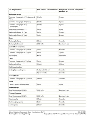
RadiologyInfo.org                                                                                             July 8, 2009


Safety: Radiation Exposure in X-ray
Examinations
This information is reviewed by a physician with expertise in the area presented and is further reviewed by committees
from the American College of Radiology (ACR) and the Radiological Society of North America (RSNA), comprising
physicians with expertise in several radiologic areas.


What are x-rays and what do                                       Naturally-occurring
they do?                                                          "background" radiation
X-rays are forms of radiant energy, like light or radio
                                                                  exposure
waves. Unlike light, x-rays can penetrate the body, which         We are exposed to radiation from natural sources all the
allows a radiologist to produce pictures of internal              time. The average person in the U.S. receives an
structures. The radiologist can view these on                     effective dose of about 3 mSv per year from naturally
photographic film or on a TV or computer monitor.                 occurring radioactive materials and cosmic radiation
X-ray examinations provide valuable information about             from outer space. These natural "background" doses vary
your health and play an important role in helping your            throughout the country.
doctor make an accurate diagnosis. In some cases x-rays           People living in the plateaus of Colorado or New Mexico
are used to assist with the placement of tubes or other           receive about 1.5 mSv more per year than those living
devices in the body or with other therapeutic procedures.         near sea level. The added dose from cosmic rays during a
                                                                  coast-to-coast round trip flight in a commercial airplane
Measuring radiation dosage                                        is about 0.03 mSv. Altitude plays a big role, but the
                                                                  largest source of background radiation comes from radon
The scientific unit of measurement for radiation dose,            gas in our homes (about 2 mSv per year). Like other
commonly referred to as effective dose, is the
                                                                  sources of background radiation, exposure to radon
millisievert (mSv). Other radiation dose measurement
                                                                  varies widely from one part of the country to another.
units include rad, rem, Roentgen, Sievert, and Gray.
                                                                  To explain it in simple terms, we can compare the
Because different tissues and organs have varying                 radiation exposure from one chest x-ray as equivalent to
sensitivity to radiation exposure, the actual radiation risk
                                                                  the amount of radiation exposure one experiences from
to different parts of the body from an x-ray procedure
                                                                  our natural surroundings in 10 days.
varies. The term effective dose is used when referring to
the radiation risk averaged over the entire body. The             Following are comparisons of effective radiation dose
effective dose accounts for the relative sensitivities of the     with background radiation exposure for several
different tissues exposed. More importantly, it allows for        radiological procedures described within this Web site:
quantification of risk and comparison to more familiar
sources of exposure that range from natural background
radiation to radiographic medical procedures.


July 8, 2009                                                Safety: Radiation Exposure in X-ray Examinations; 1 of 4
Copyright © 2009 Radiological Society of North America (RSNA)                     RadiologyInfo: RadiologyInfo.org
For this procedure:                        Your effective radiation dose is: Comparable to natural background
                                                                             radiation for:
Abdominal region:
Computed Tomography (CT)-Abdomen &         10 mSv                            3 years
Pelvis
Computed Tomography (CT)-Body              10 mSv                            3 years
Computed Tomography (CT)-                  10 mSv                            3 years
Colonography
Intravenous Pyelogram (IVP)                3 mSv                             1 year
Radiography-Lower GI Tract                 8 mSv                             3 years
Radiography-Upper GI Tract                 6 mSv                             2 years
Bone:
Radiography-Spine                          1.5 mSv                           6 months
Radiography-Extremity                      0.001 mSv                         Less than 1 day
Central Nervous system:
Computed Tomography (CT)-Head              2 mSv                             8 months
Computed Tomography (CT)-Spine             6 mSv                             2 years
Myelography                                4 mSv                             16 months
Chest:
Computed Tomography (CT)-Chest             7 mSv                             2 years
Radiography-Chest                          0.1 mSv                           10 days
Children's imaging:
Voiding Cystourethrogram                   5-10 yr. old: 1.6 mSv             6 months
                                           Infant: 0.8 mSv                   3 months
Face and neck:
Computed Tomography (CT)-Sinuses           0.6 mSv                           2 months
Heart:
Cardiac CT for Calcium Scoring             3 mSv                             1 year
Men's Imaging:
Bone Densitometry (DEXA)                   0.001 mSv                         Less than 1 day
Women's Imaging:
Bone Densitometry (DEXA)                   0.001 mSv                         Less than 1 day
Galactography                              0.7 mSv                           3 months
Hysterosalpingography                      1 mSv                             4 months
Mammography                                0.7 mSv                           3 months


Safety: Radiation Exposure in X-ray Examinations; 2 of 4                                                  July 8, 2009
RadiologyInfo: RadiologyInfo.org                        Copyright © 2009 Radiological Society of North America (RSNA)
When studies of the abdomen or pelvis are required, the
X-ray safety                                                  physician may prefer to order a different type of exam
                                                              for a pregnant patient or reduce the number of x-rays
As with other medical procedures, x-rays are safe when        from that which are normally acquired.
used with care. Radiologists and x-ray technologists have
been trained to use the minimum amount of radiation           Therefore it is important that you inform your physician
necessary to obtain the needed results. The amount of         or the x-ray technologist about your reproductive status
radiation used in most examinations is very small and the     before the x-ray study is performed.
benefits greatly outweigh the risk of harm.
                                                              Most standard x-ray examinations of the abdomen are
X-rays are produced only when a switch is momentarily         not likely to pose a serious risk to the child. Some
turned on. As with visible light, no radiation remains        abdominal and pelvic studies such as CT deliver greater
after the switch is turned off.                               amounts of radiation to a developing pregnancy.
                                                              Informing the radiologist that you are or might be
X-rays over your lifetime                                     pregnant is important so that your medical care can be
The decision to have an x-ray exam is a medical one,          planned with both you and your baby in mind.
based on the likelihood of benefit from the exam and the      Remember, this is done to optimize medical care by
potential risk from radiation. For low dose examinations,     reducing any potential risk.
usually those that involve only films taken by a
                                                              Radionuclide exams, also known as nuclear medicine,
technologist, this is generally an easy decision. For
                                                              use an x-ray-like radiation. The method of use, however,
higher dose exams such as computed tomography (CT)
                                                              is quite different from x-rays and produce very different
scans and those involving the use of contrast materials
                                                              looking images. The same advice for informing your
(dyes) such as barium or iodine, the radiologist may want
                                                              physician or the nuclear medicine technologist about any
to consider your past history of exposure to x-rays. If you
                                                              possible pregnancy before the examination begins is
have had frequent x-ray exams and change healthcare
                                                              important.
providers, it is a good idea to keep a record of your x-ray
history for yourself. This can help your doctor make an       However, in nuclear medicine another precaution is
informed decision. It is also very important to tell your     advised for women who are breast-feeding a child. Some
doctor if you are pregnant before having an exam that         of the pharmaceuticals that are used for the study can
involves the abdomen or pelvic region.                        pass into the mother's milk and subsequently the child
                                                              will consume them. To avoid this possibility, it is
Pregnancy and x-rays                                          important that a nursing mother inform her physician and
As with any aspect of medical care, knowing that a            the nuclear medicine technologist about this before the
patient is or could be pregnant is important information.     examination begins. Usually, you will be asked to
Pregnancy, for example, might explain certain symptoms        discontinue breast-feeding for a short while, pump your
or medical findings. When a pregnant patient is ill or        breasts in the interim and discard the milk. Breast-
injured, the physician will carefully select medications to   feeding can often resume shortly afterwards.
avoid potential risks to the developing child. This is also
true of x-rays.                                               Radiation dose from
While the vast majority of medical x-rays do not pose a       interventional radiology
critical risk to a developing child, there may be a small     procedures
likelihood of causing a serious illness or other
complication. The actual risk depends on how far along        Interventional radiologic procedures use diagnostic-type
the pregnancy is and on the type of x-ray. Ultrasound         imaging equipment to assist a physician in the treatment
studies, for example, don't use x-rays and have never         of a patient's condition. These procedures frequently
demonstrated any potential risk to pregnancy. X-ray           provide favorable medical results with minimal recovery
studies of the head, arms, legs and chest do not usually      time. In some cases these procedures avoid the need for
expose the baby directly to x-rays and typically the          conventional surgery or improve the prospects for a
technologist who takes the x-rays will implement special      favorable outcome from surgery. As with any medical
precautions to ensure that the baby of a pregnant patient     procedure, there are associated risks and the nature of
is not directly exposed.                                      these risks depend on the procedure.

Sometimes patients need examinations of the abdomen           With interventional radiology procedures using x-rays,
or pelvis while they are pregnant.                            the level of risk depends on the type of procedure
                                                              because some use very little radiation, while complex
                                                              procedures use much more.


July 8, 2009                                                Safety: Radiation Exposure in X-ray Examinations; 3 of 4
Copyright © 2009 Radiological Society of North America (RSNA)                     RadiologyInfo: RadiologyInfo.org
In general, the risk of developing a cancer from the
                                                              Disclaimer:
exposure is not a major concern when compared to the
benefits of the procedure. Many of the complex
                                                              This information is copied from the RadiologyInfo Web
procedures, such as ones used to open a partially blocked
                                                              site (http://www.radiologyinfo.org) which is dedicated to
blood vessel, repair a weak area of a bulging vessel, or to
                                                              providing the highest quality information. To ensure that,
redirect blood flow through malformed vessels, use
                                                              each section is reviewed by a physician with expertise in
extensive radiation.
                                                              the area presented. All information contained in the Web
But such complex procedures are also frequently               site is further reviewed by an ACR (American College of
lifesaving in their benefit and the risks associated with     Radiology) - RSNA (Radiological Society of North
the radiation are of secondary consideration. In very rare    America) committee, comprising physicians with
cases, some patients develop skin damage as a result of       expertise in several radiologic areas.
the procedure. As with any surgical procedure, these rare
events are important possibilities to consider when           However, it is not possible to assure that this Web site
procedures are difficult and extensive. Since the risk for    contains complete, up-to-date information on any
such complications depends on the individual                  particular subject. Therefore, ACR and RSNA make no
circumstances, the physician should discuss these             representations or warranties about the suitability of this
possibilities with the patient as is appropriate.             information for use for any particular purpose. All
                                                              information is provided "as is" without express or
Ultrasound imaging is sometimes used for interventional       implied warranty.
radiology procedures. Ultrasound uses acoustic radiation
and, at current intensities, no risk is known to exist for    Please visit the RadiologyInfo Web site at
this type of imaging procedure. Magnetic resonance            http://www.radiologyinfo.org to view or download the
imaging is used for other interventional radiology            latest information.
procedures. For these procedures, a careful screening is
performed prior to admission to the scanner room. This        Note: Images are shown for illustrative purposes. Do not
screening is to make sure that you have not had previous      attempt to draw conclusions or make diagnoses by
medical or cosmetic procedures that might make the            comparing these images to other medical images,
procedure hazardous.                                          particularly your own. Only qualified physicians should
                                                              interpret images; the radiologist is the physician expert
                                                              trained in medical imaging.

                                                              Copyright © 2009 Radiological Society of North America, Inc.
                                                              Send comments via email to: webmast2@rsna.org


Safety: Radiation Exposure in X-ray Examinations; 4 of 4                                                  July 8, 2009
RadiologyInfo: RadiologyInfo.org                        Copyright © 2009 Radiological Society of North America (RSNA)

  • 1.
    RadiologyInfo.org July 8, 2009 Safety: Radiation Exposure in X-ray Examinations This information is reviewed by a physician with expertise in the area presented and is further reviewed by committees from the American College of Radiology (ACR) and the Radiological Society of North America (RSNA), comprising physicians with expertise in several radiologic areas. What are x-rays and what do Naturally-occurring they do? "background" radiation X-rays are forms of radiant energy, like light or radio exposure waves. Unlike light, x-rays can penetrate the body, which We are exposed to radiation from natural sources all the allows a radiologist to produce pictures of internal time. The average person in the U.S. receives an structures. The radiologist can view these on effective dose of about 3 mSv per year from naturally photographic film or on a TV or computer monitor. occurring radioactive materials and cosmic radiation X-ray examinations provide valuable information about from outer space. These natural "background" doses vary your health and play an important role in helping your throughout the country. doctor make an accurate diagnosis. In some cases x-rays People living in the plateaus of Colorado or New Mexico are used to assist with the placement of tubes or other receive about 1.5 mSv more per year than those living devices in the body or with other therapeutic procedures. near sea level. The added dose from cosmic rays during a coast-to-coast round trip flight in a commercial airplane Measuring radiation dosage is about 0.03 mSv. Altitude plays a big role, but the largest source of background radiation comes from radon The scientific unit of measurement for radiation dose, gas in our homes (about 2 mSv per year). Like other commonly referred to as effective dose, is the sources of background radiation, exposure to radon millisievert (mSv). Other radiation dose measurement varies widely from one part of the country to another. units include rad, rem, Roentgen, Sievert, and Gray. To explain it in simple terms, we can compare the Because different tissues and organs have varying radiation exposure from one chest x-ray as equivalent to sensitivity to radiation exposure, the actual radiation risk the amount of radiation exposure one experiences from to different parts of the body from an x-ray procedure our natural surroundings in 10 days. varies. The term effective dose is used when referring to the radiation risk averaged over the entire body. The Following are comparisons of effective radiation dose effective dose accounts for the relative sensitivities of the with background radiation exposure for several different tissues exposed. More importantly, it allows for radiological procedures described within this Web site: quantification of risk and comparison to more familiar sources of exposure that range from natural background radiation to radiographic medical procedures. July 8, 2009 Safety: Radiation Exposure in X-ray Examinations; 1 of 4 Copyright © 2009 Radiological Society of North America (RSNA) RadiologyInfo: RadiologyInfo.org
  • 2.
    For this procedure: Your effective radiation dose is: Comparable to natural background radiation for: Abdominal region: Computed Tomography (CT)-Abdomen & 10 mSv 3 years Pelvis Computed Tomography (CT)-Body 10 mSv 3 years Computed Tomography (CT)- 10 mSv 3 years Colonography Intravenous Pyelogram (IVP) 3 mSv 1 year Radiography-Lower GI Tract 8 mSv 3 years Radiography-Upper GI Tract 6 mSv 2 years Bone: Radiography-Spine 1.5 mSv 6 months Radiography-Extremity 0.001 mSv Less than 1 day Central Nervous system: Computed Tomography (CT)-Head 2 mSv 8 months Computed Tomography (CT)-Spine 6 mSv 2 years Myelography 4 mSv 16 months Chest: Computed Tomography (CT)-Chest 7 mSv 2 years Radiography-Chest 0.1 mSv 10 days Children's imaging: Voiding Cystourethrogram 5-10 yr. old: 1.6 mSv 6 months Infant: 0.8 mSv 3 months Face and neck: Computed Tomography (CT)-Sinuses 0.6 mSv 2 months Heart: Cardiac CT for Calcium Scoring 3 mSv 1 year Men's Imaging: Bone Densitometry (DEXA) 0.001 mSv Less than 1 day Women's Imaging: Bone Densitometry (DEXA) 0.001 mSv Less than 1 day Galactography 0.7 mSv 3 months Hysterosalpingography 1 mSv 4 months Mammography 0.7 mSv 3 months Safety: Radiation Exposure in X-ray Examinations; 2 of 4 July 8, 2009 RadiologyInfo: RadiologyInfo.org Copyright © 2009 Radiological Society of North America (RSNA)
  • 3.
    When studies ofthe abdomen or pelvis are required, the X-ray safety physician may prefer to order a different type of exam for a pregnant patient or reduce the number of x-rays As with other medical procedures, x-rays are safe when from that which are normally acquired. used with care. Radiologists and x-ray technologists have been trained to use the minimum amount of radiation Therefore it is important that you inform your physician necessary to obtain the needed results. The amount of or the x-ray technologist about your reproductive status radiation used in most examinations is very small and the before the x-ray study is performed. benefits greatly outweigh the risk of harm. Most standard x-ray examinations of the abdomen are X-rays are produced only when a switch is momentarily not likely to pose a serious risk to the child. Some turned on. As with visible light, no radiation remains abdominal and pelvic studies such as CT deliver greater after the switch is turned off. amounts of radiation to a developing pregnancy. Informing the radiologist that you are or might be X-rays over your lifetime pregnant is important so that your medical care can be The decision to have an x-ray exam is a medical one, planned with both you and your baby in mind. based on the likelihood of benefit from the exam and the Remember, this is done to optimize medical care by potential risk from radiation. For low dose examinations, reducing any potential risk. usually those that involve only films taken by a Radionuclide exams, also known as nuclear medicine, technologist, this is generally an easy decision. For use an x-ray-like radiation. The method of use, however, higher dose exams such as computed tomography (CT) is quite different from x-rays and produce very different scans and those involving the use of contrast materials looking images. The same advice for informing your (dyes) such as barium or iodine, the radiologist may want physician or the nuclear medicine technologist about any to consider your past history of exposure to x-rays. If you possible pregnancy before the examination begins is have had frequent x-ray exams and change healthcare important. providers, it is a good idea to keep a record of your x-ray history for yourself. This can help your doctor make an However, in nuclear medicine another precaution is informed decision. It is also very important to tell your advised for women who are breast-feeding a child. Some doctor if you are pregnant before having an exam that of the pharmaceuticals that are used for the study can involves the abdomen or pelvic region. pass into the mother's milk and subsequently the child will consume them. To avoid this possibility, it is Pregnancy and x-rays important that a nursing mother inform her physician and As with any aspect of medical care, knowing that a the nuclear medicine technologist about this before the patient is or could be pregnant is important information. examination begins. Usually, you will be asked to Pregnancy, for example, might explain certain symptoms discontinue breast-feeding for a short while, pump your or medical findings. When a pregnant patient is ill or breasts in the interim and discard the milk. Breast- injured, the physician will carefully select medications to feeding can often resume shortly afterwards. avoid potential risks to the developing child. This is also true of x-rays. Radiation dose from While the vast majority of medical x-rays do not pose a interventional radiology critical risk to a developing child, there may be a small procedures likelihood of causing a serious illness or other complication. The actual risk depends on how far along Interventional radiologic procedures use diagnostic-type the pregnancy is and on the type of x-ray. Ultrasound imaging equipment to assist a physician in the treatment studies, for example, don't use x-rays and have never of a patient's condition. These procedures frequently demonstrated any potential risk to pregnancy. X-ray provide favorable medical results with minimal recovery studies of the head, arms, legs and chest do not usually time. In some cases these procedures avoid the need for expose the baby directly to x-rays and typically the conventional surgery or improve the prospects for a technologist who takes the x-rays will implement special favorable outcome from surgery. As with any medical precautions to ensure that the baby of a pregnant patient procedure, there are associated risks and the nature of is not directly exposed. these risks depend on the procedure. Sometimes patients need examinations of the abdomen With interventional radiology procedures using x-rays, or pelvis while they are pregnant. the level of risk depends on the type of procedure because some use very little radiation, while complex procedures use much more. July 8, 2009 Safety: Radiation Exposure in X-ray Examinations; 3 of 4 Copyright © 2009 Radiological Society of North America (RSNA) RadiologyInfo: RadiologyInfo.org
  • 4.
    In general, therisk of developing a cancer from the Disclaimer: exposure is not a major concern when compared to the benefits of the procedure. Many of the complex This information is copied from the RadiologyInfo Web procedures, such as ones used to open a partially blocked site (http://www.radiologyinfo.org) which is dedicated to blood vessel, repair a weak area of a bulging vessel, or to providing the highest quality information. To ensure that, redirect blood flow through malformed vessels, use each section is reviewed by a physician with expertise in extensive radiation. the area presented. All information contained in the Web But such complex procedures are also frequently site is further reviewed by an ACR (American College of lifesaving in their benefit and the risks associated with Radiology) - RSNA (Radiological Society of North the radiation are of secondary consideration. In very rare America) committee, comprising physicians with cases, some patients develop skin damage as a result of expertise in several radiologic areas. the procedure. As with any surgical procedure, these rare events are important possibilities to consider when However, it is not possible to assure that this Web site procedures are difficult and extensive. Since the risk for contains complete, up-to-date information on any such complications depends on the individual particular subject. Therefore, ACR and RSNA make no circumstances, the physician should discuss these representations or warranties about the suitability of this possibilities with the patient as is appropriate. information for use for any particular purpose. All information is provided "as is" without express or Ultrasound imaging is sometimes used for interventional implied warranty. radiology procedures. Ultrasound uses acoustic radiation and, at current intensities, no risk is known to exist for Please visit the RadiologyInfo Web site at this type of imaging procedure. Magnetic resonance http://www.radiologyinfo.org to view or download the imaging is used for other interventional radiology latest information. procedures. For these procedures, a careful screening is performed prior to admission to the scanner room. This Note: Images are shown for illustrative purposes. Do not screening is to make sure that you have not had previous attempt to draw conclusions or make diagnoses by medical or cosmetic procedures that might make the comparing these images to other medical images, procedure hazardous. particularly your own. Only qualified physicians should interpret images; the radiologist is the physician expert trained in medical imaging. Copyright © 2009 Radiological Society of North America, Inc. Send comments via email to: webmast2@rsna.org Safety: Radiation Exposure in X-ray Examinations; 4 of 4 July 8, 2009 RadiologyInfo: RadiologyInfo.org Copyright © 2009 Radiological Society of North America (RSNA)