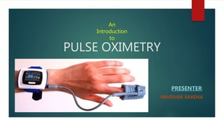
Pulse oximetry is a non-invasive method to measure oxygen saturation in the blood. It uses light transmitted through tissue to determine the ratio of oxygenated to deoxygenated hemoglobin. A pulse oximeter reading below 90% may indicate hypoxemia. While generally accurate, readings can be affected by factors like low perfusion, dyshemoglobinemias, or artificial nail finishes. Immediate actions for extremely low saturations include checking ABCs. A reading that improves with supplemental oxygen but remains low implies severe hypoxemia. Readings are lost after cardiac arrest and saturations decrease after respiratory arrest until cardiac arrest occurs.
Pulse oximetry is a noninvasive method for monitoring a person's oxygen saturation. Peripheral oxygen saturation (SpO2) readings are typically within 2% accuracy (within 4% accuracy in the worst 5% of cases) of the more desirable (and invasive) reading of arterial oxygen saturation (SaO2) from arterial blood gas analysis.
An arterial blood gas (ABG) test measures the acidity (PH) and levels of oxygen and carbon dioxide in the blood from an artery.
This test used to check how well your lungs are able to move oxygen into the blood and remove carbon dioxide from the blood
Pulse oximetry is a noninvasive method for monitoring a person's oxygen saturation. Peripheral oxygen saturation (SpO2) readings are typically within 2% accuracy (within 4% accuracy in the worst 5% of cases) of the more desirable (and invasive) reading of arterial oxygen saturation (SaO2) from arterial blood gas analysis.
An arterial blood gas (ABG) test measures the acidity (PH) and levels of oxygen and carbon dioxide in the blood from an artery.
This test used to check how well your lungs are able to move oxygen into the blood and remove carbon dioxide from the blood
Design of the Pulse Oximetry Measurement Circuit and Its Sensing System Based...IOSRJEEE
The pulse oximetry circuit and its sensing system is designed based on the standard CMOS technology of 0.18um. The reflection oxygen sensor is used to collect the pulse oximeter signal of human body, then the collected physiological signals are processed by the data processing circuit The data processing circuit is composed of two parts: the amplifying circuit and the band-pass filter circuit, and the pulse oximeter data processed by the data processing circuit is written into the tag through the SPI communication The RFID reader read the data in the RFID tag through wireless communication, and display the data . The experimental results show that the maximum error is ±1%. The maximum error of the pulse is ±1.9%. The stability and feasibility of pulse blood oxygen sensing system is demonstrated in this paper and it will have a good application prospect in the direction of wearable medical wisdom research
SpO2 stands for peripheral capillary oxygen saturation, an estimate of the amount of oxygen in the blood. More specifically, it is the percentage of oxygenated haemoglobin (haemoglobin containing oxygen) compared to the total amount of haemoglobin in the blood (oxygenated and non-oxygenated haemoglobin).SpO2 is an estimate of arterial oxygen saturation, or SaO2, which refers to the amount of oxygenated haemoglobin in the blood.
A very short, but effective presentation given by me on the topic, ' THE POWER OF SUBCONSCIOUS MIND ' at Jetking Regional Center, New Delhi. PPT, along with my words left a great impact on the minds of the listeners. And I received lots of appreciation in the end.
- Video recording of this lecture in English language: https://youtu.be/lK81BzxMqdo
- Video recording of this lecture in Arabic language: https://youtu.be/Ve4P0COk9OI
- Link to download the book free: https://nephrotube.blogspot.com/p/nephrotube-nephrology-books.html
- Link to NephroTube website: www.NephroTube.com
- Link to NephroTube social media accounts: https://nephrotube.blogspot.com/p/join-nephrotube-on-social-media.html
Factory Supply Best Quality Pmk Oil CAS 28578–16–7 PMK Powder in Stockrebeccabio
Factory Supply Best Quality Pmk Oil CAS 28578–16–7 PMK Powder in Stock
Telegram: bmksupplier
signal: +85264872720
threema: TUD4A6YC
You can contact me on Telegram or Threema
Communicate promptly and reply
Free of customs clearance, Double Clearance 100% pass delivery to USA, Canada, Spain, Germany, Netherland, Poland, Italy, Sweden, UK, Czech Republic, Australia, Mexico, Russia, Ukraine, Kazakhstan.Door to door service
Hot Selling Organic intermediates
Couples presenting to the infertility clinic- Do they really have infertility...Sujoy Dasgupta
Dr Sujoy Dasgupta presented the study on "Couples presenting to the infertility clinic- Do they really have infertility? – The unexplored stories of non-consummation" in the 13th Congress of the Asia Pacific Initiative on Reproduction (ASPIRE 2024) at Manila on 24 May, 2024.
Ozempic: Preoperative Management of Patients on GLP-1 Receptor Agonists Saeid Safari
Preoperative Management of Patients on GLP-1 Receptor Agonists like Ozempic and Semiglutide
ASA GUIDELINE
NYSORA Guideline
2 Case Reports of Gastric Ultrasound
Report Back from SGO 2024: What’s the Latest in Cervical Cancer?bkling
Are you curious about what’s new in cervical cancer research or unsure what the findings mean? Join Dr. Emily Ko, a gynecologic oncologist at Penn Medicine, to learn about the latest updates from the Society of Gynecologic Oncology (SGO) 2024 Annual Meeting on Women’s Cancer. Dr. Ko will discuss what the research presented at the conference means for you and answer your questions about the new developments.
HOT NEW PRODUCT! BIG SALES FAST SHIPPING NOW FROM CHINA!! EU KU DB BK substit...GL Anaacs
Contact us if you are interested:
Email / Skype : kefaya1771@gmail.com
Threema: PXHY5PDH
New BATCH Ku !!! MUCH IN DEMAND FAST SALE EVERY BATCH HAPPY GOOD EFFECT BIG BATCH !
Contact me on Threema or skype to start big business!!
Hot-sale products:
NEW HOT EUTYLONE WHITE CRYSTAL!!
5cl-adba precursor (semi finished )
5cl-adba raw materials
ADBB precursor (semi finished )
ADBB raw materials
APVP powder
5fadb/4f-adb
Jwh018 / Jwh210
Eutylone crystal
Protonitazene (hydrochloride) CAS: 119276-01-6
Flubrotizolam CAS: 57801-95-3
Metonitazene CAS: 14680-51-4
Payment terms: Western Union,MoneyGram,Bitcoin or USDT.
Deliver Time: Usually 7-15days
Shipping method: FedEx, TNT, DHL,UPS etc.Our deliveries are 100% safe, fast, reliable and discreet.
Samples will be sent for your evaluation!If you are interested in, please contact me, let's talk details.
We specializes in exporting high quality Research chemical, medical intermediate, Pharmaceutical chemicals and so on. Products are exported to USA, Canada, France, Korea, Japan,Russia, Southeast Asia and other countries.
Title: Sense of Taste
Presenter: Dr. Faiza, Assistant Professor of Physiology
Qualifications:
MBBS (Best Graduate, AIMC Lahore)
FCPS Physiology
ICMT, CHPE, DHPE (STMU)
MPH (GC University, Faisalabad)
MBA (Virtual University of Pakistan)
Learning Objectives:
Describe the structure and function of taste buds.
Describe the relationship between the taste threshold and taste index of common substances.
Explain the chemical basis and signal transduction of taste perception for each type of primary taste sensation.
Recognize different abnormalities of taste perception and their causes.
Key Topics:
Significance of Taste Sensation:
Differentiation between pleasant and harmful food
Influence on behavior
Selection of food based on metabolic needs
Receptors of Taste:
Taste buds on the tongue
Influence of sense of smell, texture of food, and pain stimulation (e.g., by pepper)
Primary and Secondary Taste Sensations:
Primary taste sensations: Sweet, Sour, Salty, Bitter, Umami
Chemical basis and signal transduction mechanisms for each taste
Taste Threshold and Index:
Taste threshold values for Sweet (sucrose), Salty (NaCl), Sour (HCl), and Bitter (Quinine)
Taste index relationship: Inversely proportional to taste threshold
Taste Blindness:
Inability to taste certain substances, particularly thiourea compounds
Example: Phenylthiocarbamide
Structure and Function of Taste Buds:
Composition: Epithelial cells, Sustentacular/Supporting cells, Taste cells, Basal cells
Features: Taste pores, Taste hairs/microvilli, and Taste nerve fibers
Location of Taste Buds:
Found in papillae of the tongue (Fungiform, Circumvallate, Foliate)
Also present on the palate, tonsillar pillars, epiglottis, and proximal esophagus
Mechanism of Taste Stimulation:
Interaction of taste substances with receptors on microvilli
Signal transduction pathways for Umami, Sweet, Bitter, Sour, and Salty tastes
Taste Sensitivity and Adaptation:
Decrease in sensitivity with age
Rapid adaptation of taste sensation
Role of Saliva in Taste:
Dissolution of tastants to reach receptors
Washing away the stimulus
Taste Preferences and Aversions:
Mechanisms behind taste preference and aversion
Influence of receptors and neural pathways
Impact of Sensory Nerve Damage:
Degeneration of taste buds if the sensory nerve fiber is cut
Abnormalities of Taste Detection:
Conditions: Ageusia, Hypogeusia, Dysgeusia (parageusia)
Causes: Nerve damage, neurological disorders, infections, poor oral hygiene, adverse drug effects, deficiencies, aging, tobacco use, altered neurotransmitter levels
Neurotransmitters and Taste Threshold:
Effects of serotonin (5-HT) and norepinephrine (NE) on taste sensitivity
Supertasters:
25% of the population with heightened sensitivity to taste, especially bitterness
Increased number of fungiform papillae
Tom Selleck Health: A Comprehensive Look at the Iconic Actor’s Wellness Journeygreendigital
Tom Selleck, an enduring figure in Hollywood. has captivated audiences for decades with his rugged charm, iconic moustache. and memorable roles in television and film. From his breakout role as Thomas Magnum in Magnum P.I. to his current portrayal of Frank Reagan in Blue Bloods. Selleck's career has spanned over 50 years. But beyond his professional achievements. fans have often been curious about Tom Selleck Health. especially as he has aged in the public eye.
Follow us on: Pinterest
Introduction
Many have been interested in Tom Selleck health. not only because of his enduring presence on screen but also because of the challenges. and lifestyle choices he has faced and made over the years. This article delves into the various aspects of Tom Selleck health. exploring his fitness regimen, diet, mental health. and the challenges he has encountered as he ages. We'll look at how he maintains his well-being. the health issues he has faced, and his approach to ageing .
Early Life and Career
Childhood and Athletic Beginnings
Tom Selleck was born on January 29, 1945, in Detroit, Michigan, and grew up in Sherman Oaks, California. From an early age, he was involved in sports, particularly basketball. which played a significant role in his physical development. His athletic pursuits continued into college. where he attended the University of Southern California (USC) on a basketball scholarship. This early involvement in sports laid a strong foundation for his physical health and disciplined lifestyle.
Transition to Acting
Selleck's transition from an athlete to an actor came with its physical demands. His first significant role in "Magnum P.I." required him to perform various stunts and maintain a fit appearance. This role, which he played from 1980 to 1988. necessitated a rigorous fitness routine to meet the show's demands. setting the stage for his long-term commitment to health and wellness.
Fitness Regimen
Workout Routine
Tom Selleck health and fitness regimen has evolved. adapting to his changing roles and age. During his "Magnum, P.I." days. Selleck's workouts were intense and focused on building and maintaining muscle mass. His routine included weightlifting, cardiovascular exercises. and specific training for the stunts he performed on the show.
Selleck adjusted his fitness routine as he aged to suit his body's needs. Today, his workouts focus on maintaining flexibility, strength, and cardiovascular health. He incorporates low-impact exercises such as swimming, walking, and light weightlifting. This balanced approach helps him stay fit without putting undue strain on his joints and muscles.
Importance of Flexibility and Mobility
In recent years, Selleck has emphasized the importance of flexibility and mobility in his fitness regimen. Understanding the natural decline in muscle mass and joint flexibility with age. he includes stretching and yoga in his routine. These practices help prevent injuries, improve posture, and maintain mobilit
3. Introduction
The maintenance of optimal O2 delivery is the core concern during anaesthesia
“Oxygen lacks not only stops the machinery, but wrecks the machinery” – J.S Haldane
Monitoring of oxygenation using pulse oximeter avoids many catastrophes
4. Hemoglobin
Hemoglobin (abbreviated Hb) is the iron containing oxygen transport mettaloprotein in
the red blood cells of the vertebrates.
Hemoglobin transports oxygen from the lungs to the rest of the body, where it releases
the oxygen for the cell use.
5.
6. Definition
PULSE OXIMETER is used to measure oxygen saturation in the body i.e how much of the
hemoglobin in the blood is carrying oxygen.
7. Pulse oximetry, sometimes called the fifth vital sign, is a non-invasive method of measuring
hemoglobin saturation (SpO2) by using a light signal transmitted through the tissue
A low SpO2 can provide warning of hypoxemia before other signs such as cyanosis or a change in
heart rate are observed.
It is one of the most essential monitors for routine use in anesthesia and intensive care
8. Pulse Oximetry
It measures the heart rate
Gives an idea about tissue perfusion by pulse wave form
Increased amplitude indicates vasodilation
Decreased amplitude indicates
vasoconstriction
9. Percentage of Hemoglobin (Hb) that is saturated with oxygen
The oxygen saturation (SpO2) is a measure of how much oxygen the blood is carrying as a
percentage of the maximum it could carry and is sometimes referred to colloquially as the
“sats” reading.
Heart rate refers to the number of times that the heart contracts in a period of one minute.
10. Types of Oximeters
Pulse Oximeter as a part of an anesthetic machine
A portable desktop unit
A finger/ mobile pulse oximeter
11.
12. A pulse oximeter can detect hypoxia (too little oxygen to fulfill the needs of the brain and
body) before the patient shows signs of becoming cyanotic (bluish discoloration of the
skin and the mucous membranes due to not enough oxygen in the blood)
Pulse oximeters may be used in patients:
a) Undergoing surgical procedure under
general anesthesia
b) Undergoing surgical procedure under
conscious sedation
13. c) Emergency situations like loss of consciousness,
Trauma etc
d) After surgery during the recovery phase
e) Monitoring the blood oxygen saturation in various
Aviation situations
f) Sports applications – e.g. Mountaineering
14. Pulse oximeters reading may not be accurate:
A) Reduced peripheral pulsatile blood flow
B) Various congestion (partial obstruction of the veins) of an arm or the leg
C) Bright overhead lights, such as in an operating theatre
D) Shivering or significant, repeated movement of sensor
E) Distinguishing between different forms of hemoglobin, such as carboxy Hb
F) Nail varnish may cause falsely low readings with most pulse oximeters, especially the
ones with color as blue or black.
16. Normal Reading
A fit, healthy person should have an oxygen saturation level between 95% - 99%
Results lower than this, and specially below 90% may be caused by problems including lung
diseases, such as COPD, breathing difficulties, cigarette smoking or circulatory problems
such as excessive bleeding or blood circulation problem.
17. History
Karl Matthes – 1935
First oximeter to measure O2 saturation
Subsequent oximeters developed by Hewlett Packardwere bulky and expensive ($10,000)
1972 – Takuo Aoyagi
Pulsatile changes in absorption of red and infra-red light to measure arterial oxygen saturation
BiOx, Nellcor (started by anesthesiologist Bill New) and Novametrix began manufacturing in 1980’s
1987 – ASA recommends inclusion of pulse oximetry and capnography into operating room as standard of care
Prior to this, morbidity and mortalityrelated to hypoxemia estimated at 1/2000 – 1/7000 cases
19. Beer-Lambert Law
The combination of both Beer’s Law and Lambert’s Law
Beer’s Law – the absorption of light is proportional to
the concentration of a sample
Lambert’s Law – absorption is proportional to the
thickness of a sample
20. Hemoglobin has a quaternary structure
Most commonly – Hgb A – 2 alpha and 2 beta subunits
Infants – Hgb F – 2 alpha and 2 gamma subunits
Within each of these subunits is a Heme group with a bound Iron atom
Fe2+ can bind oxygen. Upon binding oxygen, Fe2+ is oxidized to Fe3+ (methemoglobin), which can
not bind to oxygen
Upon binding a molecule of oxygen, hemoglobin undergoes a conformational change
This change enables further oxygen molecules to be bound more easily
21. This conformational change makes oxygen binding a cooperative process,
and is responsible for the shape of the oxygen-hemoglobin saturation curve
Pulse oximetry is based on the differential absorption
of light by oxyhemoglobin and deoxyhemoglobin
The oxygenated hemoglobin allows red light to transmit
through and absorbsmore infrared light while the deoxygenated
hemoglobin allows infrared to transmit through and absorbsmore
red light
22. A photodetector in the
sensor measures
unabsorbed light from the
LEDs
The resulting signal is
inverted and resembles
the diagram below
23. At each site, there are constant light absorbers
present
Tissue, venous blood and non pulsatile
arterial blood
Surge in arterial blood with each heartbeat
results in more light absorbed.
So the troughs of lower light absorption are
subtracted from the peaks, leaving only arterial
bloods light absorption being measured
Hence “pulse oximetry”
24. After the photodetector, the Red/Infrared ratio is calculated
This is compared to an algorithm that is specific to each company/device
and is based on measurements obtained in healthy volunteers
This ratio corresponds to Sp02
0.5 is approx 100%
1 is approx 85%
2 is approx 0%
25. Uses
Arterial oxygenation, and, because the variation in light
absorption is proportional to the volume of arterial blood with
each heart beat, it can be used to estimate volume status (with
some computational help)
26. Sources of Error
Strength of Arterial Pulse
Any factor that reduces arterial pulsations will reduce the ability of the instrument to
obtain and analyze the signal
Hypothermia
Hypotension
Vasopressor use
Body Movement
Extraneous movements can cause intermittent changes in absorbance
Shivering
Parkinsonian tremors
27. Dyshemoglobinemias
Carboxyhemoglobin
CO binds to heme competitively with 250 times the affinity of oxygen
COHgb has same absorption pattern of 660nm light as O2Hgb
Readings are artificially high
Describes the oxidized form of hemoglobin (Fe3+)
Methemoglobin absorbs as much 660nm red light as it does the 940nm infrared
Sats approach 85%
Falsely low at high Sp02, falsely high at low SpO2
Methemoglobin
28. Methylene Blue, indigo carmine, indocyanin green
Cause drop in Sp02
Color Interference
Pulse oximetry not affected by skin color
Is affected by artificial or opaque nail finishes that may interfere
with transmission of light
29. Physical Factors
Electrocautery
Interferes with signal
BP Cuff
Don’t place it on the same arm (and forget…)
High intensity light
Can interfere with signal
(make sure the probe is covered)
30. Venous Pulsations
Secondary to AV fistulas
Saturations below 80% are inferred, and not based on
measurement
The R/IR ratio and its correlation to oxygen saturation is based on
measurements made on healthy volunteers
Only a genocidal IRB would allow for measurements of both to be
made at sats < 70%
31. Q1.
If a patient’s saturation is extremely low, what are your immediate actions?
Check Airways, Breathing and Circulation [ABC]
32. Q2.
An elderly patient is admitted with pneumonia and has a pulse oximetry reading
of 75% breathing air. With oxygen 6L / min, saturation improves to 85%. What are
the implications of this oximetry reading?
The patient is severely hypoxic [Lack of Oxygen]
“INCREASE OXYGEN LEVELS”
33. Q3.
What happens to the pulse oximeter reading of a patient immediately after a
cardiac arrest?
The pulse would be lost (causing the alarm to sound)
And the saturations will decrease
34. Q4.
What happens to the Pulse Oximeter reading of a patient immediately after a
respiratory arrest?
The saturations decrease until cardiac arrest occurs