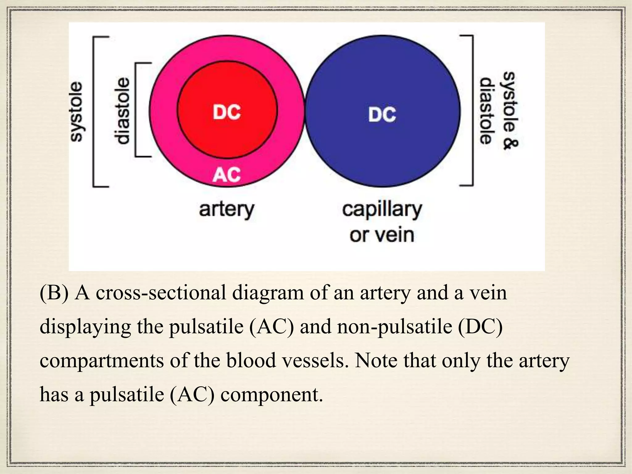
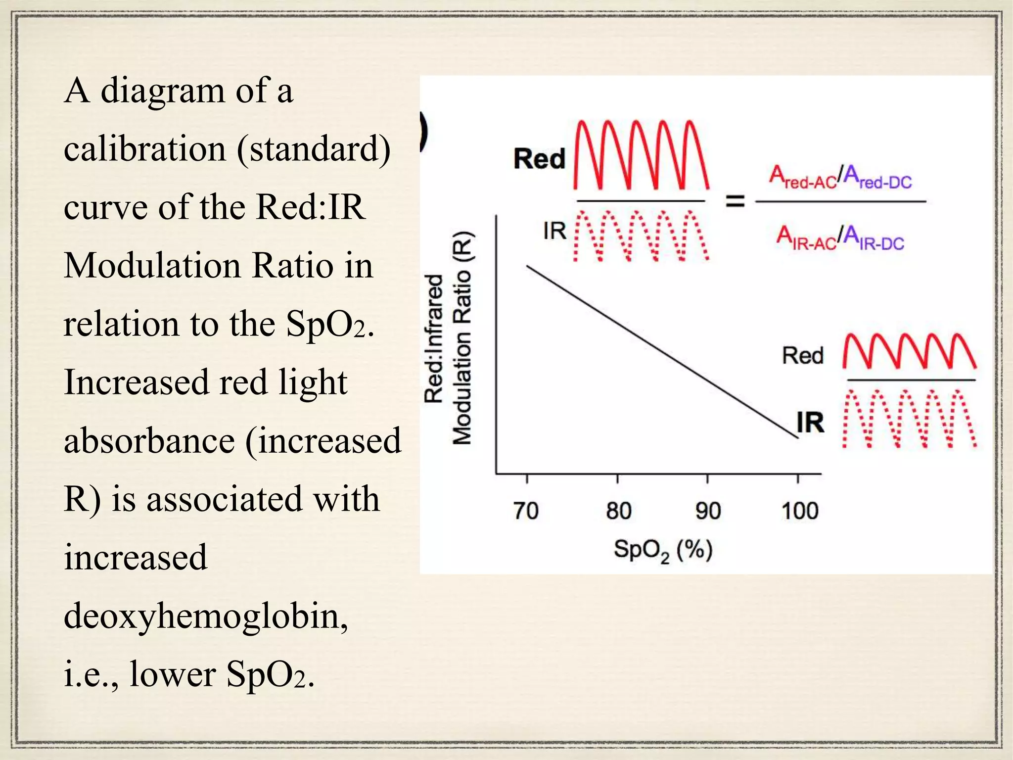
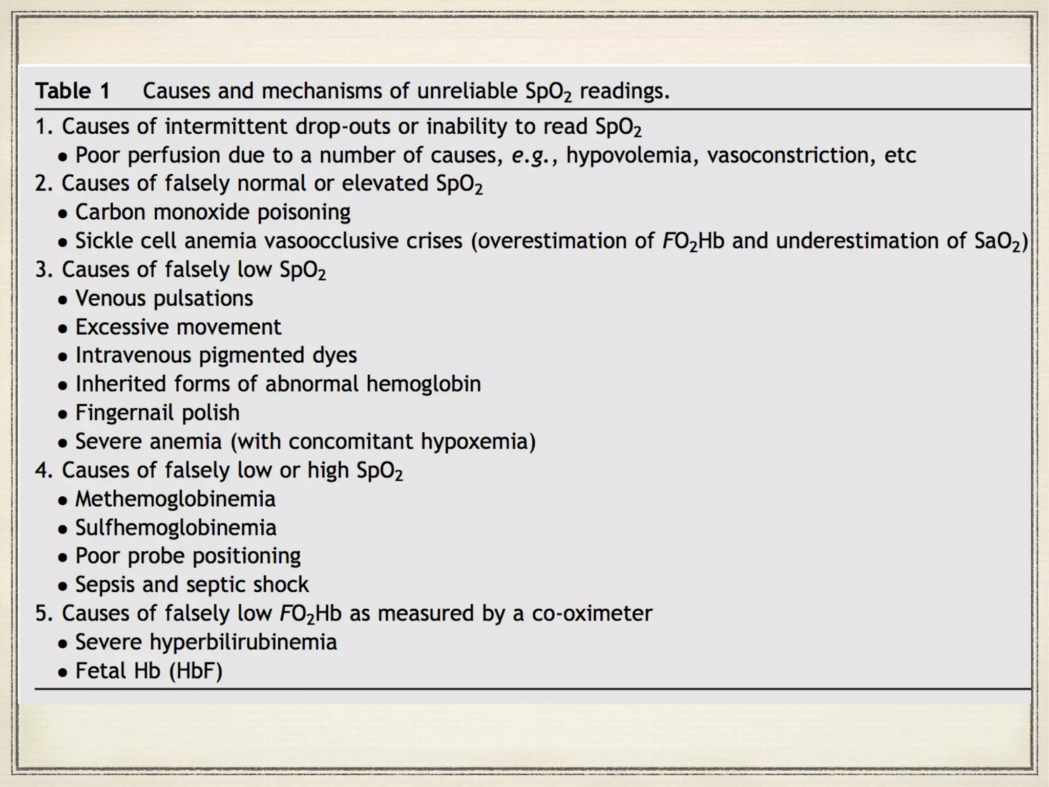
PULSE OXIMETRY: UNDERSTANDING
ITS BASIC PRINCIPLES FACILITATES
APPRECIATION OF ITS LIMITATIONS
Edward D. Chan a,b,c,d,*, Michael M. Chan e
, Mallory M. Chan f
DR. AMIT SINGH
BASIC PRINCIPLES OF FUNCTION
The pulse oximeter is a device with its ability to continuously and
transcutaneously & monitor the functional oxygen saturation of
hemoglobin in arterial blood.
It is important to understand the functions as well as its
limitations because erroneous readings can lead to unnecessary
testing.
Frequent false alarms in the intensive care unit can also
undermine patient safety by distracting caregivers.
2 basic principles of pulse oximetry is required:
(i) how oxyhemoglobin (O2Hb) is distinguished from
deoxyhemoglobin (HHb) and
(ii) how the SpO2 is calculated only from the arterial
compartment of blood.
Pulse oximetry is based on the principle that O2Hb and HHb
differentially absorb red and near-infrared (IR) light.
O2Hb absorbs greater amounts of IR light and lower amounts
of red light than does HHb.
oxygenated blood with its higher concentrations of O2Hb
appears bright red to the eye because it scatters more red
light than does HHb.
On the other hand, HHb absorb more red light and appears
less red.
pulse oximeters emit two wavelengths of light, red at 660 nm
and near-IR at 940 nm from a pair of small light-emitting
diodes located in one arm of the finger probe.
The light that is transmitted through the finger is then detected
by a photodiode on the opposite arm of the probe to ultimately
determine the proportion of Hb bound to oxygen.
(A) In a person with good
cardiac function, the onset of the
cardiac systole, as denoted by
the onset of the QRS complex
coincides with the onset in the
increase of the arterial blood
volume. The amount of red and
IR light absorbed in the arterial
compartment also rises and fall
with systole and diastole,
respectively, due to the increase
and decrease in blood volume.
The volume that increases with
systole is also known as the
pulsatile or “alternating current”
(AC) compartment and the
compartment in which the blood
volume does not change with the
cardiac cycle is known as the
non-pulsatile or “direct current”
(DC) compartment.
(B) A cross-sectional diagram of an artery and a vein
displaying the pulsatile (AC) and non-pulsatile (DC)
compartments of the blood vessels. Note that only the artery
has a pulsatile (AC) component.
The ability of pulse oximetry is to detect SpO2 of only arterial blood is based on
the principle that the amount of red and IR light absorbed fluctuates with the
cardiac cycle, as the arterial blood volume increases during systole and
decreases during diastole
An adequate pulse is necessary for pulse oximeters to work and is the basis for
the well-known fact that attempting to measure SpO2 in regions with little or no
blood perfusion will result in absent or inaccurate readings.
R is a double-ratio of the pulsatile and non-pulsatile components of red light
absorption to IR light absorption.
At low arterial oxygen saturations :
higher R value ,higher oxygen saturations,
and the R value is lower ..
A microprocessor in pulse oximeters uses this ratio
(calculated over a series of pulses) to determine the SpO2
based on a calibration curve that was generated empirically
by measuring R in healthy volunteers whose saturations
were altered from 100% to approximately 70%.
Thus, SpO2readings below 70% should not be considered
quantitatively reliable although it is unlikely any clinical
decisions would be altered based on any differences in
SpO2 measured below 70%.
A diagram of a
calibration (standard)
curve of the Red:IR
Modulation Ratio in
relation to the SpO2.
Increased red light
absorbance (increased
R) is associated with
increased
deoxyhemoglobin,
i.e., lower SpO2.
PROS AND CONS OF DIFFERENT TYPES OF
PULSE OXIMETER PROBES
The pulse oximeter probes interrogate the finger, nose, ear lobe,
and forehead as in these areas have a much higher vascular
density
2 types of phrobes are used:
Reusable clip probes (finger, nasal, ear)
{repidity, cost-effectivenesss, ease to use, opd pt, multiple pt,
and low amplitude waves}
Single- patient adhesive probes (finger, forehead)
{less transmission of nosocomial infections , more secure
placement when there is excessive pt. movement}
Even after optimizing the type of probe, false readings of SpO22 may
occur in various settings and medical conditions.
Causes of intermittent dropouts or inability to
read SpO2
It is common knowledge that an attenuated and inconsistent wave tracing
generated by pulse oximeters displayed on intensive care unit monitors is
an indication that the SpO2 reading is unreliable.
The amplitude of such a pulse oximeter waveform reflects the amount of
cardiac induced light modulation .
Although portable pulse oximeters do not typically have such wave
tracings.
They often have a pulse signal bar that shows the level of change in light
absorbance
Low-amplitude pulse oximetry wave tracings may be due to poor finger
perfusion resulting from vasoconstriction and/or hypotension from a
number of causes including distributive or hypovoluemic shock,
hypothermia, use of vasoconstrictor agents, and poor cardiac output due
to pump failure or dysrhythmia.
Arterial compression or arterial blockage proximal to probe placement can
Causes of falsely normal or elevated
SpO22
Carbon monoxide (CO) is an odorless, colorless, tasteless, and
non-irritating gas.
The principal pathogenic mechanism of CO poisoning is the strong
avidity of CO for Hb (240 greater than O2), forming carboxy-Hb
(COHb), reducing the O2 carrying capacity of Hb and precipitating
tissue hypoxemia.
Because O2Hb and COHb absorb red light (660 nm) similarly and
COHb absorbs very little near-IR light (940 nm), the photodiode of
standard pulse oximeters that only emit red and near-IR light
cannot differentiate between O2Hb and COHb.
COHb will decrease both HHb and O2Hb concentrations, the
normally greater red light absorption by HHb will be decreased but
the red light absorption of the combined O2Hb and COHb will be
Carbon monoxide poisoning
Because of HHb still absorbs red light better than either COHb or
O2Hb, the reduction in HHb in the presence of
carboxyhemoglobinemia results in a net effect of decreased red
light absorption.
Thus, in significant carboxyhemoglobinemia, standard pulse
oximeters can give a false normal (or elevated) SpO2
Complications of sickle cell disease, such as vasoocclusive crisis and
acute chest syndrome, are often precipitated or exacerbated by
hypoxemia, and can result in a vicious cycle of additional sickling and
vasoocclusive crises.
The accuracy of pulse oximetry in monitoring oxygenation in sickle cell
disease has been debated.
In a study of 17 patients with sickle cell disease, SpO2 was found to
overestimate FO2Hb content by an average of 3.4% and to
underestimate SaO2 by an average of 1.1%.
Similarly, a study of 24 patients with sickle cell disease determined that
SpO2 underestimated SaO2 readings by 1.6%
FO2Hb may be a more useful measurement, as some individuals with
sickle cell disease may have elevated COHb (due to the metabolism of
heme to bilirubin and CO)
In these instances, SpO2 over- estimates the FO2Hb
Sickle cell anemia vasoocclusive crises
Another important aspect with oxygen saturation
determination in sickle cell patients is that under
normoxic conditions, where there is little HbS
polymerization, HbS affinity for oxygen is similar to the
normal HbA.
However, in the presence of conditions that precipitates
sickling, including hypoxemia, HbS has less affinity for
oxygen and the oxygen-Hb dissociation curve is shifted
to the right, i.e., for any given PaO2, the SaO2 is lower.
Teleologically, this shift is advantageous to increase
oxygen unloading to tissues.
Causes of falsely low SpO2
Venous pulsations, causing a significant change in venous
volume with each cardiac cycle, can contribute to a falsely
low SpO2 reading
Venous pulsations can occur when adhesive fingerprobes are
placed too tightly around the finger, in severe tricuspid
regurgitation, when the location of the probes are in
dependent positions
And possibly in distributive shock where wide- spread
vasodilation results in physiologic arteriovenous shunting.
When venous pulsations is considered to be occurring with a
forehead sensor in a patient requiring the supine or
Trendelenburg position, an elastic-tensioned headband
Venous pulsations
Excessive movement such as tremor or convulsions
has been documented to cause spuriously low SpO2,
with desaturations below 50% sometimes observed,
though less commonly SpO2 overestimations can also
occur.
In theory, motion can cause the normally static tissues
in relation to the sensor position to change over the
time frame of the arterial pulses.
At times, this motion can augment or mimic the
cardiac-induced signals as the blood in the veins (and
other previously stationary tissues) are now moving,
further modulating the red and IR light attenuation in the
probed tissue
Excessive movement :
Pigmented dyes are used medically or as agents in various medical
diagnostic tests; e.g., the principal medicinal use of methylene blue is in
methemoglobinemia where it serves as a reducing agent.
Methylene blue — endoscopic polypectomy
Indigo carmine based dye —to detect amniotic fluid leaks as well
as to detect leaks in the urinary system during surgery
Iodocyanine —determination of hepatic function and liver blood
flow, and for ophthalmic angiography.
Peak light absorption of methylene blue is 668 nm , very close to the
strong red light absorption by HHb, resulting in a higher R value and
mimicking HHb.
Hence, administration of methylene blue into the blood stream can
falsely reduce SpO2
readings.
Intravenous pigmented dyes:
Earlier reports of pulse oximeters noted that
fingernail polish, particularly black, blue, and
green color, can lower SpO2 by up to 10%.
Black and brown fingernail polish displayed the
greatest reduction in the SpO2 reading but by an
average decrease of 2%.
Fingernail polish:
Causes of falsely low or high SpO2
Methemoglobin (MetHb) is formed when the iron (Fe) in the heme
moiety is oxidized from Fe+2 to Fe+3.
MetHb impairs oxygen delivery to tissues by two distinct mechanisms:
(i) MetHb itself is less able to bind oxygen and ,
(ii) MetHb causes the oxygen dissociation curve of normal Hb to shift
to the left which impairs unloading of oxygen at the tissue level.
Most cases of methemoglobinemia are usually due to oxidizing
chemicals or drugs such as nitrites, nitrates, aniline dyes, aniline
derivatives (phenacetin, dapsone), sulfonamides, and lidocaine.
Only 1.5-2 gm MetHb/dL of blood (a 10-20% of total Hb) can cause
cyanosis.
Methemoglobinemia
MetHb levels between 20 and 45% can produce
headaches, weakness, breathlessness, and altered
mental status.
MetHb levels between 50 and 60% are often fatal,
presenting with hemodynamic instability, generalized
seizures, and coma.
MetHb absorbs more IR than either O2Hb or HHb .
Since its absorbance of red light is similar to HHb.
Moreover, because MetHb absorbs both red and IR
light equally well , high MetHb levels result in an R
value that approaches 1, which happens to be
equivalent to a SpO2 of 80-85%.
Sulfhemoglobin (SulfHb) is formed by the irreversible oxidation of Feþ2
in the
porphyrin ring of Hb to Feþ3
(similar to MetHb) but additionally has a sulfur atom
incorporated into the porphyrin ring.
SulfHb may be formed by aniline dyes, acetanilid, phenacetin
phenozapyridine, nitrates, sulfonamides, metoclopramide (structurally similar
to aniline dyes), flutamide, dapsone, dimethyl sulfoxide (DMSO) or by
exposure to sulfurated chemicals such as hydroxylamine sulfate used in
various cleaning solutions.42e45
SulfHb cannot carry oxygen.
The major clinical manifestation of sulfhemoglobinemia is the presence of
cyanosis. A mean capillary concentration of only 0.5 gm/dL
SulfHb has greater red light (660 nm) absorbance than HHb or MetHb.43
Although it is not known precisely, SulfHb most likely has a similar degree of
absorbance at red and near-IR light because there have been several reports
that the SpO2 of persons with severe sulfhemoglobinemia are w85%, a sign that
absorbance of red and near-IR light are similar with SulfHb, resulting in a R
value that approaches 1 (as seen with methemoglobinemia).
Sulfhemoglobinemia:
In older models of pulse oximeters, ambient light
complicated SpO2
measurements by directly hitting the
photodetector and/or by increasing the amount light
going through the tissues, making the SpO2
measurements unreliable.
However, most modern pulse oximeters are able to
“subtract” the ambient light signals.
obe positioning resulting in decrease absorption of red and/or
Secker and Spierscompared 80 paired SpO2 and SaO2 readings in
patients with septic shock and showed that only in those with low
systemic vascular resistance, the SpO2 underestimated the SaO2 by
a mean of 1.4% .
A hypothesis to explain this phenomenon is that vasodilation from
the sepsis resulted in creation of arteriovenous shunts that
culminated in venous pulsations and spurious detection by the pulse
oximeter of some venous blood as arterial.
A retrospective review of 88 patients with severe sepsis and septic
shock found that in those with hypoxemia (defined as SaO2 < 90%),
SpO2 significantly overestimated SaO2 by nearly 5%.
In several studies examining critically ill patients with various acute
illnesses, some found SpO2 underestimated SaO2 while others found
the opposite.
Sepsis and septic shock
Even in those studies that examined only septic
patients, factors that may account for a difference in
the direction in which SpO2 is biased include
differences in:
(i) The extent of fluid resuscitation and tissue
perfusion,
(ii) Sepsis-induced cardiac dysfunction,
(iii) Sites interrogated by the pulse oximeter probe,
(iv) Types of pulse oximeters and probes used,
(v) Vasoconstrictor use, and
(vi) The presence of other comorbid conditions that
may spuriously affect SpO2 values unpredictably.
Causes of falsely low FO2
Hb as measured by
a co-oximeter
With metabolism of each heme molecule, one molecule of
bilirubin and one molecule of CO is produced. Bilirubin
itself has minimal effect on SpO2 measurements on
standard pulse oximeters since this pigment has little or
no absorbance of red and near-IR light.
However, very high bilirubin levels can make oximeter
measurements of FO2Hb falsely low by increasing MetHb
and COHb levels; thus, pulse oximetry measurements of
oxygen saturation (SpO2) may actually be more accurate
than FO2Hb in the presence of severe hyperbilirubinemia.
to increased heme metabolism (hemolysis) or decreased biliru
Summary
Pulse oximetry is based on the principle that O2Hb absorbs more
near-IR light than HHb, and HHb absorbs more red light than
O2Hb. Under optimal conditions, pulse oximeters do not calculate
SpO2 of venous blood.
Since pulse oximeters are calibrated for SpO2 between 70 and
100%, displayed values below 70% should only be considered
qualitatively accurate and not quantitatively.
Inaccurate SpO2 readings can occur with conditions that decrease
arterial blood perfusion.
The SpO2 can overestimate FO2Hb content in blood that contains
increased amount of CO.
In sickle cell anemia-associated vasoocclusive crises,
SpO2 can overestimate the FO2Hb (due likely to the
presence of increased endogenous production of CO) but
could underestimate the SaO2 (due to vasoocclusion-
induced underperfusion).
SpO2 can either overestimate or underestimate the SaO2
with methemoglobinemia, sulfhemoglobinemia, and poor
probe positioning.
Finally, it is important to emphasize that even in the most
optimal patient-pulse oximeter interface and settings, a
completely normal SpO2 does not rule out gas exchange
problems in the lungs or the adequacy of ventilation since
the alveolar-arterial oxygen difference and PaCO2 are not
measured by pulse oximetry.
1. Neff T. Routine oximetry: a fifth vital sign? Chest 1988;94:227. 2. Stoneham MD, Saville GM, Wilson IH. Knowledge about pulse oximetry among medical and nursing
staff. Lancet 1994;344:
1339e42.
3. Sinex JE. Pulse oximetry: principles and limitations. Am J
Emerg Med 1999;17:59e67.
4. Mannheimer PD. The light-tissue interaction of pulse oximetry.
Anesth Analg 2007;105:S10e7.
5. Schnapp LM, Cohen NH. Pulse oximetry: uses and abuses. Chest
1990;98:1244e50.
6. Kelleher JF. Pulse oximetry. J Clin Monit 1989;5:37e62.
7. EvansML,GeddesLA.Anassessmentofbloodvesselvasoactivity
using photoplethysmography. Med Instrum 1988;22:29e32.
8. Berkenbosch JW, Tobias JD. Comparison of a new forehead reflectance pulse oximeter sensor with a conventional digit
sensor in pediatric patients. Respir Care 2006;51:726e31.
9. MacLeod DB, Cortinez LI, Keifer JC, Cameron D, Wright DR, White WD, et al. The desaturation response time of finger pulse oximeters during mild hypothermia.
Anaesthesia 2005;60:
65e71.
10. Tremper KK, Barker SJ. Pulse oximetry. Anesthesiology 1989;
70:98e108.
11.Gladwin MT, Vichinsky E. Pulmonary complications of sickle cell disease. N Engl J Med 2008;359:2254e65.
12.Ortiz FO, Aldrich TK, Nagel RL, Benjamin LJ. Accuracy of pulse oximetry in sickle cell disease. Am J Respir Crit Care Med 1999; 159:447e51.
13.Fitzgerald RK, Johnson A. Pulse oximetry in sickle cell anemia. Crit Care Med 2001;29:1803e6.
14.Ahmed S, Siddiqui AK, Sison CP, Shahid RK, Mattana J. Hemo- globin oxygen saturation discrepancy using various methods in patients with sickle cell vaso-
occlusive painful crisis. Eur J Haematol 2005;74:309e14.
15.Comber JT, Lopez BL. Evaluation of pulse oximetry in sickle cell anemia patients presenting to the emergency department in acute vasoocclusive crisis. Am J
Emerg Med 1996;14:16e8.
16.Abdu A, Go ́mez-Ma ́rquez J, Aldrich TK. The oxygen affinity of sickle hemoglobin. Respir Physiol Neurobiol 2008;161:92e4.
17.Lonsdorfer J, Bogui P, Otayeck A, Bursaux A, Poyart C, Cabannes R. Cardiorespiratory adjustments in chronic sickle cell anemia. Bull Eur Physiopathol Respir
1983;19:339e44.
18.Secker C, Spiers P. Accuracy of pulse oximetry in patients with low systemic vascular resistance. Anaesthesia 1997;52:127e30.
19.Stewart KG, Rowbottom SJ. Inaccuracy of pulse oximetry in patients with severe tricuspid regurgitation. Anaesthesia 1991;
46:668e70.
20.Agashe GS, Coakley J, Mannheimer PD. Forehead pulse oxim-
40. Perkins GD, McAuley DF, Giles S, Routledge H, Gao F. Do changes in pulse oximeter oxygen saturation predict equiva- lent changes in arterial oxygen saturation?
Crit Care Med 2003;
7:R67e71.
41. Janssen WJ, Dhaliwal G, Collard HR, Saint S. Clinical problem-
solving. Why “why” matters. N Engl J Med 2004;351:2429e34. 42. Lu HC, Shih RD, Marcus S, Ruck B, Jennis T. Pseudomethemo- globinemia: a case report and review
of sulfhemoglobinemia.
References :
21.Petterson MT, Begnoche VL, Graybeal JM. The effect of motion on pulse oximetry and its clinical significance. Anesth Analg
2007;105:S78e84.
22.Ortega R, Hansen CJ, Elterman K, Woo A. Videos in clinical medicine. Pulse oximetry. N Engl J Med 2011;364:e33.
23.Scheller MS, Unger RJ, Kelner MJ. Effects of intravenously administered dyes on pulse oximetry readings. Anesthesiology
1986;65:550e2.
24.Sarikonda KV, Ribeiro RS, Herrick JL, Hoyer JD. Hemoglobin lansing: a novel hemoglobin variant causing falsely decreased oxygen
saturation by pulse oximetry. Am J Hematol 2009;84:541.
25.Zur B, Hornung A, Breuer J, Doll U, Bernhardt C, Ludwig M, et al. A novel hemoglobin, Bonn, causes falsely decreased oxygen
saturation measurements in pulse oximetry. Clin Chem 2008;54:594e6.
26.Gottschalk A, Silverberg M. An unexpected finding with pulse oximetry in a patient with hemoglobin K ̈oln. Anesthesiology
1994;80:474e6.
27.Katoh R, Miyake T, Arai T. Unexpectedly low pulse oximeter readings in a boy with unstable hemoglobin K ̈oln. Anesthesi- ology
1994;80:472e4.
28.Mariette S, Leteurtre S, Lambilliotte A, Leclerc F. Pulse ox- imetry and genetic hemoglobinopathies. Intensive Care Med 2005;31:1597.
29.Hohl RJ, Sherburne AR, Feeley JE, Huisman TH, Burns CP. Low pulse oximeter-measured hemoglobin oxygen saturations with
hemoglobin Cheverly. Am J Hematol 1998;59.
30.Cote ́ CJ, Goldstein A, Fuchsman WH, Hoaglin DC. The effect of nail polish on pulse oximetry. Anesth Analg 1988;67:683e6.
31.Rubin AS. Nail polish color can affect pulse oximeter satura- tion. Anesthesiology 1988;68:825.
32.Chan MM, Chan MM, Chan ED. What is the effect of fingernail polish on pulse oximetry? Chest 2003;123:2163e4.
33.Hinkelbein J, Genzwuerker HV, Sogl R, Fiedler F. Effect of nail polish on oxygen saturation determined by pulse oximetry in critically ill
patients. Resuscitation 2007;72:82e91.
34. McGuire TJ, Pointer JE. Evaluation of a pulse oximeter in the prehospital setting. Ann Emerg Med 1988;17:1058e62.
35. New W. Pulse oximetry. J Clin Monit 1985;1:126e9.
36. Schmitt JM. Simple photon diffusion analysis of the effects of multiple scattering on pulse oximetry. IEEE Trans Biomed Eng
1991;38:1194e203.
37. Lee S, Tremper KK, Barker SJ. Effects of anemia on pulse ox-
imetry and continuous mixed venous hemoglobin saturation
monitoring in dogs. Anesthesiology 1991;75:118e22.
38. Severinghaus JW, Koh SO. Effect of anemia on pulse oximeter
accuracy at low saturation. J Clin Monit 1990;6:85e8.
39. Jay GD, Hughes L, Renzi FP. Pulse oximetry is accurate in acute
Pulse oximetry journal

Pulse oximetry journal

  • 1.
    PULSE OXIMETRY: UNDERSTANDING ITSBASIC PRINCIPLES FACILITATES APPRECIATION OF ITS LIMITATIONS Edward D. Chan a,b,c,d,*, Michael M. Chan e , Mallory M. Chan f DR. AMIT SINGH
  • 2.
    BASIC PRINCIPLES OFFUNCTION The pulse oximeter is a device with its ability to continuously and transcutaneously & monitor the functional oxygen saturation of hemoglobin in arterial blood. It is important to understand the functions as well as its limitations because erroneous readings can lead to unnecessary testing. Frequent false alarms in the intensive care unit can also undermine patient safety by distracting caregivers. 2 basic principles of pulse oximetry is required: (i) how oxyhemoglobin (O2Hb) is distinguished from deoxyhemoglobin (HHb) and (ii) how the SpO2 is calculated only from the arterial compartment of blood.
  • 3.
    Pulse oximetry isbased on the principle that O2Hb and HHb differentially absorb red and near-infrared (IR) light. O2Hb absorbs greater amounts of IR light and lower amounts of red light than does HHb. oxygenated blood with its higher concentrations of O2Hb appears bright red to the eye because it scatters more red light than does HHb. On the other hand, HHb absorb more red light and appears less red. pulse oximeters emit two wavelengths of light, red at 660 nm and near-IR at 940 nm from a pair of small light-emitting diodes located in one arm of the finger probe. The light that is transmitted through the finger is then detected by a photodiode on the opposite arm of the probe to ultimately determine the proportion of Hb bound to oxygen.
  • 4.
    (A) In aperson with good cardiac function, the onset of the cardiac systole, as denoted by the onset of the QRS complex coincides with the onset in the increase of the arterial blood volume. The amount of red and IR light absorbed in the arterial compartment also rises and fall with systole and diastole, respectively, due to the increase and decrease in blood volume. The volume that increases with systole is also known as the pulsatile or “alternating current” (AC) compartment and the compartment in which the blood volume does not change with the cardiac cycle is known as the non-pulsatile or “direct current” (DC) compartment.
  • 5.
    (B) A cross-sectionaldiagram of an artery and a vein displaying the pulsatile (AC) and non-pulsatile (DC) compartments of the blood vessels. Note that only the artery has a pulsatile (AC) component.
  • 6.
    The ability ofpulse oximetry is to detect SpO2 of only arterial blood is based on the principle that the amount of red and IR light absorbed fluctuates with the cardiac cycle, as the arterial blood volume increases during systole and decreases during diastole An adequate pulse is necessary for pulse oximeters to work and is the basis for the well-known fact that attempting to measure SpO2 in regions with little or no blood perfusion will result in absent or inaccurate readings. R is a double-ratio of the pulsatile and non-pulsatile components of red light absorption to IR light absorption. At low arterial oxygen saturations : higher R value ,higher oxygen saturations, and the R value is lower ..
  • 7.
    A microprocessor inpulse oximeters uses this ratio (calculated over a series of pulses) to determine the SpO2 based on a calibration curve that was generated empirically by measuring R in healthy volunteers whose saturations were altered from 100% to approximately 70%. Thus, SpO2readings below 70% should not be considered quantitatively reliable although it is unlikely any clinical decisions would be altered based on any differences in SpO2 measured below 70%.
  • 8.
    A diagram ofa calibration (standard) curve of the Red:IR Modulation Ratio in relation to the SpO2. Increased red light absorbance (increased R) is associated with increased deoxyhemoglobin, i.e., lower SpO2.
  • 9.
    PROS AND CONSOF DIFFERENT TYPES OF PULSE OXIMETER PROBES The pulse oximeter probes interrogate the finger, nose, ear lobe, and forehead as in these areas have a much higher vascular density 2 types of phrobes are used: Reusable clip probes (finger, nasal, ear) {repidity, cost-effectivenesss, ease to use, opd pt, multiple pt, and low amplitude waves} Single- patient adhesive probes (finger, forehead) {less transmission of nosocomial infections , more secure placement when there is excessive pt. movement} Even after optimizing the type of probe, false readings of SpO22 may occur in various settings and medical conditions.
  • 11.
    Causes of intermittentdropouts or inability to read SpO2 It is common knowledge that an attenuated and inconsistent wave tracing generated by pulse oximeters displayed on intensive care unit monitors is an indication that the SpO2 reading is unreliable. The amplitude of such a pulse oximeter waveform reflects the amount of cardiac induced light modulation . Although portable pulse oximeters do not typically have such wave tracings. They often have a pulse signal bar that shows the level of change in light absorbance Low-amplitude pulse oximetry wave tracings may be due to poor finger perfusion resulting from vasoconstriction and/or hypotension from a number of causes including distributive or hypovoluemic shock, hypothermia, use of vasoconstrictor agents, and poor cardiac output due to pump failure or dysrhythmia. Arterial compression or arterial blockage proximal to probe placement can
  • 13.
    Causes of falselynormal or elevated SpO22 Carbon monoxide (CO) is an odorless, colorless, tasteless, and non-irritating gas. The principal pathogenic mechanism of CO poisoning is the strong avidity of CO for Hb (240 greater than O2), forming carboxy-Hb (COHb), reducing the O2 carrying capacity of Hb and precipitating tissue hypoxemia. Because O2Hb and COHb absorb red light (660 nm) similarly and COHb absorbs very little near-IR light (940 nm), the photodiode of standard pulse oximeters that only emit red and near-IR light cannot differentiate between O2Hb and COHb. COHb will decrease both HHb and O2Hb concentrations, the normally greater red light absorption by HHb will be decreased but the red light absorption of the combined O2Hb and COHb will be Carbon monoxide poisoning
  • 14.
    Because of HHbstill absorbs red light better than either COHb or O2Hb, the reduction in HHb in the presence of carboxyhemoglobinemia results in a net effect of decreased red light absorption. Thus, in significant carboxyhemoglobinemia, standard pulse oximeters can give a false normal (or elevated) SpO2
  • 15.
    Complications of sicklecell disease, such as vasoocclusive crisis and acute chest syndrome, are often precipitated or exacerbated by hypoxemia, and can result in a vicious cycle of additional sickling and vasoocclusive crises. The accuracy of pulse oximetry in monitoring oxygenation in sickle cell disease has been debated. In a study of 17 patients with sickle cell disease, SpO2 was found to overestimate FO2Hb content by an average of 3.4% and to underestimate SaO2 by an average of 1.1%. Similarly, a study of 24 patients with sickle cell disease determined that SpO2 underestimated SaO2 readings by 1.6% FO2Hb may be a more useful measurement, as some individuals with sickle cell disease may have elevated COHb (due to the metabolism of heme to bilirubin and CO) In these instances, SpO2 over- estimates the FO2Hb Sickle cell anemia vasoocclusive crises
  • 16.
    Another important aspectwith oxygen saturation determination in sickle cell patients is that under normoxic conditions, where there is little HbS polymerization, HbS affinity for oxygen is similar to the normal HbA. However, in the presence of conditions that precipitates sickling, including hypoxemia, HbS has less affinity for oxygen and the oxygen-Hb dissociation curve is shifted to the right, i.e., for any given PaO2, the SaO2 is lower. Teleologically, this shift is advantageous to increase oxygen unloading to tissues.
  • 17.
    Causes of falselylow SpO2 Venous pulsations, causing a significant change in venous volume with each cardiac cycle, can contribute to a falsely low SpO2 reading Venous pulsations can occur when adhesive fingerprobes are placed too tightly around the finger, in severe tricuspid regurgitation, when the location of the probes are in dependent positions And possibly in distributive shock where wide- spread vasodilation results in physiologic arteriovenous shunting. When venous pulsations is considered to be occurring with a forehead sensor in a patient requiring the supine or Trendelenburg position, an elastic-tensioned headband Venous pulsations
  • 18.
    Excessive movement suchas tremor or convulsions has been documented to cause spuriously low SpO2, with desaturations below 50% sometimes observed, though less commonly SpO2 overestimations can also occur. In theory, motion can cause the normally static tissues in relation to the sensor position to change over the time frame of the arterial pulses. At times, this motion can augment or mimic the cardiac-induced signals as the blood in the veins (and other previously stationary tissues) are now moving, further modulating the red and IR light attenuation in the probed tissue Excessive movement :
  • 19.
    Pigmented dyes areused medically or as agents in various medical diagnostic tests; e.g., the principal medicinal use of methylene blue is in methemoglobinemia where it serves as a reducing agent. Methylene blue — endoscopic polypectomy Indigo carmine based dye —to detect amniotic fluid leaks as well as to detect leaks in the urinary system during surgery Iodocyanine —determination of hepatic function and liver blood flow, and for ophthalmic angiography. Peak light absorption of methylene blue is 668 nm , very close to the strong red light absorption by HHb, resulting in a higher R value and mimicking HHb. Hence, administration of methylene blue into the blood stream can falsely reduce SpO2 readings. Intravenous pigmented dyes:
  • 20.
    Earlier reports ofpulse oximeters noted that fingernail polish, particularly black, blue, and green color, can lower SpO2 by up to 10%. Black and brown fingernail polish displayed the greatest reduction in the SpO2 reading but by an average decrease of 2%. Fingernail polish:
  • 21.
    Causes of falselylow or high SpO2 Methemoglobin (MetHb) is formed when the iron (Fe) in the heme moiety is oxidized from Fe+2 to Fe+3. MetHb impairs oxygen delivery to tissues by two distinct mechanisms: (i) MetHb itself is less able to bind oxygen and , (ii) MetHb causes the oxygen dissociation curve of normal Hb to shift to the left which impairs unloading of oxygen at the tissue level. Most cases of methemoglobinemia are usually due to oxidizing chemicals or drugs such as nitrites, nitrates, aniline dyes, aniline derivatives (phenacetin, dapsone), sulfonamides, and lidocaine. Only 1.5-2 gm MetHb/dL of blood (a 10-20% of total Hb) can cause cyanosis. Methemoglobinemia
  • 22.
    MetHb levels between20 and 45% can produce headaches, weakness, breathlessness, and altered mental status. MetHb levels between 50 and 60% are often fatal, presenting with hemodynamic instability, generalized seizures, and coma. MetHb absorbs more IR than either O2Hb or HHb . Since its absorbance of red light is similar to HHb. Moreover, because MetHb absorbs both red and IR light equally well , high MetHb levels result in an R value that approaches 1, which happens to be equivalent to a SpO2 of 80-85%.
  • 23.
    Sulfhemoglobin (SulfHb) isformed by the irreversible oxidation of Feþ2 in the porphyrin ring of Hb to Feþ3 (similar to MetHb) but additionally has a sulfur atom incorporated into the porphyrin ring. SulfHb may be formed by aniline dyes, acetanilid, phenacetin phenozapyridine, nitrates, sulfonamides, metoclopramide (structurally similar to aniline dyes), flutamide, dapsone, dimethyl sulfoxide (DMSO) or by exposure to sulfurated chemicals such as hydroxylamine sulfate used in various cleaning solutions.42e45 SulfHb cannot carry oxygen. The major clinical manifestation of sulfhemoglobinemia is the presence of cyanosis. A mean capillary concentration of only 0.5 gm/dL SulfHb has greater red light (660 nm) absorbance than HHb or MetHb.43 Although it is not known precisely, SulfHb most likely has a similar degree of absorbance at red and near-IR light because there have been several reports that the SpO2 of persons with severe sulfhemoglobinemia are w85%, a sign that absorbance of red and near-IR light are similar with SulfHb, resulting in a R value that approaches 1 (as seen with methemoglobinemia). Sulfhemoglobinemia:
  • 24.
    In older modelsof pulse oximeters, ambient light complicated SpO2 measurements by directly hitting the photodetector and/or by increasing the amount light going through the tissues, making the SpO2 measurements unreliable. However, most modern pulse oximeters are able to “subtract” the ambient light signals. obe positioning resulting in decrease absorption of red and/or
  • 25.
    Secker and Spierscompared80 paired SpO2 and SaO2 readings in patients with septic shock and showed that only in those with low systemic vascular resistance, the SpO2 underestimated the SaO2 by a mean of 1.4% . A hypothesis to explain this phenomenon is that vasodilation from the sepsis resulted in creation of arteriovenous shunts that culminated in venous pulsations and spurious detection by the pulse oximeter of some venous blood as arterial. A retrospective review of 88 patients with severe sepsis and septic shock found that in those with hypoxemia (defined as SaO2 < 90%), SpO2 significantly overestimated SaO2 by nearly 5%. In several studies examining critically ill patients with various acute illnesses, some found SpO2 underestimated SaO2 while others found the opposite. Sepsis and septic shock
  • 26.
    Even in thosestudies that examined only septic patients, factors that may account for a difference in the direction in which SpO2 is biased include differences in: (i) The extent of fluid resuscitation and tissue perfusion, (ii) Sepsis-induced cardiac dysfunction, (iii) Sites interrogated by the pulse oximeter probe, (iv) Types of pulse oximeters and probes used, (v) Vasoconstrictor use, and (vi) The presence of other comorbid conditions that may spuriously affect SpO2 values unpredictably.
  • 27.
    Causes of falselylow FO2 Hb as measured by a co-oximeter With metabolism of each heme molecule, one molecule of bilirubin and one molecule of CO is produced. Bilirubin itself has minimal effect on SpO2 measurements on standard pulse oximeters since this pigment has little or no absorbance of red and near-IR light. However, very high bilirubin levels can make oximeter measurements of FO2Hb falsely low by increasing MetHb and COHb levels; thus, pulse oximetry measurements of oxygen saturation (SpO2) may actually be more accurate than FO2Hb in the presence of severe hyperbilirubinemia. to increased heme metabolism (hemolysis) or decreased biliru
  • 28.
    Summary Pulse oximetry isbased on the principle that O2Hb absorbs more near-IR light than HHb, and HHb absorbs more red light than O2Hb. Under optimal conditions, pulse oximeters do not calculate SpO2 of venous blood. Since pulse oximeters are calibrated for SpO2 between 70 and 100%, displayed values below 70% should only be considered qualitatively accurate and not quantitatively. Inaccurate SpO2 readings can occur with conditions that decrease arterial blood perfusion. The SpO2 can overestimate FO2Hb content in blood that contains increased amount of CO.
  • 29.
    In sickle cellanemia-associated vasoocclusive crises, SpO2 can overestimate the FO2Hb (due likely to the presence of increased endogenous production of CO) but could underestimate the SaO2 (due to vasoocclusion- induced underperfusion). SpO2 can either overestimate or underestimate the SaO2 with methemoglobinemia, sulfhemoglobinemia, and poor probe positioning. Finally, it is important to emphasize that even in the most optimal patient-pulse oximeter interface and settings, a completely normal SpO2 does not rule out gas exchange problems in the lungs or the adequacy of ventilation since the alveolar-arterial oxygen difference and PaCO2 are not measured by pulse oximetry.
  • 30.
    1. Neff T.Routine oximetry: a fifth vital sign? Chest 1988;94:227. 2. Stoneham MD, Saville GM, Wilson IH. Knowledge about pulse oximetry among medical and nursing staff. Lancet 1994;344: 1339e42. 3. Sinex JE. Pulse oximetry: principles and limitations. Am J Emerg Med 1999;17:59e67. 4. Mannheimer PD. The light-tissue interaction of pulse oximetry. Anesth Analg 2007;105:S10e7. 5. Schnapp LM, Cohen NH. Pulse oximetry: uses and abuses. Chest 1990;98:1244e50. 6. Kelleher JF. Pulse oximetry. J Clin Monit 1989;5:37e62. 7. EvansML,GeddesLA.Anassessmentofbloodvesselvasoactivity using photoplethysmography. Med Instrum 1988;22:29e32. 8. Berkenbosch JW, Tobias JD. Comparison of a new forehead reflectance pulse oximeter sensor with a conventional digit sensor in pediatric patients. Respir Care 2006;51:726e31. 9. MacLeod DB, Cortinez LI, Keifer JC, Cameron D, Wright DR, White WD, et al. The desaturation response time of finger pulse oximeters during mild hypothermia. Anaesthesia 2005;60: 65e71. 10. Tremper KK, Barker SJ. Pulse oximetry. Anesthesiology 1989; 70:98e108. 11.Gladwin MT, Vichinsky E. Pulmonary complications of sickle cell disease. N Engl J Med 2008;359:2254e65. 12.Ortiz FO, Aldrich TK, Nagel RL, Benjamin LJ. Accuracy of pulse oximetry in sickle cell disease. Am J Respir Crit Care Med 1999; 159:447e51. 13.Fitzgerald RK, Johnson A. Pulse oximetry in sickle cell anemia. Crit Care Med 2001;29:1803e6. 14.Ahmed S, Siddiqui AK, Sison CP, Shahid RK, Mattana J. Hemo- globin oxygen saturation discrepancy using various methods in patients with sickle cell vaso- occlusive painful crisis. Eur J Haematol 2005;74:309e14. 15.Comber JT, Lopez BL. Evaluation of pulse oximetry in sickle cell anemia patients presenting to the emergency department in acute vasoocclusive crisis. Am J Emerg Med 1996;14:16e8. 16.Abdu A, Go ́mez-Ma ́rquez J, Aldrich TK. The oxygen affinity of sickle hemoglobin. Respir Physiol Neurobiol 2008;161:92e4. 17.Lonsdorfer J, Bogui P, Otayeck A, Bursaux A, Poyart C, Cabannes R. Cardiorespiratory adjustments in chronic sickle cell anemia. Bull Eur Physiopathol Respir 1983;19:339e44. 18.Secker C, Spiers P. Accuracy of pulse oximetry in patients with low systemic vascular resistance. Anaesthesia 1997;52:127e30. 19.Stewart KG, Rowbottom SJ. Inaccuracy of pulse oximetry in patients with severe tricuspid regurgitation. Anaesthesia 1991; 46:668e70. 20.Agashe GS, Coakley J, Mannheimer PD. Forehead pulse oxim- 40. Perkins GD, McAuley DF, Giles S, Routledge H, Gao F. Do changes in pulse oximeter oxygen saturation predict equiva- lent changes in arterial oxygen saturation? Crit Care Med 2003; 7:R67e71. 41. Janssen WJ, Dhaliwal G, Collard HR, Saint S. Clinical problem- solving. Why “why” matters. N Engl J Med 2004;351:2429e34. 42. Lu HC, Shih RD, Marcus S, Ruck B, Jennis T. Pseudomethemo- globinemia: a case report and review of sulfhemoglobinemia. References :
  • 31.
    21.Petterson MT, BegnocheVL, Graybeal JM. The effect of motion on pulse oximetry and its clinical significance. Anesth Analg 2007;105:S78e84. 22.Ortega R, Hansen CJ, Elterman K, Woo A. Videos in clinical medicine. Pulse oximetry. N Engl J Med 2011;364:e33. 23.Scheller MS, Unger RJ, Kelner MJ. Effects of intravenously administered dyes on pulse oximetry readings. Anesthesiology 1986;65:550e2. 24.Sarikonda KV, Ribeiro RS, Herrick JL, Hoyer JD. Hemoglobin lansing: a novel hemoglobin variant causing falsely decreased oxygen saturation by pulse oximetry. Am J Hematol 2009;84:541. 25.Zur B, Hornung A, Breuer J, Doll U, Bernhardt C, Ludwig M, et al. A novel hemoglobin, Bonn, causes falsely decreased oxygen saturation measurements in pulse oximetry. Clin Chem 2008;54:594e6. 26.Gottschalk A, Silverberg M. An unexpected finding with pulse oximetry in a patient with hemoglobin K ̈oln. Anesthesiology 1994;80:474e6. 27.Katoh R, Miyake T, Arai T. Unexpectedly low pulse oximeter readings in a boy with unstable hemoglobin K ̈oln. Anesthesi- ology 1994;80:472e4. 28.Mariette S, Leteurtre S, Lambilliotte A, Leclerc F. Pulse ox- imetry and genetic hemoglobinopathies. Intensive Care Med 2005;31:1597. 29.Hohl RJ, Sherburne AR, Feeley JE, Huisman TH, Burns CP. Low pulse oximeter-measured hemoglobin oxygen saturations with hemoglobin Cheverly. Am J Hematol 1998;59. 30.Cote ́ CJ, Goldstein A, Fuchsman WH, Hoaglin DC. The effect of nail polish on pulse oximetry. Anesth Analg 1988;67:683e6. 31.Rubin AS. Nail polish color can affect pulse oximeter satura- tion. Anesthesiology 1988;68:825. 32.Chan MM, Chan MM, Chan ED. What is the effect of fingernail polish on pulse oximetry? Chest 2003;123:2163e4. 33.Hinkelbein J, Genzwuerker HV, Sogl R, Fiedler F. Effect of nail polish on oxygen saturation determined by pulse oximetry in critically ill patients. Resuscitation 2007;72:82e91. 34. McGuire TJ, Pointer JE. Evaluation of a pulse oximeter in the prehospital setting. Ann Emerg Med 1988;17:1058e62. 35. New W. Pulse oximetry. J Clin Monit 1985;1:126e9. 36. Schmitt JM. Simple photon diffusion analysis of the effects of multiple scattering on pulse oximetry. IEEE Trans Biomed Eng 1991;38:1194e203. 37. Lee S, Tremper KK, Barker SJ. Effects of anemia on pulse ox- imetry and continuous mixed venous hemoglobin saturation monitoring in dogs. Anesthesiology 1991;75:118e22. 38. Severinghaus JW, Koh SO. Effect of anemia on pulse oximeter accuracy at low saturation. J Clin Monit 1990;6:85e8. 39. Jay GD, Hughes L, Renzi FP. Pulse oximetry is accurate in acute