Downloaded 43 times


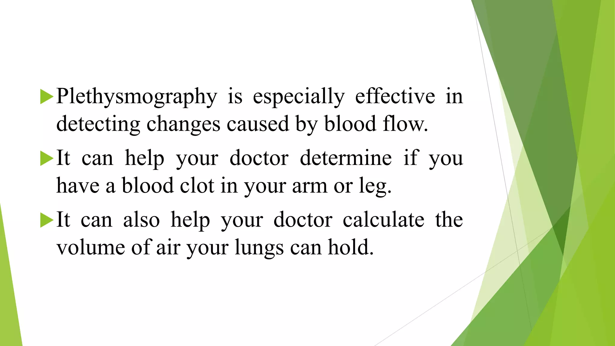


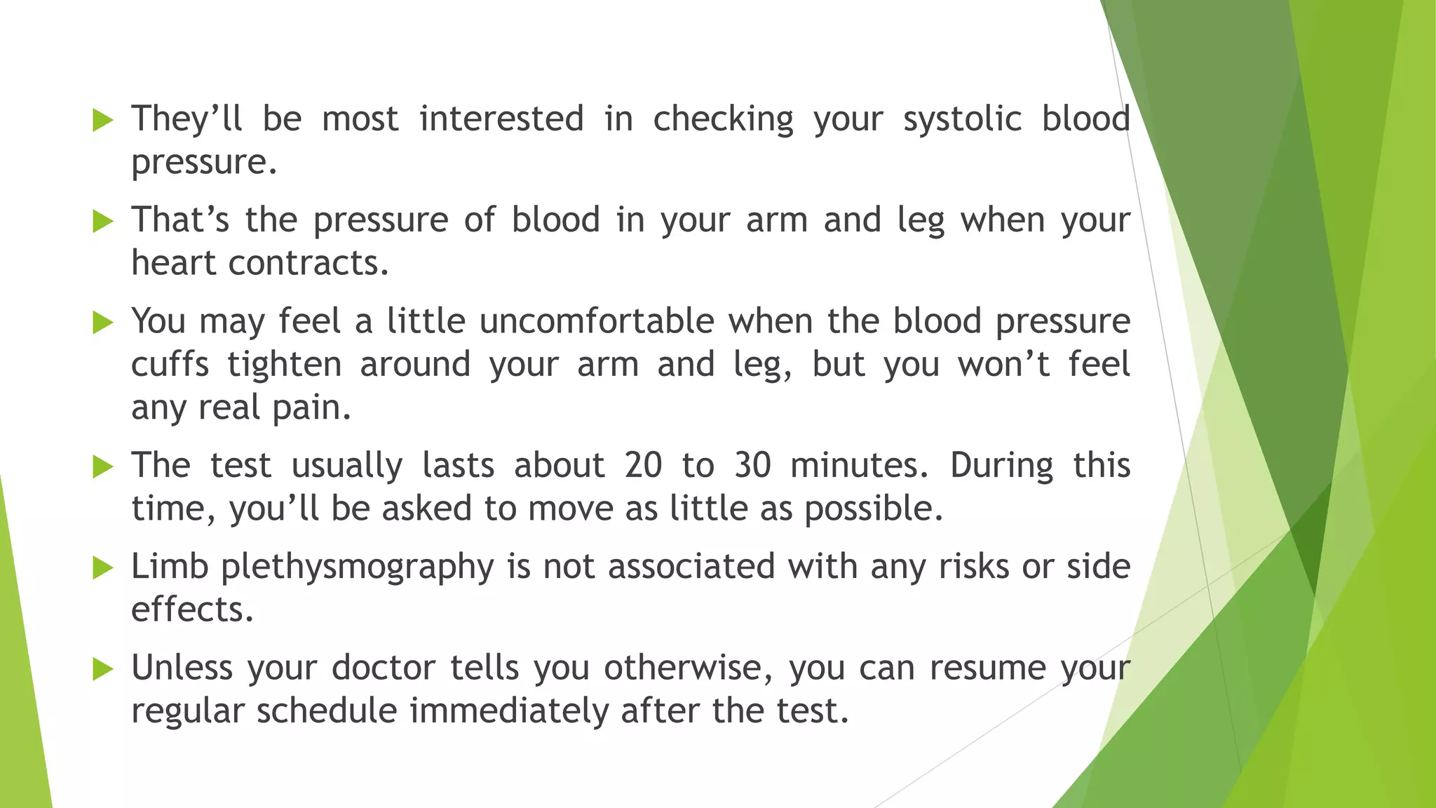


Plethysmography is a technique that measures changes in volume in different areas of the body using blood pressure cuffs or other sensors attached to a machine called a plethysmograph. It is effective at detecting changes caused by blood flow and can help doctors determine if a patient has blood clots or calculate lung volume. The document describes the procedures for limb and lung plethysmography tests and how they are interpreted to assess conditions like blood clots or respiratory issues. Common uses of plethysmography are listed in clinical settings like operating rooms and ICUs.
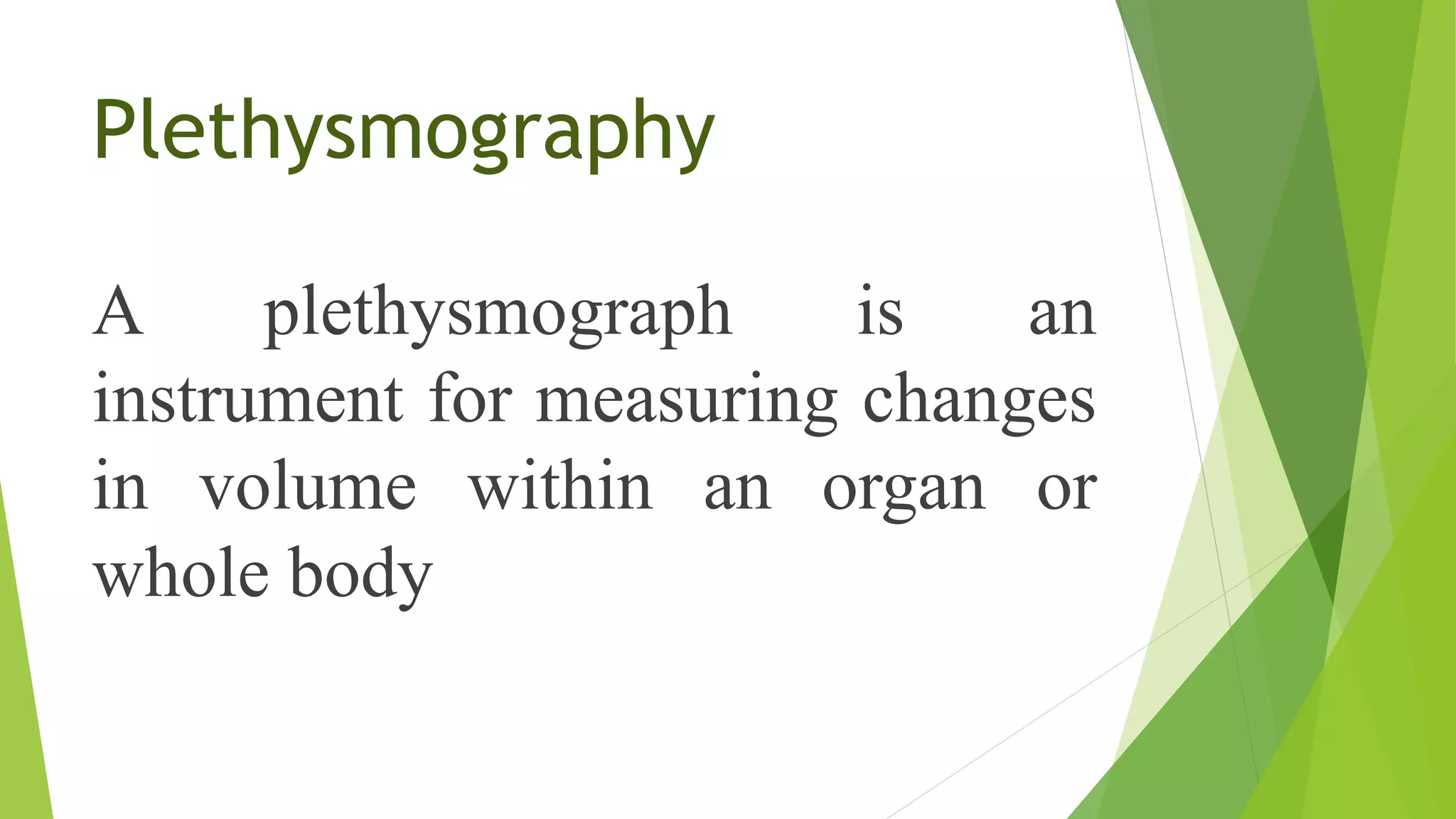
Introduction to plethysmography, its uses in measuring blood flow and lung volume, and its procedures.
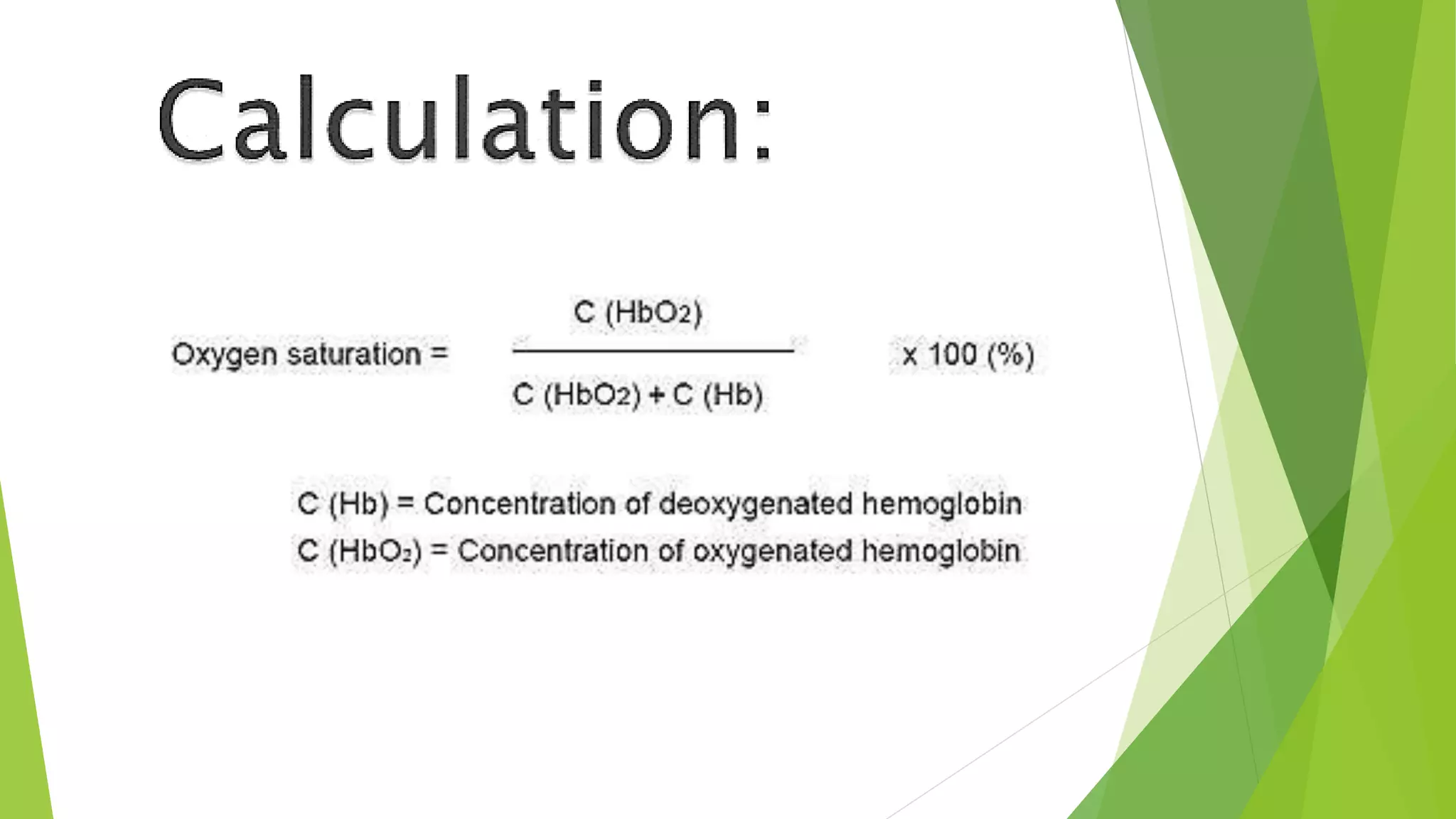
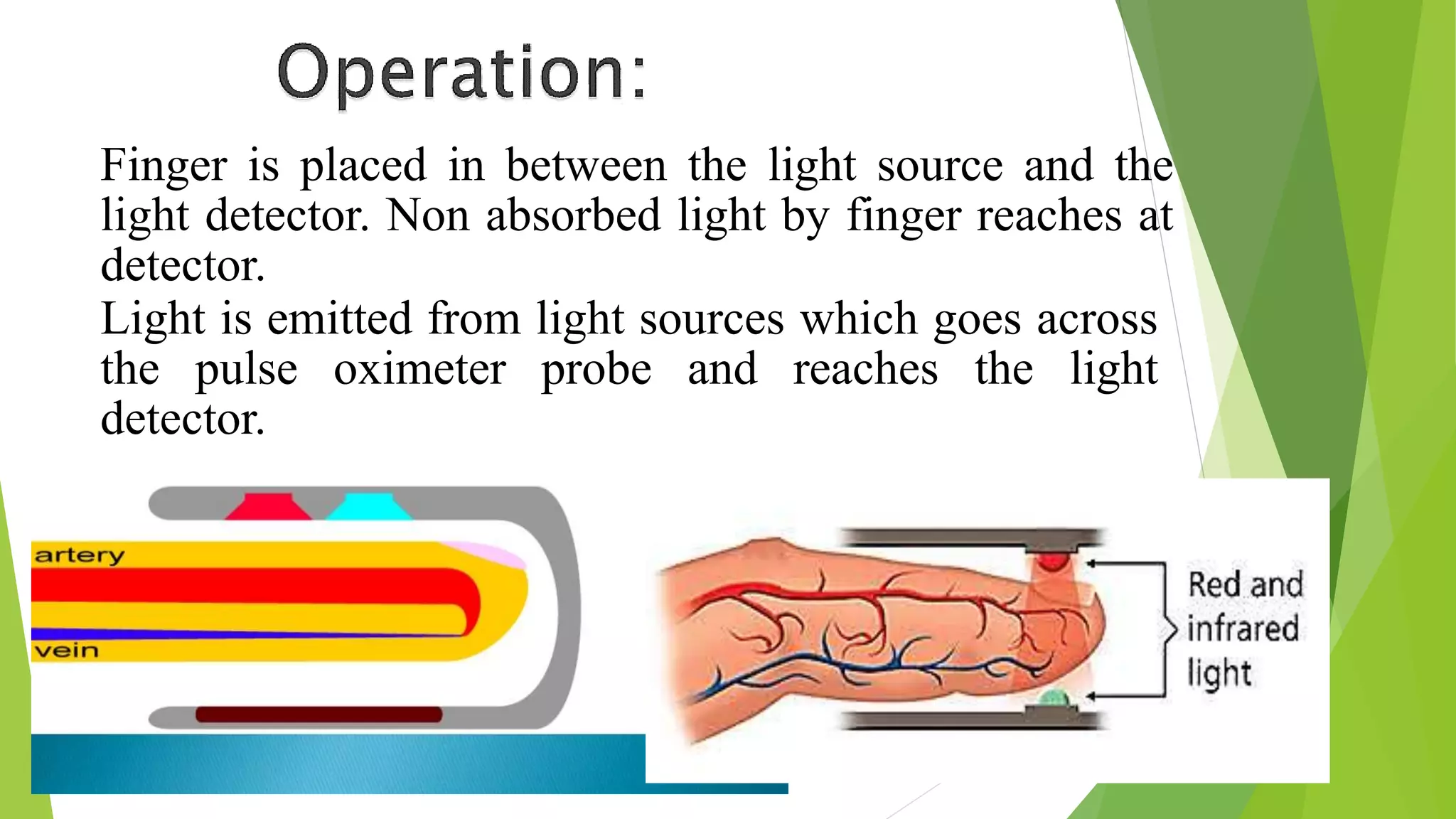
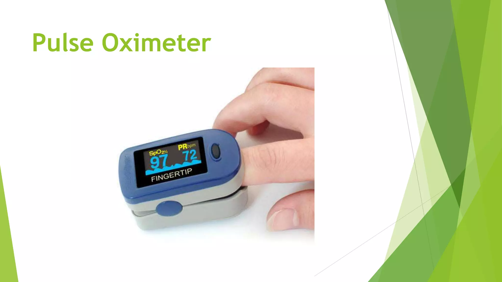
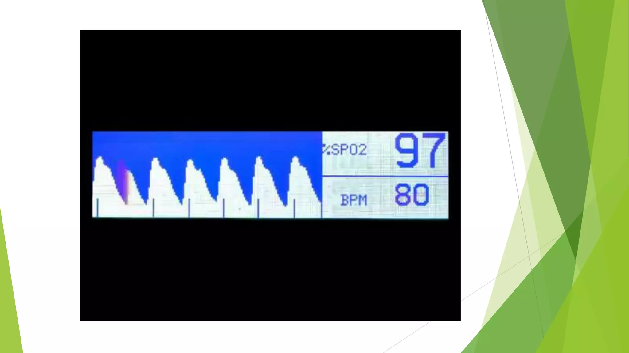
Mechanism of pulse oximeters in measuring blood oxygen saturation through light absorption.
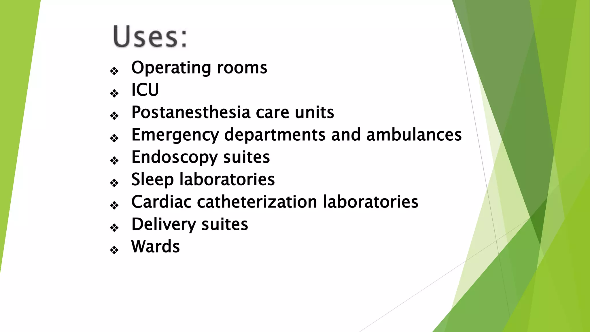
Various medical settings where pulse oximeters are used effectively.
Summary of safety precautions for using the pulse oximeter and listing of references.


