SlideShare a Scribd company logo
JOURNAL PRESENTATION
PULMONARY PATHOLOGY OF COVID-19
Sansar Babu Tiwari, MBBS, PGY I
Department of Pathology
TUTH
3rd May 2020
1
Pulmonary post-mortem findings in
a large series of COVID-19 cases
from Northern Italy
• Aurelio Sonzogni, MD et. al.
• Pathology Unit, L. Sacco Hospital, Milan, Italy
• Department of Pathology- Papa Giovanni XXIII
Hospital - Bergamo, Italy
INTRODUCTION:
• Italy has been the first country in Europe to be
reached by the epidemic and Lombardy was
devastated in just one month.
• L. Sacco Hospital, Milan and Papa Giovanni XXIII,
Bergamo have been the first hospitals in this
region to manage the epidemic crisis.
• The clinical spectrum of SARS-CoV-2 disease
(COVID-19) is reported to include mild
asymptomatic infection, mild upper respiratory
disease with fever and cough, and severe
pneumonia that can lead to ARDS in 15% of the
hospitalized cases.
INTRODUCTION:
• They described the first available large series of
lung histopathological findings in patients died
from COVID-19 in Northern Italy, with the aim to
report the main microscopic pulmonary lesions in
SARS-CoV-2 infection and severe respiratory failure.
METHODS:
• Histological analysis of post-mortem lung tissues
from 38 cases who died for COVID-19 between
February and March in two referral centres for the
management of the COVID-19 outbreak in
Northern Italy, Luigi Sacco Hospital, Milan, and
Papa Giovanni XXIII, Bergamo (patient permission
given by the Ethics Committees of the two hospitals
to use personal and sensitive data for scientific
research related to the disease was collected).
METHODS:
• The autopsies were performed in Airborne Infection
Isolation Autopsy Rooms and the personnel used the
correct Personal Protection Equipment (PPE), according
to “Engineering control and PPE recommendations for
autopsies”.
• A medium of 7 tissue blocks were taken from each lung
(range 5-9), selecting the most representative areas at
macroscopic examination. Tissues were fixed in 10%
buffered formalin for >48 hours. Three-μm paraffin
sections were stained by hematoxylin-eosin.
Immunohistochemistry reactions were performed on
selected cases (CD45, CD68, CD61, TTF1, p40, Ki67,
Masson Trichome) to better characterize inflammatory
infiltrate, epithelial cells and fibrosis.
METHODS:
• Histological evaluation was performed blindly by
two pathologists from each hospital, with expertise
in the field. Histological features of the cellular and
interstitial damage were described and graded by
using a semi-quantitative scale.
• Additional samples from selected cases were fixed
in glutaraldehyde for electron microscopy and
examined by EM-109 ZEISS and CCD-Megaview G2
(I-TEM imaging platform software).
RESULTS:
• Patients were 33 males and 5 females, average age
of 69 years (range 32-86); the time that the
patients spent in the Subintensive/Intensive Care
Unit ranged from 1 to 23 days (6.87 days).
• Regarding past comorbidities, data were available
in 31/38 patients: 9 diabetes, 18 hypertension, 4
past malignancies, 11 cardiovascular disorders, 3
mild chronic obstructive pulmonary disorders.
RESULTS:
• At the time of hospitalization, all the patients had
throat swab sample positive for SARS-CoV-2
infection and had clinical and radiological features
of interstitial pneumonia. D-dimer was available in
26/38 patients, with high value in all of them (>10 x
the upper reference limit).
• Patients died after a median time of 16.27 days
(range 5-31) from the onset of symptoms.
RESULTS:
• Macroscopic examination of the lungs revealed heavy,
congested and oedematous organs, with spotty
involvement.
• At histological examination, the features of Diffuse
Alveolar Disease (DAD) were found, corresponding to
those observable in the exudative and
early/intermediate proliferative phases of the disease.
• Both phases often overlapped in the different areas of
the lungs, with pluri-focal pattern of distribution. The
fibrotic phase was rarely observed, possibly due to the
short duration of the disease. Moreover, five patients
also had bacterial (4) and fungal (1) abscesses.
RESULTS:
Hyaline membrane Microthrombus
Megakaryocyte
entrapment
Type II pneumocyte
hyperplasia
RESULTS:
• Capillary congestion, interstitial edema, dilated
alveolar ducts, hyaline membranes composed of
serum proteins and condensed fibrin, loss of
pneumocytes were the histological patterns of the
exudative phase mostly observed in all the cases.
• Platelet-fibrin thrombi in small arterial vessels
(<1mm in diameter) were found in 33 cases.
• Moreover, type II pneumocyte hyperplasia showing
reactive atypia, myofibroblast proliferation, alveolar
granulation tissue and obliterating fibrosis were
present in half of the patients but were focal.
RESULTS:
Luminal organizing
fibrosis
Interstitial myofibroblastic
reaction
RESULTS:
• Microcystic honeycombing and mural fibrosis were
occasionally present.
• Inflammatory component was represented by a few
CD45 positive lymphocytes located in the
interstitial space; a large number of CD68 positive
macrophages were mainly localized in the alveolar
lumens.
• Immunohistochemistry with anti CD61 antibody
identified increased number of megakaryocytes in
lung capillaries
RESULTS:
CD68 macrophages CD45 Interstitial
lymphocytes
RESULTS:
• Ultrastructural examination
revealed viral particles,
with morphology typical of
the family of Coronaviridae
and localized along
plasmalemmal membranes
and within cytoplasmic
vacuoles of pneumocytes.
• Virions had an average
diameter of 82nm and viral
projection about 13nm in
length.
DISCUSSIONS:
• The peculiar histopathological findings were
atypical pneumocytes (reactive atypia) and diffuse
thrombosis of the peripheral small vessels.
• In their study, fibrin thrombi of small arterial
vessels (diameter < 1mm) were observed in 33 /38
patients, half of them with >25% of tissue
involvement and associated with high levels of D-
dimer in blood.
• These findings might explain the severe hypoxemia
which characterizes the clinical feature of ARDS in
SARS-CoV-2 patients.
DISCUSSIONS:
• Their data strongly support the hypothesis
proposed by recent clinical studies, that COVID-19
is complicated or anyway strictly related to
coagulopathy and thrombosis.
• Moreover detection of D-dimer values >1 μg/ml
have been associated with fatal outcome of COVID-
19.
• For these reasons, the use of anticoagulants has
been recently suggested as potentially beneficial in
patients with severe COVID-19, albeit its efficacy
and safety have not been demonstrated.
DISCUSSIONS:
• Finally, the search for viral particles was carried out
in a subset of patients and highlighted the
presence of rare virions in the cytoplasm of
pneumocytes. Despite the low number of cases,
these findings may suggest that the virus remains
in lung tissue for many days, even if in small
quantities, possibly being the trigger of the
mechanism that leads to and feeds lung damage.
Histopathology and genetic
susceptibility in COVID-19
pneumonia.
• von der Thüsen, MD, PhD
• Menno van der Eerden, MD PhD
• Dept. of Pathology and Dept. of Pulmonology,
Erasmus MC, Rotterdam, The Netherlands
INFLUENZA VIRUS:
• The respiratory tract histology of influenza
reflects its cellular tropism, as influenza virus
replicates in respiratory epithelial cells
throughout the respiratory tree, with non-fatal
infections predominantly involving the upper
respiratory tract and trachea, but fatal cases of
influenza usually result from pneumonia.
• Changes in the smaller airways include necrosis
and complete loss of the epithelial layer (both
ciliated and goblet cells), often resulting in the
formation of hyaline membranes at these sites.
Neutrophilic inflammation may be present in
the lumen, and extend into surrounding alveoli.
INFLUENZA VIRUS:
• In addition, a more chronic lymphoplasma-
histiocytic infiltrate is often seen in influenza-
infected airways. The parenchyma shows
evidence of acute injury with interstitial
congestion, edema, and inflammation
(predominantly neutrophilic with some
eosinophils), as well as desquamated alveolar
epithelial cells, intra-alveolar edema, intra-
alveolar hemorrhage, fibrin, hyaline membrane
formation, and sometimes necrosis of the
alveolar septa (necrotizing alveolitis), the latter
possibly resulting from frequently observed
vascular changes with capillary congestion and
thrombosis.
COVID-19:
• They have recently also observed similar
changes in COVID-19 autopsy cases, with
evidence of extensive microvascular damage
and thrombotic occlusion as the foremost
pattern of injury, resulting in intra-alveolar
fibrinous exudates akin to acute fibrinous
and organizing pneumonia (AFOP).
COVID-19 :
• This spectrum of abnormalities is highly
reminiscent of changes seen in the context of
the chronic lung allograft rejection (CLAD) of the
restrictive phenotype (restrictive allograft
syndrome (RAS)), which is presumed to occur
secondary to antibody-mediated endothelial
damage and complement activation of humoral
rejection episodes.
• Such vascular changes are likely to play a central
role in the pathogenesis of COVID-19, and may
in part be attributable to dysregulation of the
endothelial ACE2 receptor with ensuing
bradykinin-dependent local lung edema, as well
as a highly pro-thrombotic state, which can
manifest itself in the lung as well as in extra-
pulmonary tissues.
COVID-19 :
• Direct infection of endothelial cells may play a central
role in this process. More chronic changes included
intra-alveolar organization with fibroblastic
proliferation and diffuse fibrotic thickening of
alveolar walls, consisting of proliferating interstitial
fibroblasts.
• In RAS, we have found this to eventually result in
chronic fibrotic patterns of intra-alveolar
fibroelastosis and non-specific interstitial pneumonia
(NSIP).
• Time will tell if a similar progression to fibrosis will
take place in a subset of survivors of severe COVID-
19, and whether this is indeed secondary to vascular
damage, primarily related to the initial epithelial
infection, or a combination of these response
patterns.
OTHERS CORONA VIRUS:
• Apart from the prominent microvascular
changes, the pathological features of COVID-
19 thereby appear to resemble those seen in
SARS and Middle Eastern respiratory
syndrome (MERS) coronavirus infection.
• Early SARS-CoV infections were found to be
typified by acute diffuse alveolar damage,
and late stages by a combination of diffuse
alveolar damage and acute fibrinous and
organizing pneumonia.
OTHERS CORONA VIRUS:
• MERS-CoV infections also showed exudative
diffuse alveolar damage with hyaline
membranes, pulmonary edema, type II
pneumocyte hyperplasia, (lymphocytic)
interstitial pneumonia, and multinucleated
syncytial cells.
FUTURE GUIDANCE:
• Poor oxygenation requiring ventilation and oxygen
administration in influenza as well as coronavirus infection is
likely to be in part directly related to decreased diffusion
capacity due to parenchymal destruction as well as
increased diffusion distances due to
1) intra-alveolar aggregates of edema, fibrin, hyaline
membranes, cells and organization
2) widening of alveolar septa due to edema and
inflammation,
3) vascular changes with capillary congestion and
thrombosis and
4) eventually, intrabronchiolar, intra-alveolar and interstitial
fibrosis.
FUTURE GUIDANCE:
• While regeneration and reversal of the first 3 phenomena is
theoretically possible, and as in influenza may be related to
the regenerative capacity of the host, it may occur at lower
rates in SARS-CoV-2 infection as compared to other viral
pneumonias.
• Intra-alveolar and interstitial fibrosis, however, are most
likely irreversible, and may have contributed to the
remarkably high number of patients who cannot be weaned
off ventilation in the current COVID-19 outbreak.
FUTURE GUIDANCE:
• Also, in survivors we may eventually see a significant number
of cases with irreversible fibrotic lung damage and limited
pulmonary function, which has previously been termed COLD
(‘Corona Obstructive Lung Disease’).
• To which extent this term truly reflects the underlying
physiology remains to be seen, as the interstitial fibrotic
changes which have thus far been observed are likely to also
lead to a restrictive lung defect as well as permanently
decreased diffusion capacity, and are thus more akin to other
types of (idiopathic) interstitial lung fibrosis, such as forms of
the abovementioned intra-alveolar fibroelastosis and fibrotic
NSIP, and could therefore perhaps be more accurately termed
Corona-Associated Lung Disease (‘CALD’). This will require
multi-center studies which correlate histopathological findings
with clinical and radiological data.
WHY COVID-19 AFFECTS MALE:
• Interestingly, as an X-linked phenotype, the effectiveness of
interaction-booster and interaction-inhibitor variants of
ACE2 can be more definite in males than females, and could
contribute towards a higher mortality rate in males,
accounting for up to ∼70% of death caused by SARS-CoV2,
SARS-CoV or MERS-CoV.
• ACE inhibitors or ARB increases the ACE2 receptor thereby
increasing the viral entry.
• However it would to premature to say that ACE2 inhibitors
should be stopped.
• In the lung tissue of patients who died of respiratory failure
due to COVID19 pneumonia, there can be evidence of
epithelial infection with
• cytopathic effects of pneumocytes (A, box); denudation of
bronchiolar epithelium (B), evidence of diffuse alveolar
damage (DAD) with hyaline membrane formation with
organization (C).
• Observed vascular changes include extensive bilateral and
diffuse (micro)vascular damage and its sequelae, with
arterial thrombosis with organization (D), microvascular
fibrinoid change with hyaline thrombi (E, fibrin-Lendrum
(MSB) stain, arrows) and edema (**), and extensive intra-
alveolar fibrinous aggregates (F) with an acute fibrinous and
organizing pneumonia (AFOP) pattern (F, *).
COVID-19 :
• Fibrotic changes vary in appearance and include
organizing pneumonia with progression to fibrosis
(G), intra-alveolar fibro-elastosis (Elastic-van
Gieson, H) and fibrotic non-specific interstitial
pneumonia (F-NSIP; I).
• There is often relatively sparse interstitial chronic
inflammation with an acute interstitial pneumonia
pattern (AIP; J), foci of lymphocytic vasculitis (K)
and acute inflammation, especially in relation to
areas of necrosis and possible secondary infection
(L).
COVID-19 :
Thank You!!

More Related Content

What's hot

necrotizing pneumonia.ppt
necrotizing pneumonia.pptnecrotizing pneumonia.ppt
necrotizing pneumonia.ppt
farhanashafie3
 
atypical pneumonia.pptx
atypical pneumonia.pptxatypical pneumonia.pptx
atypical pneumonia.pptx
Rasika Deshmukh
 
NON RESOLVING PNEUMONIA
NON RESOLVING PNEUMONIANON RESOLVING PNEUMONIA
NON RESOLVING PNEUMONIA
SURABHI SUSHMA REDDY
 
MCQs in male reproductive pathology ss1
MCQs in male reproductive pathology ss1MCQs in male reproductive pathology ss1
MCQs in male reproductive pathology ss1
Dewan Raja
 
Hypersensitivity pneumonitis
Hypersensitivity pneumonitisHypersensitivity pneumonitis
Rheumatology MCQs Practice questions with explanation
Rheumatology MCQs Practice questions with explanationRheumatology MCQs Practice questions with explanation
Rheumatology MCQs Practice questions with explanation
Dr. Almas A
 
Lung Cancer Pathology & Clinical
Lung Cancer Pathology & Clinical Lung Cancer Pathology & Clinical
Lung Cancer Pathology & Clinical
Shahd Al Ali
 
MCQs KIDNEY PATHOLOGY
MCQs KIDNEY PATHOLOGYMCQs KIDNEY PATHOLOGY
MCQs KIDNEY PATHOLOGY
Dr. Roopam Jain
 
Bethesda Cervical CYtology
Bethesda Cervical CYtologyBethesda Cervical CYtology
Bethesda Cervical CYtology
Sansar Babu Tiwari
 
Pulmonary vasculitis
Pulmonary vasculitisPulmonary vasculitis
Pulmonary vasculitis
Arvind Ghongane
 
Management of cytokine storm during Covid 19
Management of cytokine storm during Covid 19Management of cytokine storm during Covid 19
Management of cytokine storm during Covid 19
Dr.Mahmoud Abbas
 
Atypical pneumonia
Atypical pneumoniaAtypical pneumonia
Atypical pneumonia
Narendra Patil
 
Pathology of Biliary & Pancreatic Disorders - Quiz
Pathology of Biliary & Pancreatic Disorders - QuizPathology of Biliary & Pancreatic Disorders - Quiz
Pathology of Biliary & Pancreatic Disorders - Quiz
Shashidhar Venkatesh Murthy
 
Vasculitis - Wegners, churg strauss,PAN, Temporal arteritis, Buerger's disea...
Vasculitis - Wegners, churg strauss,PAN, Temporal arteritis,  Buerger's disea...Vasculitis - Wegners, churg strauss,PAN, Temporal arteritis,  Buerger's disea...
Vasculitis - Wegners, churg strauss,PAN, Temporal arteritis, Buerger's disea...
Subhash Thakur
 
Sarcoidosis
SarcoidosisSarcoidosis
Sarcoidosis
Opeyemi Muyiwa
 
Idiopathic interstitial pneumonias
Idiopathic interstitial pneumoniasIdiopathic interstitial pneumonias
Idiopathic interstitial pneumonias
Arvind Ghongane
 
Pulmonary sarcoidosis
Pulmonary sarcoidosisPulmonary sarcoidosis
Pulmonary sarcoidosis
airwave12
 
Hepatitis C
Hepatitis C Hepatitis C
Hepatitis C
Melaku Yetbarek,MD
 

What's hot (20)

necrotizing pneumonia.ppt
necrotizing pneumonia.pptnecrotizing pneumonia.ppt
necrotizing pneumonia.ppt
 
atypical pneumonia.pptx
atypical pneumonia.pptxatypical pneumonia.pptx
atypical pneumonia.pptx
 
NON RESOLVING PNEUMONIA
NON RESOLVING PNEUMONIANON RESOLVING PNEUMONIA
NON RESOLVING PNEUMONIA
 
MCQs in male reproductive pathology ss1
MCQs in male reproductive pathology ss1MCQs in male reproductive pathology ss1
MCQs in male reproductive pathology ss1
 
Hypersensitivity pneumonitis
Hypersensitivity pneumonitisHypersensitivity pneumonitis
Hypersensitivity pneumonitis
 
Rheumatology MCQs Practice questions with explanation
Rheumatology MCQs Practice questions with explanationRheumatology MCQs Practice questions with explanation
Rheumatology MCQs Practice questions with explanation
 
Lung pathology
Lung pathologyLung pathology
Lung pathology
 
Lung Cancer Pathology & Clinical
Lung Cancer Pathology & Clinical Lung Cancer Pathology & Clinical
Lung Cancer Pathology & Clinical
 
MCQs KIDNEY PATHOLOGY
MCQs KIDNEY PATHOLOGYMCQs KIDNEY PATHOLOGY
MCQs KIDNEY PATHOLOGY
 
Bethesda Cervical CYtology
Bethesda Cervical CYtologyBethesda Cervical CYtology
Bethesda Cervical CYtology
 
Pulmonary vasculitis
Pulmonary vasculitisPulmonary vasculitis
Pulmonary vasculitis
 
Management of cytokine storm during Covid 19
Management of cytokine storm during Covid 19Management of cytokine storm during Covid 19
Management of cytokine storm during Covid 19
 
Atypical pneumonia
Atypical pneumoniaAtypical pneumonia
Atypical pneumonia
 
Pathology of Biliary & Pancreatic Disorders - Quiz
Pathology of Biliary & Pancreatic Disorders - QuizPathology of Biliary & Pancreatic Disorders - Quiz
Pathology of Biliary & Pancreatic Disorders - Quiz
 
Vasculitis - Wegners, churg strauss,PAN, Temporal arteritis, Buerger's disea...
Vasculitis - Wegners, churg strauss,PAN, Temporal arteritis,  Buerger's disea...Vasculitis - Wegners, churg strauss,PAN, Temporal arteritis,  Buerger's disea...
Vasculitis - Wegners, churg strauss,PAN, Temporal arteritis, Buerger's disea...
 
Sarcoidosis
SarcoidosisSarcoidosis
Sarcoidosis
 
Idiopathic interstitial pneumonias
Idiopathic interstitial pneumoniasIdiopathic interstitial pneumonias
Idiopathic interstitial pneumonias
 
Hcv
HcvHcv
Hcv
 
Pulmonary sarcoidosis
Pulmonary sarcoidosisPulmonary sarcoidosis
Pulmonary sarcoidosis
 
Hepatitis C
Hepatitis C Hepatitis C
Hepatitis C
 

Similar to Pulmonary pathology of corona virus

PATHOLOGY OF COVID-19
PATHOLOGY OF COVID-19PATHOLOGY OF COVID-19
PATHOLOGY OF COVID-19
DrRashmiBudha
 
Pathology of Acute Lungi Injury- Recent advances
Pathology of Acute Lungi Injury- Recent advancesPathology of Acute Lungi Injury- Recent advances
Pathology of Acute Lungi Injury- Recent advances
Dr Snehal Kosale
 
pneumonia.pptx
pneumonia.pptxpneumonia.pptx
pneumonia.pptx
ssuserc0ce211
 
RS Lecture 3.ppt
RS Lecture 3.pptRS Lecture 3.ppt
RS Lecture 3.ppt
ssuser9dd35a
 
suppurative lung syndromes.ppt
suppurative lung syndromes.pptsuppurative lung syndromes.ppt
suppurative lung syndromes.ppt
MostafaAdly7
 
J of thrombosis haemost 2021 - khismatullin - pathology of lung‐specific th...
J of thrombosis haemost   2021 - khismatullin - pathology of lung‐specific th...J of thrombosis haemost   2021 - khismatullin - pathology of lung‐specific th...
J of thrombosis haemost 2021 - khismatullin - pathology of lung‐specific th...
IuliaLupascu3
 
Pulmonary_inections[1].pptx
Pulmonary_inections[1].pptxPulmonary_inections[1].pptx
Pulmonary_inections[1].pptx
khaalidmohamed6
 
Pulmonary inections.pptx
Pulmonary inections.pptxPulmonary inections.pptx
Pulmonary inections.pptx
yusufArashid
 
Pneumonia
PneumoniaPneumonia
Pneumonia
asadullah ansari
 
Pneumonia.pptx
Pneumonia.pptxPneumonia.pptx
Pneumonia.pptx
SanthoshNS8
 
antifibrotic drugs
antifibrotic drugsantifibrotic drugs
antifibrotic drugs
DrVeereshDhanni
 
bronchiectasis Presentation1.pptx
bronchiectasis Presentation1.pptxbronchiectasis Presentation1.pptx
bronchiectasis Presentation1.pptx
devanshi92
 
Lrti punemococcal pneumonia and bordetella pertussis
Lrti punemococcal pneumonia and bordetella pertussisLrti punemococcal pneumonia and bordetella pertussis
Lrti punemococcal pneumonia and bordetella pertussis
Prasad Gunjal
 
Pneumonia and case studies for medical students
Pneumonia and case studies for medical studentsPneumonia and case studies for medical students
Pneumonia and case studies for medical students
GokulnathMbbs
 
pneumonia.pptx
pneumonia.pptxpneumonia.pptx
pneumonia.pptx
PrashantJadav5
 
pneumonia for C-1.pptx
pneumonia for C-1.pptxpneumonia for C-1.pptx
pneumonia for C-1.pptx
IbsaAli1
 
Cavernous Sinus Thrombosis:Current Therapy
Cavernous Sinus Thrombosis:Current TherapyCavernous Sinus Thrombosis:Current Therapy
Cavernous Sinus Thrombosis:Current Therapy
DrKamini Dadsena
 
Respiratory pathology harshmohan
Respiratory pathology harshmohanRespiratory pathology harshmohan
Respiratory pathology harshmohan
utsav parmar
 
Intrestitial lung disease 16 5-2016
Intrestitial  lung disease 16 5-2016Intrestitial  lung disease 16 5-2016
Intrestitial lung disease 16 5-2016
pathologydept
 

Similar to Pulmonary pathology of corona virus (20)

PATHOLOGY OF COVID-19
PATHOLOGY OF COVID-19PATHOLOGY OF COVID-19
PATHOLOGY OF COVID-19
 
Pathology of Acute Lungi Injury- Recent advances
Pathology of Acute Lungi Injury- Recent advancesPathology of Acute Lungi Injury- Recent advances
Pathology of Acute Lungi Injury- Recent advances
 
pneumonia.pptx
pneumonia.pptxpneumonia.pptx
pneumonia.pptx
 
RS Lecture 3.ppt
RS Lecture 3.pptRS Lecture 3.ppt
RS Lecture 3.ppt
 
suppurative lung syndromes.ppt
suppurative lung syndromes.pptsuppurative lung syndromes.ppt
suppurative lung syndromes.ppt
 
J of thrombosis haemost 2021 - khismatullin - pathology of lung‐specific th...
J of thrombosis haemost   2021 - khismatullin - pathology of lung‐specific th...J of thrombosis haemost   2021 - khismatullin - pathology of lung‐specific th...
J of thrombosis haemost 2021 - khismatullin - pathology of lung‐specific th...
 
Pulmonary_inections[1].pptx
Pulmonary_inections[1].pptxPulmonary_inections[1].pptx
Pulmonary_inections[1].pptx
 
Pulmonary inections.pptx
Pulmonary inections.pptxPulmonary inections.pptx
Pulmonary inections.pptx
 
Pneumonia
PneumoniaPneumonia
Pneumonia
 
Pneumonia.pptx
Pneumonia.pptxPneumonia.pptx
Pneumonia.pptx
 
antifibrotic drugs
antifibrotic drugsantifibrotic drugs
antifibrotic drugs
 
bronchiectasis Presentation1.pptx
bronchiectasis Presentation1.pptxbronchiectasis Presentation1.pptx
bronchiectasis Presentation1.pptx
 
Lrti punemococcal pneumonia and bordetella pertussis
Lrti punemococcal pneumonia and bordetella pertussisLrti punemococcal pneumonia and bordetella pertussis
Lrti punemococcal pneumonia and bordetella pertussis
 
Pneumonia and case studies for medical students
Pneumonia and case studies for medical studentsPneumonia and case studies for medical students
Pneumonia and case studies for medical students
 
pneumonia.pptx
pneumonia.pptxpneumonia.pptx
pneumonia.pptx
 
pneumonia for C-1.pptx
pneumonia for C-1.pptxpneumonia for C-1.pptx
pneumonia for C-1.pptx
 
Cavernous Sinus Thrombosis:Current Therapy
Cavernous Sinus Thrombosis:Current TherapyCavernous Sinus Thrombosis:Current Therapy
Cavernous Sinus Thrombosis:Current Therapy
 
Lung abscess
Lung abscessLung abscess
Lung abscess
 
Respiratory pathology harshmohan
Respiratory pathology harshmohanRespiratory pathology harshmohan
Respiratory pathology harshmohan
 
Intrestitial lung disease 16 5-2016
Intrestitial  lung disease 16 5-2016Intrestitial  lung disease 16 5-2016
Intrestitial lung disease 16 5-2016
 

More from Sansar Babu Tiwari

Liver cytology
Liver cytologyLiver cytology
Liver cytology
Sansar Babu Tiwari
 
Kidney transplant
Kidney transplantKidney transplant
Kidney transplant
Sansar Babu Tiwari
 
Olga medicine
Olga medicineOlga medicine
Olga medicine
Sansar Babu Tiwari
 
Acute Leukemia Cytogenetics
Acute Leukemia CytogeneticsAcute Leukemia Cytogenetics
Acute Leukemia Cytogenetics
Sansar Babu Tiwari
 
ALD, Cirrhosis, PBC and PSC
ALD, Cirrhosis, PBC and PSCALD, Cirrhosis, PBC and PSC
ALD, Cirrhosis, PBC and PSC
Sansar Babu Tiwari
 
Chronic lymphocytic leukemia
Chronic lymphocytic leukemiaChronic lymphocytic leukemia
Chronic lymphocytic leukemia
Sansar Babu Tiwari
 
Pericardial fluid eosinophilia
Pericardial fluid eosinophiliaPericardial fluid eosinophilia
Pericardial fluid eosinophilia
Sansar Babu Tiwari
 
Corona virus
Corona virusCorona virus
Corona virus
Sansar Babu Tiwari
 
Stains
StainsStains
Grossing thyroid gland
Grossing thyroid glandGrossing thyroid gland
Grossing thyroid gland
Sansar Babu Tiwari
 
Journal prostate and crispr
Journal prostate and crisprJournal prostate and crispr
Journal prostate and crispr
Sansar Babu Tiwari
 
Pancreas cytology
Pancreas cytologyPancreas cytology
Pancreas cytology
Sansar Babu Tiwari
 
Eosinophilia
EosinophiliaEosinophilia
Eosinophilia
Sansar Babu Tiwari
 
Journal article review
Journal article reviewJournal article review
Journal article review
Sansar Babu Tiwari
 

More from Sansar Babu Tiwari (14)

Liver cytology
Liver cytologyLiver cytology
Liver cytology
 
Kidney transplant
Kidney transplantKidney transplant
Kidney transplant
 
Olga medicine
Olga medicineOlga medicine
Olga medicine
 
Acute Leukemia Cytogenetics
Acute Leukemia CytogeneticsAcute Leukemia Cytogenetics
Acute Leukemia Cytogenetics
 
ALD, Cirrhosis, PBC and PSC
ALD, Cirrhosis, PBC and PSCALD, Cirrhosis, PBC and PSC
ALD, Cirrhosis, PBC and PSC
 
Chronic lymphocytic leukemia
Chronic lymphocytic leukemiaChronic lymphocytic leukemia
Chronic lymphocytic leukemia
 
Pericardial fluid eosinophilia
Pericardial fluid eosinophiliaPericardial fluid eosinophilia
Pericardial fluid eosinophilia
 
Corona virus
Corona virusCorona virus
Corona virus
 
Stains
StainsStains
Stains
 
Grossing thyroid gland
Grossing thyroid glandGrossing thyroid gland
Grossing thyroid gland
 
Journal prostate and crispr
Journal prostate and crisprJournal prostate and crispr
Journal prostate and crispr
 
Pancreas cytology
Pancreas cytologyPancreas cytology
Pancreas cytology
 
Eosinophilia
EosinophiliaEosinophilia
Eosinophilia
 
Journal article review
Journal article reviewJournal article review
Journal article review
 

Recently uploaded

Flu Vaccine Alert in Bangalore Karnataka
Flu Vaccine Alert in Bangalore KarnatakaFlu Vaccine Alert in Bangalore Karnataka
Flu Vaccine Alert in Bangalore Karnataka
addon Scans
 
Basavarajeeyam - Ayurvedic heritage book of Andhra pradesh
Basavarajeeyam - Ayurvedic heritage book of Andhra pradeshBasavarajeeyam - Ayurvedic heritage book of Andhra pradesh
Basavarajeeyam - Ayurvedic heritage book of Andhra pradesh
Dr. Madduru Muni Haritha
 
Triangles of Neck and Clinical Correlation by Dr. RIG.pptx
Triangles of Neck and Clinical Correlation by Dr. RIG.pptxTriangles of Neck and Clinical Correlation by Dr. RIG.pptx
Triangles of Neck and Clinical Correlation by Dr. RIG.pptx
Dr. Rabia Inam Gandapore
 
Thyroid Gland- Gross Anatomy by Dr. Rabia Inam Gandapore.pptx
Thyroid Gland- Gross Anatomy by Dr. Rabia Inam Gandapore.pptxThyroid Gland- Gross Anatomy by Dr. Rabia Inam Gandapore.pptx
Thyroid Gland- Gross Anatomy by Dr. Rabia Inam Gandapore.pptx
Dr. Rabia Inam Gandapore
 
Ozempic: Preoperative Management of Patients on GLP-1 Receptor Agonists
Ozempic: Preoperative Management of Patients on GLP-1 Receptor Agonists  Ozempic: Preoperative Management of Patients on GLP-1 Receptor Agonists
Ozempic: Preoperative Management of Patients on GLP-1 Receptor Agonists
Saeid Safari
 
Pictures of Superficial & Deep Fascia.ppt.pdf
Pictures of Superficial & Deep Fascia.ppt.pdfPictures of Superficial & Deep Fascia.ppt.pdf
Pictures of Superficial & Deep Fascia.ppt.pdf
Dr. Rabia Inam Gandapore
 
ANATOMY AND PHYSIOLOGY OF URINARY SYSTEM.pptx
ANATOMY AND PHYSIOLOGY OF URINARY SYSTEM.pptxANATOMY AND PHYSIOLOGY OF URINARY SYSTEM.pptx
ANATOMY AND PHYSIOLOGY OF URINARY SYSTEM.pptx
Swetaba Besh
 
planning for change nursing Management ppt
planning for change nursing Management pptplanning for change nursing Management ppt
planning for change nursing Management ppt
Thangamjayarani
 
Evaluation of antidepressant activity of clitoris ternatea in animals
Evaluation of antidepressant activity of clitoris ternatea in animalsEvaluation of antidepressant activity of clitoris ternatea in animals
Evaluation of antidepressant activity of clitoris ternatea in animals
Shweta
 
HOT NEW PRODUCT! BIG SALES FAST SHIPPING NOW FROM CHINA!! EU KU DB BK substit...
HOT NEW PRODUCT! BIG SALES FAST SHIPPING NOW FROM CHINA!! EU KU DB BK substit...HOT NEW PRODUCT! BIG SALES FAST SHIPPING NOW FROM CHINA!! EU KU DB BK substit...
HOT NEW PRODUCT! BIG SALES FAST SHIPPING NOW FROM CHINA!! EU KU DB BK substit...
GL Anaacs
 
Phone Us ❤85270-49040❤ #ℂall #gIRLS In Surat By Surat @ℂall @Girls Hotel With...
Phone Us ❤85270-49040❤ #ℂall #gIRLS In Surat By Surat @ℂall @Girls Hotel With...Phone Us ❤85270-49040❤ #ℂall #gIRLS In Surat By Surat @ℂall @Girls Hotel With...
Phone Us ❤85270-49040❤ #ℂall #gIRLS In Surat By Surat @ℂall @Girls Hotel With...
Savita Shen $i11
 
Sex determination from mandible pelvis and skull
Sex determination from mandible pelvis and skullSex determination from mandible pelvis and skull
Sex determination from mandible pelvis and skull
ShashankRoodkee
 
ARTHROLOGY PPT NCISM SYLLABUS AYURVEDA STUDENTS
ARTHROLOGY PPT NCISM SYLLABUS AYURVEDA STUDENTSARTHROLOGY PPT NCISM SYLLABUS AYURVEDA STUDENTS
ARTHROLOGY PPT NCISM SYLLABUS AYURVEDA STUDENTS
Dr. Vinay Pareek
 
For Better Surat #ℂall #Girl Service ❤85270-49040❤ Surat #ℂall #Girls
For Better Surat #ℂall #Girl Service ❤85270-49040❤ Surat #ℂall #GirlsFor Better Surat #ℂall #Girl Service ❤85270-49040❤ Surat #ℂall #Girls
For Better Surat #ℂall #Girl Service ❤85270-49040❤ Surat #ℂall #Girls
Savita Shen $i11
 
Surat @ℂall @Girls ꧁❤8527049040❤꧂@ℂall @Girls Service Vip Top Model Safe
Surat @ℂall @Girls ꧁❤8527049040❤꧂@ℂall @Girls Service Vip Top Model SafeSurat @ℂall @Girls ꧁❤8527049040❤꧂@ℂall @Girls Service Vip Top Model Safe
Surat @ℂall @Girls ꧁❤8527049040❤꧂@ℂall @Girls Service Vip Top Model Safe
Savita Shen $i11
 
How STIs Influence the Development of Pelvic Inflammatory Disease.pptx
How STIs Influence the Development of Pelvic Inflammatory Disease.pptxHow STIs Influence the Development of Pelvic Inflammatory Disease.pptx
How STIs Influence the Development of Pelvic Inflammatory Disease.pptx
FFragrant
 
Tom Selleck Health: A Comprehensive Look at the Iconic Actor’s Wellness Journey
Tom Selleck Health: A Comprehensive Look at the Iconic Actor’s Wellness JourneyTom Selleck Health: A Comprehensive Look at the Iconic Actor’s Wellness Journey
Tom Selleck Health: A Comprehensive Look at the Iconic Actor’s Wellness Journey
greendigital
 
Maxilla, Mandible & Hyoid Bone & Clinical Correlations by Dr. RIG.pptx
Maxilla, Mandible & Hyoid Bone & Clinical Correlations by Dr. RIG.pptxMaxilla, Mandible & Hyoid Bone & Clinical Correlations by Dr. RIG.pptx
Maxilla, Mandible & Hyoid Bone & Clinical Correlations by Dr. RIG.pptx
Dr. Rabia Inam Gandapore
 
heat stroke and heat exhaustion in children
heat stroke and heat exhaustion in childrenheat stroke and heat exhaustion in children
heat stroke and heat exhaustion in children
SumeraAhmad5
 
ARTIFICIAL INTELLIGENCE IN HEALTHCARE.pdf
ARTIFICIAL INTELLIGENCE IN  HEALTHCARE.pdfARTIFICIAL INTELLIGENCE IN  HEALTHCARE.pdf
ARTIFICIAL INTELLIGENCE IN HEALTHCARE.pdf
Anujkumaranit
 

Recently uploaded (20)

Flu Vaccine Alert in Bangalore Karnataka
Flu Vaccine Alert in Bangalore KarnatakaFlu Vaccine Alert in Bangalore Karnataka
Flu Vaccine Alert in Bangalore Karnataka
 
Basavarajeeyam - Ayurvedic heritage book of Andhra pradesh
Basavarajeeyam - Ayurvedic heritage book of Andhra pradeshBasavarajeeyam - Ayurvedic heritage book of Andhra pradesh
Basavarajeeyam - Ayurvedic heritage book of Andhra pradesh
 
Triangles of Neck and Clinical Correlation by Dr. RIG.pptx
Triangles of Neck and Clinical Correlation by Dr. RIG.pptxTriangles of Neck and Clinical Correlation by Dr. RIG.pptx
Triangles of Neck and Clinical Correlation by Dr. RIG.pptx
 
Thyroid Gland- Gross Anatomy by Dr. Rabia Inam Gandapore.pptx
Thyroid Gland- Gross Anatomy by Dr. Rabia Inam Gandapore.pptxThyroid Gland- Gross Anatomy by Dr. Rabia Inam Gandapore.pptx
Thyroid Gland- Gross Anatomy by Dr. Rabia Inam Gandapore.pptx
 
Ozempic: Preoperative Management of Patients on GLP-1 Receptor Agonists
Ozempic: Preoperative Management of Patients on GLP-1 Receptor Agonists  Ozempic: Preoperative Management of Patients on GLP-1 Receptor Agonists
Ozempic: Preoperative Management of Patients on GLP-1 Receptor Agonists
 
Pictures of Superficial & Deep Fascia.ppt.pdf
Pictures of Superficial & Deep Fascia.ppt.pdfPictures of Superficial & Deep Fascia.ppt.pdf
Pictures of Superficial & Deep Fascia.ppt.pdf
 
ANATOMY AND PHYSIOLOGY OF URINARY SYSTEM.pptx
ANATOMY AND PHYSIOLOGY OF URINARY SYSTEM.pptxANATOMY AND PHYSIOLOGY OF URINARY SYSTEM.pptx
ANATOMY AND PHYSIOLOGY OF URINARY SYSTEM.pptx
 
planning for change nursing Management ppt
planning for change nursing Management pptplanning for change nursing Management ppt
planning for change nursing Management ppt
 
Evaluation of antidepressant activity of clitoris ternatea in animals
Evaluation of antidepressant activity of clitoris ternatea in animalsEvaluation of antidepressant activity of clitoris ternatea in animals
Evaluation of antidepressant activity of clitoris ternatea in animals
 
HOT NEW PRODUCT! BIG SALES FAST SHIPPING NOW FROM CHINA!! EU KU DB BK substit...
HOT NEW PRODUCT! BIG SALES FAST SHIPPING NOW FROM CHINA!! EU KU DB BK substit...HOT NEW PRODUCT! BIG SALES FAST SHIPPING NOW FROM CHINA!! EU KU DB BK substit...
HOT NEW PRODUCT! BIG SALES FAST SHIPPING NOW FROM CHINA!! EU KU DB BK substit...
 
Phone Us ❤85270-49040❤ #ℂall #gIRLS In Surat By Surat @ℂall @Girls Hotel With...
Phone Us ❤85270-49040❤ #ℂall #gIRLS In Surat By Surat @ℂall @Girls Hotel With...Phone Us ❤85270-49040❤ #ℂall #gIRLS In Surat By Surat @ℂall @Girls Hotel With...
Phone Us ❤85270-49040❤ #ℂall #gIRLS In Surat By Surat @ℂall @Girls Hotel With...
 
Sex determination from mandible pelvis and skull
Sex determination from mandible pelvis and skullSex determination from mandible pelvis and skull
Sex determination from mandible pelvis and skull
 
ARTHROLOGY PPT NCISM SYLLABUS AYURVEDA STUDENTS
ARTHROLOGY PPT NCISM SYLLABUS AYURVEDA STUDENTSARTHROLOGY PPT NCISM SYLLABUS AYURVEDA STUDENTS
ARTHROLOGY PPT NCISM SYLLABUS AYURVEDA STUDENTS
 
For Better Surat #ℂall #Girl Service ❤85270-49040❤ Surat #ℂall #Girls
For Better Surat #ℂall #Girl Service ❤85270-49040❤ Surat #ℂall #GirlsFor Better Surat #ℂall #Girl Service ❤85270-49040❤ Surat #ℂall #Girls
For Better Surat #ℂall #Girl Service ❤85270-49040❤ Surat #ℂall #Girls
 
Surat @ℂall @Girls ꧁❤8527049040❤꧂@ℂall @Girls Service Vip Top Model Safe
Surat @ℂall @Girls ꧁❤8527049040❤꧂@ℂall @Girls Service Vip Top Model SafeSurat @ℂall @Girls ꧁❤8527049040❤꧂@ℂall @Girls Service Vip Top Model Safe
Surat @ℂall @Girls ꧁❤8527049040❤꧂@ℂall @Girls Service Vip Top Model Safe
 
How STIs Influence the Development of Pelvic Inflammatory Disease.pptx
How STIs Influence the Development of Pelvic Inflammatory Disease.pptxHow STIs Influence the Development of Pelvic Inflammatory Disease.pptx
How STIs Influence the Development of Pelvic Inflammatory Disease.pptx
 
Tom Selleck Health: A Comprehensive Look at the Iconic Actor’s Wellness Journey
Tom Selleck Health: A Comprehensive Look at the Iconic Actor’s Wellness JourneyTom Selleck Health: A Comprehensive Look at the Iconic Actor’s Wellness Journey
Tom Selleck Health: A Comprehensive Look at the Iconic Actor’s Wellness Journey
 
Maxilla, Mandible & Hyoid Bone & Clinical Correlations by Dr. RIG.pptx
Maxilla, Mandible & Hyoid Bone & Clinical Correlations by Dr. RIG.pptxMaxilla, Mandible & Hyoid Bone & Clinical Correlations by Dr. RIG.pptx
Maxilla, Mandible & Hyoid Bone & Clinical Correlations by Dr. RIG.pptx
 
heat stroke and heat exhaustion in children
heat stroke and heat exhaustion in childrenheat stroke and heat exhaustion in children
heat stroke and heat exhaustion in children
 
ARTIFICIAL INTELLIGENCE IN HEALTHCARE.pdf
ARTIFICIAL INTELLIGENCE IN  HEALTHCARE.pdfARTIFICIAL INTELLIGENCE IN  HEALTHCARE.pdf
ARTIFICIAL INTELLIGENCE IN HEALTHCARE.pdf
 

Pulmonary pathology of corona virus

  • 1. JOURNAL PRESENTATION PULMONARY PATHOLOGY OF COVID-19 Sansar Babu Tiwari, MBBS, PGY I Department of Pathology TUTH 3rd May 2020 1
  • 2. Pulmonary post-mortem findings in a large series of COVID-19 cases from Northern Italy • Aurelio Sonzogni, MD et. al. • Pathology Unit, L. Sacco Hospital, Milan, Italy • Department of Pathology- Papa Giovanni XXIII Hospital - Bergamo, Italy
  • 3. INTRODUCTION: • Italy has been the first country in Europe to be reached by the epidemic and Lombardy was devastated in just one month. • L. Sacco Hospital, Milan and Papa Giovanni XXIII, Bergamo have been the first hospitals in this region to manage the epidemic crisis. • The clinical spectrum of SARS-CoV-2 disease (COVID-19) is reported to include mild asymptomatic infection, mild upper respiratory disease with fever and cough, and severe pneumonia that can lead to ARDS in 15% of the hospitalized cases.
  • 4. INTRODUCTION: • They described the first available large series of lung histopathological findings in patients died from COVID-19 in Northern Italy, with the aim to report the main microscopic pulmonary lesions in SARS-CoV-2 infection and severe respiratory failure.
  • 5. METHODS: • Histological analysis of post-mortem lung tissues from 38 cases who died for COVID-19 between February and March in two referral centres for the management of the COVID-19 outbreak in Northern Italy, Luigi Sacco Hospital, Milan, and Papa Giovanni XXIII, Bergamo (patient permission given by the Ethics Committees of the two hospitals to use personal and sensitive data for scientific research related to the disease was collected).
  • 6. METHODS: • The autopsies were performed in Airborne Infection Isolation Autopsy Rooms and the personnel used the correct Personal Protection Equipment (PPE), according to “Engineering control and PPE recommendations for autopsies”. • A medium of 7 tissue blocks were taken from each lung (range 5-9), selecting the most representative areas at macroscopic examination. Tissues were fixed in 10% buffered formalin for >48 hours. Three-μm paraffin sections were stained by hematoxylin-eosin. Immunohistochemistry reactions were performed on selected cases (CD45, CD68, CD61, TTF1, p40, Ki67, Masson Trichome) to better characterize inflammatory infiltrate, epithelial cells and fibrosis.
  • 7. METHODS: • Histological evaluation was performed blindly by two pathologists from each hospital, with expertise in the field. Histological features of the cellular and interstitial damage were described and graded by using a semi-quantitative scale. • Additional samples from selected cases were fixed in glutaraldehyde for electron microscopy and examined by EM-109 ZEISS and CCD-Megaview G2 (I-TEM imaging platform software).
  • 8. RESULTS: • Patients were 33 males and 5 females, average age of 69 years (range 32-86); the time that the patients spent in the Subintensive/Intensive Care Unit ranged from 1 to 23 days (6.87 days). • Regarding past comorbidities, data were available in 31/38 patients: 9 diabetes, 18 hypertension, 4 past malignancies, 11 cardiovascular disorders, 3 mild chronic obstructive pulmonary disorders.
  • 9. RESULTS: • At the time of hospitalization, all the patients had throat swab sample positive for SARS-CoV-2 infection and had clinical and radiological features of interstitial pneumonia. D-dimer was available in 26/38 patients, with high value in all of them (>10 x the upper reference limit). • Patients died after a median time of 16.27 days (range 5-31) from the onset of symptoms.
  • 10. RESULTS: • Macroscopic examination of the lungs revealed heavy, congested and oedematous organs, with spotty involvement. • At histological examination, the features of Diffuse Alveolar Disease (DAD) were found, corresponding to those observable in the exudative and early/intermediate proliferative phases of the disease. • Both phases often overlapped in the different areas of the lungs, with pluri-focal pattern of distribution. The fibrotic phase was rarely observed, possibly due to the short duration of the disease. Moreover, five patients also had bacterial (4) and fungal (1) abscesses.
  • 12. RESULTS: • Capillary congestion, interstitial edema, dilated alveolar ducts, hyaline membranes composed of serum proteins and condensed fibrin, loss of pneumocytes were the histological patterns of the exudative phase mostly observed in all the cases. • Platelet-fibrin thrombi in small arterial vessels (<1mm in diameter) were found in 33 cases. • Moreover, type II pneumocyte hyperplasia showing reactive atypia, myofibroblast proliferation, alveolar granulation tissue and obliterating fibrosis were present in half of the patients but were focal.
  • 14. RESULTS: • Microcystic honeycombing and mural fibrosis were occasionally present. • Inflammatory component was represented by a few CD45 positive lymphocytes located in the interstitial space; a large number of CD68 positive macrophages were mainly localized in the alveolar lumens. • Immunohistochemistry with anti CD61 antibody identified increased number of megakaryocytes in lung capillaries
  • 15. RESULTS: CD68 macrophages CD45 Interstitial lymphocytes
  • 16. RESULTS: • Ultrastructural examination revealed viral particles, with morphology typical of the family of Coronaviridae and localized along plasmalemmal membranes and within cytoplasmic vacuoles of pneumocytes. • Virions had an average diameter of 82nm and viral projection about 13nm in length.
  • 17. DISCUSSIONS: • The peculiar histopathological findings were atypical pneumocytes (reactive atypia) and diffuse thrombosis of the peripheral small vessels. • In their study, fibrin thrombi of small arterial vessels (diameter < 1mm) were observed in 33 /38 patients, half of them with >25% of tissue involvement and associated with high levels of D- dimer in blood. • These findings might explain the severe hypoxemia which characterizes the clinical feature of ARDS in SARS-CoV-2 patients.
  • 18. DISCUSSIONS: • Their data strongly support the hypothesis proposed by recent clinical studies, that COVID-19 is complicated or anyway strictly related to coagulopathy and thrombosis. • Moreover detection of D-dimer values >1 μg/ml have been associated with fatal outcome of COVID- 19. • For these reasons, the use of anticoagulants has been recently suggested as potentially beneficial in patients with severe COVID-19, albeit its efficacy and safety have not been demonstrated.
  • 19. DISCUSSIONS: • Finally, the search for viral particles was carried out in a subset of patients and highlighted the presence of rare virions in the cytoplasm of pneumocytes. Despite the low number of cases, these findings may suggest that the virus remains in lung tissue for many days, even if in small quantities, possibly being the trigger of the mechanism that leads to and feeds lung damage.
  • 20. Histopathology and genetic susceptibility in COVID-19 pneumonia. • von der Thüsen, MD, PhD • Menno van der Eerden, MD PhD • Dept. of Pathology and Dept. of Pulmonology, Erasmus MC, Rotterdam, The Netherlands
  • 21. INFLUENZA VIRUS: • The respiratory tract histology of influenza reflects its cellular tropism, as influenza virus replicates in respiratory epithelial cells throughout the respiratory tree, with non-fatal infections predominantly involving the upper respiratory tract and trachea, but fatal cases of influenza usually result from pneumonia. • Changes in the smaller airways include necrosis and complete loss of the epithelial layer (both ciliated and goblet cells), often resulting in the formation of hyaline membranes at these sites. Neutrophilic inflammation may be present in the lumen, and extend into surrounding alveoli.
  • 22. INFLUENZA VIRUS: • In addition, a more chronic lymphoplasma- histiocytic infiltrate is often seen in influenza- infected airways. The parenchyma shows evidence of acute injury with interstitial congestion, edema, and inflammation (predominantly neutrophilic with some eosinophils), as well as desquamated alveolar epithelial cells, intra-alveolar edema, intra- alveolar hemorrhage, fibrin, hyaline membrane formation, and sometimes necrosis of the alveolar septa (necrotizing alveolitis), the latter possibly resulting from frequently observed vascular changes with capillary congestion and thrombosis.
  • 23. COVID-19: • They have recently also observed similar changes in COVID-19 autopsy cases, with evidence of extensive microvascular damage and thrombotic occlusion as the foremost pattern of injury, resulting in intra-alveolar fibrinous exudates akin to acute fibrinous and organizing pneumonia (AFOP).
  • 24. COVID-19 : • This spectrum of abnormalities is highly reminiscent of changes seen in the context of the chronic lung allograft rejection (CLAD) of the restrictive phenotype (restrictive allograft syndrome (RAS)), which is presumed to occur secondary to antibody-mediated endothelial damage and complement activation of humoral rejection episodes. • Such vascular changes are likely to play a central role in the pathogenesis of COVID-19, and may in part be attributable to dysregulation of the endothelial ACE2 receptor with ensuing bradykinin-dependent local lung edema, as well as a highly pro-thrombotic state, which can manifest itself in the lung as well as in extra- pulmonary tissues.
  • 25. COVID-19 : • Direct infection of endothelial cells may play a central role in this process. More chronic changes included intra-alveolar organization with fibroblastic proliferation and diffuse fibrotic thickening of alveolar walls, consisting of proliferating interstitial fibroblasts. • In RAS, we have found this to eventually result in chronic fibrotic patterns of intra-alveolar fibroelastosis and non-specific interstitial pneumonia (NSIP). • Time will tell if a similar progression to fibrosis will take place in a subset of survivors of severe COVID- 19, and whether this is indeed secondary to vascular damage, primarily related to the initial epithelial infection, or a combination of these response patterns.
  • 26. OTHERS CORONA VIRUS: • Apart from the prominent microvascular changes, the pathological features of COVID- 19 thereby appear to resemble those seen in SARS and Middle Eastern respiratory syndrome (MERS) coronavirus infection. • Early SARS-CoV infections were found to be typified by acute diffuse alveolar damage, and late stages by a combination of diffuse alveolar damage and acute fibrinous and organizing pneumonia.
  • 27. OTHERS CORONA VIRUS: • MERS-CoV infections also showed exudative diffuse alveolar damage with hyaline membranes, pulmonary edema, type II pneumocyte hyperplasia, (lymphocytic) interstitial pneumonia, and multinucleated syncytial cells.
  • 28. FUTURE GUIDANCE: • Poor oxygenation requiring ventilation and oxygen administration in influenza as well as coronavirus infection is likely to be in part directly related to decreased diffusion capacity due to parenchymal destruction as well as increased diffusion distances due to 1) intra-alveolar aggregates of edema, fibrin, hyaline membranes, cells and organization 2) widening of alveolar septa due to edema and inflammation, 3) vascular changes with capillary congestion and thrombosis and 4) eventually, intrabronchiolar, intra-alveolar and interstitial fibrosis.
  • 29. FUTURE GUIDANCE: • While regeneration and reversal of the first 3 phenomena is theoretically possible, and as in influenza may be related to the regenerative capacity of the host, it may occur at lower rates in SARS-CoV-2 infection as compared to other viral pneumonias. • Intra-alveolar and interstitial fibrosis, however, are most likely irreversible, and may have contributed to the remarkably high number of patients who cannot be weaned off ventilation in the current COVID-19 outbreak.
  • 30. FUTURE GUIDANCE: • Also, in survivors we may eventually see a significant number of cases with irreversible fibrotic lung damage and limited pulmonary function, which has previously been termed COLD (‘Corona Obstructive Lung Disease’). • To which extent this term truly reflects the underlying physiology remains to be seen, as the interstitial fibrotic changes which have thus far been observed are likely to also lead to a restrictive lung defect as well as permanently decreased diffusion capacity, and are thus more akin to other types of (idiopathic) interstitial lung fibrosis, such as forms of the abovementioned intra-alveolar fibroelastosis and fibrotic NSIP, and could therefore perhaps be more accurately termed Corona-Associated Lung Disease (‘CALD’). This will require multi-center studies which correlate histopathological findings with clinical and radiological data.
  • 31. WHY COVID-19 AFFECTS MALE: • Interestingly, as an X-linked phenotype, the effectiveness of interaction-booster and interaction-inhibitor variants of ACE2 can be more definite in males than females, and could contribute towards a higher mortality rate in males, accounting for up to ∼70% of death caused by SARS-CoV2, SARS-CoV or MERS-CoV. • ACE inhibitors or ARB increases the ACE2 receptor thereby increasing the viral entry. • However it would to premature to say that ACE2 inhibitors should be stopped.
  • 32.
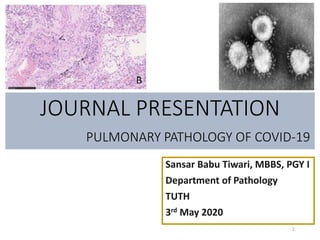
  • 33. • In the lung tissue of patients who died of respiratory failure due to COVID19 pneumonia, there can be evidence of epithelial infection with • cytopathic effects of pneumocytes (A, box); denudation of bronchiolar epithelium (B), evidence of diffuse alveolar damage (DAD) with hyaline membrane formation with organization (C). • Observed vascular changes include extensive bilateral and diffuse (micro)vascular damage and its sequelae, with arterial thrombosis with organization (D), microvascular fibrinoid change with hyaline thrombi (E, fibrin-Lendrum (MSB) stain, arrows) and edema (**), and extensive intra- alveolar fibrinous aggregates (F) with an acute fibrinous and organizing pneumonia (AFOP) pattern (F, *). COVID-19 :
  • 34. • Fibrotic changes vary in appearance and include organizing pneumonia with progression to fibrosis (G), intra-alveolar fibro-elastosis (Elastic-van Gieson, H) and fibrotic non-specific interstitial pneumonia (F-NSIP; I). • There is often relatively sparse interstitial chronic inflammation with an acute interstitial pneumonia pattern (AIP; J), foci of lymphocytic vasculitis (K) and acute inflammation, especially in relation to areas of necrosis and possible secondary infection (L). COVID-19 :