Download to read offline


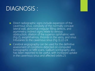


1) Cavernous sinus thrombosis is a rare but serious condition caused by infection or trauma spreading to the cavernous sinus. 2) Staphylococcus aureus and Streptococcus species are the most common infectious causes. Symptoms include fever, headache, eye signs like chemosis and proptosis. 3) Treatment involves antibiotics, surgery to drain infection sites, and controversy around the use of steroids and anticoagulants to prevent further thrombus progression while allowing antibiotic penetration.


