Bethesda Cervical CYtology
•
69 likes•20,185 views

This slide presentation summarizes a cytology case involving a 38-year-old female patient. A cervical smear shows atypical cells with dense, scanty cytoplasm and enlarged hyperchromatic nuclei throughout the smear. Background shows sheets of neutrophils, coccobacilli and hemorrhage. The specimen is categorized as an epithelial cell abnormality and interpreted as a high-grade squamous intra-epithelial lesion. Colposcopic examination is suggested. The presentation provides further details on cytology techniques, classifications of abnormal cervical cells, screening guidelines, and characteristics of low and high-grade squamous lesions.

Recommended
More Related Content
What's hot
What's hot (20)
Similar to Bethesda Cervical CYtology
Similar to Bethesda Cervical CYtology (20)
More from Sansar Babu Tiwari
More from Sansar Babu Tiwari (14)
Recently uploaded
Recently uploaded (20)
Bethesda Cervical CYtology
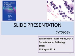
- 1. SLIDE PRESENTATION CYTOLOGY Sansar Babu Tiwari, MBBS, PGY I Department of Pathology TUTH 1st August 2019 1
- 2. Single Pap-stained Cervical Smear of 38 years female (Conventional cytology) Smear shows superficial, intermediate, and parabasal cells along with few endocervical cells. Atypical cells are found arranged singly, in sheets and in clusters throughout the smear. These cells are round to oval (parabasal) in shape with dense, scanty cytoplasm, enlarged hyperchromatic nuclei, coarse chromatin and irregular nuclear rim with high N : C ratio and inconspicuous nucleoli. SLIDE PRESENTATION 2
- 3. Background shows sheets of neutrophils and coccobacilli along with hemorrhage. Necrosis and mitosis is not seen SLIDE PRESENTATION 3
- 4. Specimen: Cervical smear Adequacy: Satisfactory for evaluation; endocervical component present General Categorization*: Epithelial cell abnormality, squamous Interpretation: High-grade squamous intra-epithelial lesion (HSIL) Adjunctive Testing*: Not done in smear Note*: Suggest colposcopic examination * Optional to mention in Report. SLIDE PRESENTATION 4
- 10. Olympus, 40x 10
- 11. Columnar lining changes into squamous in TZ TZ is a dynamic point, keep on changing. Move out towards ectocervix during puberty, pregnancy and pills. Move inwards when hormonal influences are absent like after menopause. CERVIX 11
- 12. BURDEN OF CERVICAL CANCER WHO: Cervical cancer is the second most common cancer in women living in less developed regions with an estimated 570 000 new cases in 2018 (84% of the new cases worldwide). In 2018, approximately 311 000 women died from cervical cancer; more than 85% of these deaths occurring in low- and middle- income countries. High risk HPV: 16, 18, 31, 33, 35,39, 45, 51, 52, 56, 58, 59, 68, 69, 82 Low risk HPV: 6, 11, 40, 42, 43, 44, 54, 61, 72, 81 On average, 50% of HPV infections are cleared within 8 months and 90% are cleared within 2 years. CERVICAL CANCER OVERALL BURDEN 12
- 14. HPV and cervical cancer 14 • Although HPV has been firmly established as a common cause of cervical cancer, it is not sufficient to cause cancer. • Host immune and co- carcinogen exposure influences the fate of HPV and progression to cancer.
- 16. Incidence of cervical cytology 16
- 17. United States Preventive Service Task Force (USPSTF), 2018 recommends: • Age to initiate: 21 years • Age to discontinue: 65 years • Between 21-29 years: Pap test every 3 years • After 30 years: One of these methods: • Pap test every 3 years • hrHPV testing alone every 5 years • Co-testing (Pap test and HPV testing) every 5 years Routine Screening of patients 17
- 23. TYPES OF SQUAMOUS CELLS 23
- 30. 30
- 31. 31
- 33. 33 Ovulatory Phase (14-16 days)
- 34. 34 Luteal Phase
- 35. LSIL cervical cytologic specimens that contain a few cells that are suspicious for but not diagnostic of HSIL are reported as atypical squamous cells, cannot exclude a high-grade squamous intraepithelial lesion(ASC-H). There was previously no recommendation regarding how to report these. Benign-appearing endometrial cells are reported only in women ≥45 years. This is a change from Bethesda 2001, which used an age threshold of ≥40 years. Pages: 191 324 Images: 186 370 Bethesda 2014 vs 2001 35
- 36. 1. Description of specimen type and test requested – cervical or vaginal sample, conventional Pap smear, liquid-based cytology, and/or reflex human papillomavirus (HPV) test. 2. A description of specimen adequacy 3. A general categorization (optional) – Negative, epithelial cell abnormality, or other 4. An interpretation/result – Either the specimen is NILM (although organisms and reactive changes may be present) or there is an epithelial cell abnormality as defined, or there is another finding(increased risk like endometrial cells in women ≥45 years). Bethesda 2014 Overview of Report 36
- 37. 5. A description of any ancillary testing or automated review that was performed (eg. HPV, AutoPap) 6. Educational notes and suggestions by the pathologist (optional) Bethesda 2014 Overview of Report 37
- 38. 38
- 39. 39
- 40. 1. Cellularity >5000 (LBP), 8000-12000 (CP) >2000: Low cellularity but satisfactory: Post-radiation, Post-TH vaginal) 2. Endocervical component EC/TZ is not necessary for adequate specimen, only squamous cellularity is needed Adequate TZ requires at least 10 well preserved EC or squamous metaplastic cells No relation with further diagnosis with SIL 3. Obscuring factors (Blood, lubricants) >75% of sq cells (not the slide area) : unsatisfactory 4. HPV testing on unsatisfactory specimen HPV test could be falsely negative in unsatisfactory specimen However, if HPV is positive in these specimen, follow up is still needed ADEQUACY 40
- 41. ASC refers to cytologic changes suggestive of SIL, which are qualitatively and quantitatively insufficient for definite interpretation Three essential features 1. Squamous differentiation 2. Increased ratio of nuclear to cytoplasmic area 3. Minimal nuclear hyperchromasia, chromatin clumping, irregularity, smudging or multinucleation ATYPICAL SQUAMOUS CELL 41
- 42. • Definition: cytological changes suggestive of LSIL that are quantitatively and qualitatively insufficient for a definitive diagnosis • CRITERIA – Cells resemble superficial and intermediate squamous cells in size and configuration – Nuclei are approximately 2 and half to three times the area of the nucleus of a normal intermediate squamous cell or twice the size of a squamous metaplastic cell nucleus. – Slighlty increased N:C ratio – Minimal nuclear hyperchromasia and irregularity of chromatin distribution or nuclear shape – Nuclear abnormalities associated with dense orangeophilic cytoplasm (atypical parakeratosis) ATYPICAL SQUAMOUS CELL -UNDETERMINED SIGNIFICANCE (ASC-US) 42
- 43. ATYPICAL SQUAMOUS CELL -UNDETERMINED SIGNIFICANCE (ASC-US) 43
- 44. ATYPICAL SQUAMOUS CELL -UNDETERMINED SIGNIFICANCE (ASC-US) 44
- 45. ATYPICAL SQUAMOUS CELL -UNDETERMINED SIGNIFICANCE (ASC-US) 45
- 46. • Definition: cytological changes suggestive of HSIL that are quantitatively and qualitatively insufficient for a definitive diagnosis • Patterns: A. Small cells with high N:C Ratios (Atypical immature Metaplasia) • Cells usually occur singly or in small fragments of less than 10 cells, occasionally in conventional smears, cells may stream in mucus. • Cells are size of metaplastic cells with nuclei that are about 1.5 to 2.5 times larger than normal • N/C ratio may approximate that of HSIL ASC-H Vs HSIL: Nuclear abnormalities such as hyperchromasia, chromatin irregularity and abnormal nuclear shapes with focal irregularity favor an interpretation of HSIL. ATYPICAL SQUAMOUS CELL –CANNOT EXCLUDE HSIL (ASC-H) 46
- 47. B. Crowded Sheet Pattern: • Dense cytoplasm, polygonal cell shape and fragments with sharp linear edges generally favor squamous over glandular (endocervical) differentiation. C. ASC-H Mimics: • Isolated endocervical cells • Degenerated endometrial cells • Macrophages • IUD: may shed rare cells with high N:C ratio • Pregnancy and post-partum: atypical appearing decidualized stromal cells ATYPICAL SQUAMOUS CELL –CANNOT EXCLUDE HSIL (ASC-H) 47
- 48. ATYPICAL SQUAMOUS CELL –CANNOT EXCLUDE HSIL (ASC-H) 48
- 49. • CRITERIA – Cells occur singly, in clusters, and in sheets – Cytological changes are usually confined to squamous cells with ‘mature’ intermediate or superficial squamous cell- type cytoplasm – Overall cell size is large, with fairly abundant ‘mature’ well-defined cytoplasm – Nuclear enlargement more than 3 times the area of normal intermediate nuclei results in a low but slightly increased N:C ratio Low-grade Squamous Intraepithelial Lesion (LSIL) 49
- 50. • CRITERIA – Nuclei are generally hyperchromatic but may be normochromatic – Nuclei show variable size (anisonucleosis) – Chromatin is uniformly distributed and ranges from coarsely granular to smudgy or densely opaque – Contour of nuclear membranes is variable ranging from smooth to very irregular with notches – Binucleation and multinucleation are common – Nucleoli are generally absent or inconspicuous if present Low-grade Squamous Intraepithelial Lesion (LSIL) 50
- 51. • CRITERIA – Koilocytosis or perinuclear cavitation consisting of a broad, sharply delineated clear peri-nuclear zone and a peripheral rim of densely stained cytoplasm is a characteristic viral cytopathic feature but is not required for the interpretation of LSIL – Cells may show increased keratinization with dense, eosinophilic cytoplasm with little or no evidence of koilocytosis – Cells with koilocytosis or dense orangeophilia must also show nuclear abnoramalities to be diagnostic of LSIL, perinuclear halos in the absence of nuclear abnormalities does not qualify for the interpretation of LSIL Low-grade Squamous Intraepithelial Lesion (LSIL) 51
- 52. Low-grade Squamous Intraepithelial Lesion (LSIL) 52
- 53. • Pseudokoilocytosis • Herpes Cytopathic Effects • Radiation Changes MIMICS OF LSIL 53
- 54. L S I L 54
- 57. High-grade Squamous Intraepithelial Lesion (HSIL) 57
- 58. • CRITERIA – Cells occur singly, in sheets, or in syncytial aggregates – Smaller and show less cytoplasmic maturity than cells of LSIL – Syncytial aggregates of dysplastic cells may result in hyperchromatic crowded groups – Overall cell size is variable, in general, the cells of HSIL are smaller than those of LSIL. Higher-grade lesions often contain quite small basal-type cells – Degree of nuclear enlargement is more variable than that seen in LSIL. Even if nucleus size is same as that of LSIL, cytoplasmic area is decreased leading to marked increase in N:C ratio. High-grade Squamous Intraepithelial Lesion (HSIL) 58
- 59. • CRITERIA – Nuclei are generally hyperchromatic but may be normochromatic or even hypochromatic – Chromatin may be fine or coarsely granular and is evenly distributed – Contour of the nuclear membrane is quite irregular and frequently demonstrates prominent indentations or grooves – Nucleoli are generally absent, but may occasionally be seen, particularly when HSIL extends into endocervical gland spaces or in the background of reactive or reparative changes – Cytoplasm: variable; immature, lacy or delicate or densely metaplastic ; mature and densely keratinized as in keratinizing HSIL. High-grade Squamous Intraepithelial Lesion (HSIL) 59
- 60. High-grade Squamous Intraepithelial Lesion (HSIL) 60
- 61. • Syncytial Aggregates/ Hyperchromatic Crowded Groups: • SIL with Endocervical Gland Involvement • Endometrial cells or Squamous Repair • Keratinizing High Grade Lesion • HSIL in Atrophy • LSIL with some features suggestive of concurrent HSIL Problematic Pattern in HSIL 61
- 62. Note the flattening of cells at the edge of cluster. SIL with EC gland involvement 62
- 63. • Isolated cells Reserve cells Parabasal cells Immature sq. met. cells • Histiocytes or Lymphocytes Kidney shaped Nuclear membrane notching and irregularity is absent • Decidualized Stromal cells History of pregnancy, more granular but less dense cytoplasm, prominent nucleoli. Mimics of HSIL 63
- 64. H S I L 64
- 67. • Definition: –WHO, 2014: SCC is an invasive epithelial tumor composed of squamous cells of varying degrees of differentiation Keratinizing vs Non-keratinizing SCC The Bethesda system doesnot subdivide squamous cell carcinoma per se. Squamous Cell Carcinoma 67
- 68. • CRITERIA: – Presents predominantly as isolated, single cells and less commonly in cellular aggregates – Marked variation in cellular size and shape is typical, with caudate and spindle cells that frequently contain dense orangeophilic cytoplasm – Nuclei vary markedly in area, nuclear membranes may be irregular, and numerous dense opaque nuclei are often present – Chromatin pattern, when discernible, is coarsely granular and irregularly distributed with chromatin clearing Squamous Cell Carcinoma KERATINIZING 68
- 69. • CRITERIA: – Macronucleoli may be seen but are less common in NKSCC – As keratotic changes (hyper or para) may be present but are not sufficient for the interpretation of carcinoma in the absence of nuclear abnormalities – A tumor diasthesis may be present but is usually less than that seen in NKSCC Squamous Cell Carcinoma KERATINIZING 69
- 70. Squamous Cell Carcinoma KERATINIZING 70
- 71. • CRITERIA: – Cells occur singly or in syncytial aggregates with poorly defined cell borders – Cells may be somewhat smaller than those of many HSIL, but display most of the features of HSIL – Nuclei demonstrate markedly irregular distribution of coarsely clumped chromatin with chromatin clearing – Nucleoli may be prominent – A tumor diasthesis consisting of necrotic debris and broken-down elements is often present Squamous Cell Carcinoma NON-KERATINIZING 71
- 72. Squamous Cell Carcinoma NON-KERATINIZING 72
- 73. 1. The Bethesda System of Reporting Cervical Cytopathology, 3rd edition, 2014 2. The Bethesda System and Beyond, BD Surepath 3. Robbins and Cotran Pathological Basis of Disease, 9th edition, 2015 4. Uptodate, 2019 5. www.ncbi.nlm.nih.gov/pubmed REFERENCES: 73
- 74. 74