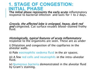
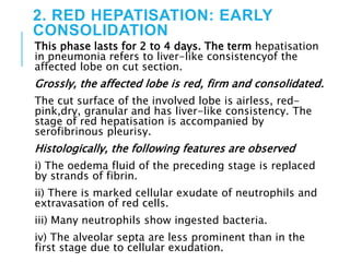
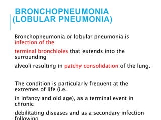
2. PNEUMONIA
Pneumonia is defined as acute inflammation of the lung
parenchyma distal to the terminal bronchioles
(consisting of
the respiratory bronchiole, alveolar ducts, alveolar sacs
and
alveoli).
The terms ‘pneumonia’ and ‘pneumonitis’ are often
used synonymously for inflammation of the lungs, while
‘consolidation’ (meaning solidification) is the term used
for
gross and radiologic appearance of the lungs in
3. PATHOGENESIS
The microorganisms gain entry into the
lungs by one of the following four routes:
1. Inhalation of the microbes present in the air.
2. Aspiration of organisms from the nasopharynx or
oropharynx.
3. Haematogenous spread from a distant focus of
infection.
4. Direct spread from an adjoining site of infection.
4. The normal lung is free of bacteria because of the
presence
of a number of lung defense mechanisms at
different levels
such as
nasopharyngeal filtering action,
mucociliary action of the lower respiratory
airways,
presence of phagocytosing alveolar macrophages
and immunoglobulins.
Failure of these defense mechanisms and
presence of certain
predisposing factors result in pneumonias.
5. ETIOLOGY
1. Altered consciousness. The oropharyngeal contents
may
be aspirated in states causing unconsciousness e.g. in
coma,
cranial trauma, seizures, cerebrovascular accidents,
drug
overdose, alcoholism etc.
2. Depressed cough and glottic reflexes. Depression of
effective cough may allow aspiration of gastric contents
e.g.
in old age, pain from trauma or thoracoabdominal
surgery,
neuromuscular disease, weakness due to malnutrition,
6. 3. Impaired mucociliary transport. The normal
protection
offered by mucus-covered ciliated epithelium in the
airways
from the larynx to the terminal bronchioles is impaired
or
destroyed in many conditions favouring passage of
bacteria
into the lung parenchyma. These conditions are
cigarette
smoking, viral respiratory infections, immotile cilia
syndrome,
inhalation of hot or corrosive gases and old age.
4. Impaired alveolar macrophage function. Pneumonias
7. 5. Endobronchial obstruction. The effective
clearance
mechanism is interfered in endobronchial
obstruction from
tumour, foreign body, cystic fibrosis and chronic
bronchitis.
6. Leucocyte dysfunctions. Disorders of
lymphocytes
including congenital and acquired
immunodeficiencies (e.g.
AIDS, immunosuppressive therapy) and granulocyte
abnormalities may predispose to pneumonia.
8. CLASSIFICATION
On the basis of the anatomic part of the
lung parenchyma involved, pneumonias are
traditionally
classified into 3 main types:
1. Lobar pneumonia
2. Bronchopneumonia (or Lobular pneumonia)
3. Interstitial pneumonia.
9. However, now that much is known about etiology
and
pathogenesis of pneumonias, current practice is to
follow the
etiologic classification which divides pneumonias
into following 3 main groups:
A. Bacterial pneumonia
B. Viral pneumonia
C. Pneumonias from other etiologies
10. A. BACTERIAL
PNEUMONIA
Bacterial infection of the lung parenchyma is the most
common cause of pneumonia or consolidation of one or
both
the lungs.
Two types of acute bacterial pneumonias are
distinguished—
lobar pneumonia and broncho-(lobular-) pneumonia,
each with distinct etiologic agent and morphologic
changes.
Another type distinguished by some workers separately
is confluent pneumonia which combines the features of
both lobar and bronchopneumonia and involves larger
(confluent) areas in both the lungs irregularly, while
11. LOBAR PNEUMONIA
Lobar pneumonia is an acute bacterial infection of a
part of a lobe,
the entire lobe,
or even two lobes of one or both the lungs.
12. ETIOLOGY.
1. Pneumococcal pneumonia. More than 90% of all lobar
pneumonias are caused by Streptococcus pneumoniae,
a lancet-shaped diplococcus.
Out of various types type 3-S. pneumoniae causes
particularly virulent form of lobar pneumonia.
Pneumococcal pneumonia in majority of cases is
community-acquired infection.
2. Staphylococcal pneumonia. Staphylococcus aureus
causes
pneumonia by haematogenous spread of infection from
another focus or after viral infections.
13. 3. Streptococcal pneumonia. β-haemolytic
streptococci may
rarely cause pneumonia such as in children after
measles or
influenza, in severely debilitated elderly patients
and in
diabetics.
4. Pneumonia by gram-negative aerobic bacteria.
Less
common causes of lobar pneumonia are gram-
negative
bacteria like Haemophilus influenzae, Klebsiella
pneumoniae
(Friedlander’s bacillus), Pseudomonas, Proteus and
Escherichia
14. MORPHOLOGIC
FEATURES
Laennec’s original description divides lobar
pneumonia into 4
Sequential pathologic phases:
stage of congestion (initial phase),
Red hepatisation (early consolidation),
grey hepatisation (late consolidation) and
resolution.
However, these classic stages seen in untreated
cases are found
much less often nowadays due to early institution
of antibiotic
Therapy and improved medical care.
15. 1. STAGE OF CONGESTION:
INITIAL PHASE
The initial phase represents the early acute inflammatory
response to bacterial infection and lasts for 1 to 2 days.
Grossly, the affected lobe is enlarged, heavy, dark red
and congested. Cut surface exudes blood-stained frothy
fluid.
Histologically, typical features of acute inflammatory
response to the organisms are seen. These are as under
i) Dilatation and congestion of the capillaries in the
alveolar walls.
ii) Pale eosinophilic oedema fluid in the air spaces.
iii) A few red cells and neutrophils in the intra-alveolar
fluid.
iv) Numerous bacteria demonstrated in the alveolar fluid
by Gram’s staining.
16.
17. 2. RED HEPATISATION: EARLY
CONSOLIDATION
This phase lasts for 2 to 4 days. The term hepatisation
in pneumonia refers to liver-like consistencyof the
affected lobe on cut section.
Grossly, the affected lobe is red, firm and consolidated.
The cut surface of the involved lobe is airless, red-
pink,dry, granular and has liver-like consistency. The
stage of red hepatisation is accompanied by
serofibrinous pleurisy.
Histologically, the following features are observed
i) The oedema fluid of the preceding stage is replaced
by strands of fibrin.
ii) There is marked cellular exudate of neutrophils and
extravasation of red cells.
iii) Many neutrophils show ingested bacteria.
iv) The alveolar septa are less prominent than in the
first stage due to cellular exudation.
18.
19. 3. GREY HEPATISATION: LATE
CONSOLIDATION
This phase lasts for 4 to 8 days.
Grossly, the affected lobe is firm and heavy. The cut
surface is dry, granular and grey in appearance with liver
like consistency
The change in colour fromred to grey begins at the
hilum and spreads towards the periphery. Fibrinous
pleurisy is prominent.
Histologically, the following changes are present
i) The fibrin strands are dense and more numerous.
ii) The cellular exudate of neutrophils is reduced due to
disintegration of many inflammatory cells as evidenced
by their pyknotic nuclei. The red cells are also fewer.
The macrophages begin to appear in the exudate.
iii) The cellular exudate is often separated from the
septal walls by a thin clear space.
iv) The organisms are less numerous and appear as
degenerated forms.
20.
21. 4. RESOLUTION
This stage begins by 8thto 9th day if no chemotherapy is
administered and is completed in 1 to 3 weeks. However,
antibiotic therapy induces resolution on about 3rd day.
Resolution proceeds in a progressive manner.
Grossly, the previously solid fibrinous constituent is
liquefied by enzymatic action, eventually restoring the
normal aeration in the affected lobe. The process of
softening begins centrally and spreads to the periphery.
The cut surface is grey-red or dirty brown and frothy
yellow, creamy fluid can be expressed on pressing. The
pleural reaction may also show resolution but ma
undergo organisation leading to fibrous obliteration of
22. Histologically, the following features are noted:
i) Macrophages are the predominant cells in the
alveolar spaces, while neutrophils diminish in
number. Many of the macrophages contain
engulfed neutrophils and debris.
ii) Granular and fragmented strands of fibrin in
thealveolar spaces are seen due to progressive
enzymatic digestion.
iii) Alveolar capillaries are engorged.
iv) There is progressive removal of fluid content as
wellas cellular exudate from the air spaces, partly
by expectoration but mainly by lymphatics,
resulting in restoration of normal lung parenchyma
23.
24. COMPLICATIONS
1. Organisation. In about 3% of cases, resolution of
the
exudate does not occur but instead it undergoes
organisation.
There is in growth of fibroblasts from the alveolar
septa resulting
in fibrosed, tough, airless leathery lung tissue. This
type
of post-pneumonic fibrosis is called carnification.
2. Pleural effusion. About 5% of treated cases of lobar
pneumonia develop inflammation of the pleura with
effusion.
The pleural effusion usually resolves but sometimes
may
25. 3. Empyema. Less than 1% of treated cases of lobar
pneumonia develop encysted pus in the pleural cavity
termed empyema.
4. Lung abscess. A rare complication of lobar
pneumonia
is formation of lung abscess, especially when there is
secondary infection by other organisms
5. Metastatic infection. Occasionally, infection in the
lungs
and pleural cavity in lobar pneumonia may extend into
the
pericardium and the heart causing purulent
pericarditis,
bacterial endocarditis and myocarditis. Other forms of
metastatic infection encountered rarely in lobar
26. CLINICAL FEATURES.
Shaking chills, fever, malaise with pleuritic chest pain,
dyspnoea and
cough with expectoration which may be mucoid, purulent
or even bloody.
The common physical findings are fever , tachycardia,
and tachypnoea, and sometimes cyanosis if the patient is
severely hypoxaemic.
There is generally a markedneutrophilic leucocytosis.
Blood cultures
are positive in about 30% of cases. Chest radiograph may
reveal
consolidation.
Culture of the organisms in the sputum and
antibioticsensitivity are most
significant investigations for institutionof specific
antibiotics. The
27. BRONCHOPNEUMONIA
(LOBULAR PNEUMONIA)
Bronchopneumonia or lobular pneumonia is
infection of the
terminal bronchioles that extends into the
surrounding
alveoli resulting in patchy consolidation of the lung.
The condition is particularly frequent at the
extremes of life (i.e.
in infancy and old age), as a terminal event in
chronic
debilitating diseases and as a secondary infection
28. ETIOLOGY
The common organisms responsible for
bronchopneumonia are staphylococci,
streptococci,
pneumococci, Klebsiella pneumoniae,
Haemophilus influenzae,
and gram-negative bacilli like
Pseudomonas and coliform bacteria.
29. EMPHYSEMA
The WHO has defined pulmonary emphysema as
combination of permanent dilatation of air spaces
distal to
the terminal bronchioles and the destruction of the
walls of
dilated air spaces. Thus, emphysema is defined
morphologically,
while chronic bronchitis is defined clinically. Since
the two
conditions coexist frequently and show
considerable overlap
in their clinical features, it is usual to label patients
as
30. a lobule is composed of about 5 acini distal to a
terminal bronchiole and that an acinus consists of 3 to 5
generations of respiratory bronchioles and a variable number
of alveolar ducts and alveolar sacs
As per WHO definition of pulmonary emphysema, it is
classified according to the portion of the acinus involved, into
5 types: centriacinar,
panacinar (panlobular),
para-septal (distal acinar),
irregular (para-cicatricial) and
mixed (unclassified)
emphysema.
A number of other conditions to which the term‘emphysema’
is loosely applied are, in fact, examples of ‘overinflation
32. ETIOPATHOGENESIS.
The commonest form of COPD is the combination of
chronic bronchitis and pulmonary emphysema.
Chronic bronchitis, however, does not always lead to
emphysema nor all cases of emphysema have changes of
chronic bronchitis.
The association of the two conditions is principally
linked to the common etiologic factors— most
importantly tobacco smoke and air pollutants.
Other less significant contributory factors are
occupational exposure, infection and somewhat poorly-
understood familial and genetic influences.
However, pathogenesis of the most significant event in
emphysema, the destruction of the alveolar walls, is not
linked to bronchial changes but is closely related to
deficiency of serum alpha-1-antitrypsin (α1-protease
inhibitor) commonly termed protease-antiprotease
hypothesis detailed below.
33. Protease-antiprotease hypothesis. Alpha-1-antitrypsin
(α- 1-AT),
also called α1-protease inhibitor (α-1-Pi), is a
glycoprotein that forms
the normal constituent of the α1- globulin fraction of the
plasma
proteins on serum electrophoresis.
The single gene locus that codes for α-1-AT is located
on the long arm of chromosome 15.
It is normally synthesised in the liver and is distributed in
the circulating
blood, tissue fluids and macrophages. The normal
function of α1-AT is
to inhibit proteases and hence its name α1-protease
inhibitor.
The proteases (mainly elastases) are derived from
34. There are several known alleles of α1-AT which have an
autosomal codominant inheritance pattern and are
classified
as normal (PiMM), deficient (PiZZ), null type (Pi null null)
having no detectable level, and dysfunctional (PiSS) type
having about half the normal level.
The most common abnormal phenotype in classic α1-
AT
deficiency is homozygous state PiZZ resulting from a
single
amino acid substitution Glu→Lys which causes
spontaneous
polymerisation of α1-AT and inhibits its release from
the
35. Clinically significant deficiency is also associated with
homozygous Pi null null and heterozygous Pi nullZ.
The heterozygote pattern of PiMZ has intermediate levels
which is not sufficient to produce clinical deficiency,
but
heterozygote individuals who smoke heavily have higher
risk of developing emphysema.
The α1-AT deficiency develops in adults and causes
pulmonary emphysema in smokers as well as in
nonsmokers,
though the smokers become symptomatic about
15 years earlier than non-smokers. The other organ showing
effects of α1-AT deficiency is the liver which may
develop
obstructive jaundice early in infancy, and cirrhosis and
hepatoma late in adulthood.
36. The mechanism of alveolar wall destruction in
emphysema by elastolytic action is based on the
imbalance
between proteases (chiefly elastase) and anti-proteases
(chiefly
anti-elastase):
By decreased anti-elastase activity i.e. deficiency of α-1
antitrypsin.
By increased activity of elastase i.e. increased
neutrophilic
infiltration in the lungs causing excessive elaboration of
neutrophil elastase.
There are enough evidences to suggest that smoking
promotes emphysema by both decreasing the amount of
antielastase
as well as by increasing the elastolytic protease in
37. 1. Oxidant in cigarette smoke has inhibitory
influence on
α-1-antitrypsin, thus lowering the level of anti-
elastase
activity.
2. Smokers have up to ten times more phagocytes
and
neutrophils in their lungs than nonsmokers; thus
they have
very high elastase activity.
38. PATHOLOGIC
FEATURES.
Grossly, the lungs are voluminous, pale with little blood.
The edges of the lungs are rounded. Mild cases show
dilatation of air spaces visible with hand lens. Advanced
cases show subpleural bullae and blebs bulging outwards
from the surface of the lungs with rib markings between
them. The bullae are air-filled cyst-like or bubble-like
structures, larger than 1 cm in diameter They
are formed by the rupture of adjacent air spaces while
blebs are the result of rupture of alveoli directly into the
subpleural interstitial tissue and are the common cause
of spontaneous pneumothorax.
39. Microscopically, depending upon the type of
emphysema,
there is dilatation of air spaces and destruction of
septal
walls of part of acinus involved i.e. respiratory
bronchioles,
alveolar ducts and alveolar sacs. Changes of
bronchitis may be present. Bullae and blebs when
present
show fibrosis and chronic inflammation of the walls
40. CLINICAL FEATURES. Cases of ‘predominant emphysema’
develop clinical features after about one-third of the
pulmonary parenchyma is damaged which occurs most
severely in panacinar emphysema. The age at the time of
diagnosis is often a decade later (about 60 years) than the
age for predominant bronchitis (about 50 years). Though
there is considerable overlap between the clinical features of
chronic bronchitis and emphysema, the following features
generally characterise ‘predominant emphysema’
(Table 17.5):
1. There is long history of slowly increasing severe
exertional dyspnoea.
2. Patient is quite distressed with obvious use of accessory
muscles of respiration
41. 3. Chest is barrel-shaped and hyperresonant.
4. Cough occurs late after dyspnoea starts and is associated
with scanty mucoid sputum.
5. Recurrent respiratory infections are not frequent.
6. Patients are called ‘pink puffers’ as they remain well
oxygenated and have tachypnoea.
7. Weight loss is common.
8. Features of right heart failure (cor pulmonale) and
hypercapneic respiratory failure are the usual terminal
events.
9. Chest X-ray shows small heart with hyperinflated lungs.
After these general comments about morphologic and
clinical features of emphysema, the specific pathologic
changes in individual types of ‘emphysema’ and
‘overinflation’ as classified in Table 17.4 are described below
42. MORPHOLOGY OF
INDIVIDUAL TYPES
OF EMPHYSEMA
1. CENTRIACINAR (CENTRILOBULAR) EMPHYSEMA.
Centriacinar or centrilobular emphysema is one of
the
common types. It is characterised by initial
involvement of
respiratory bronchioles i.e. the central or proximal
part of
the acinus (Fig. 17.19,B). This is the type of
emphysema that
usually coexists with chronic bronchitis and occurs
predominantly in smokers and in coal miners’
43. Grossly, the lesions are more common and more severe
in the upper lobes of the lungs. The characteristicappearance is
obvious in cut surface of the lung. It shows
distended air spaces in the centre of the lobules
surrounded by a rim of normal lung parenchyma in the
same lobule. The lobules are separated from each other
by fine fibrous tissue septa. Large amount of black
pigment is often present in the walls of emphysematous
spaces. In more severe cases, distal parts of acini are also
involved and the appearance may closely resemble
panacinar emphysema.
Microscopically, there is distension and destruction of the
respiratory bronchiole in the centre of lobules, surrounded
peripherally by normal uninvolved alveoli. The
terminal bronchioles supplying the acini show chronic
inflammation and are narrowed.
44. 2. PANACINAR (PANLOBULAR) EMPHYSEMA.
Panacinar or panlobular emphysema is the other
common
type. In this type, all portions of the acinus are
affected but
not of the entire lung. Panacinar emphysema
is most often associated with α1-antitrypsin
deficiency in
middle-aged smokers and is the one that produces
the most
characteristic anatomical changes in the lung in
emphysema.
45. Grossly, in contrast to centriacinar emphysema, the
panacinar emphysema involves lower zone of lungs more
frequently and more severely than the upper zone. The
involvement may be confined to a few lobules, or may be
more widespread affecting a lobe or part of a lobe of the
lung. The lungs are enlarged and overinflated.
Microscopically, usually all the alveoli within a lobule
are affected to the same degree. All portions of acini are
distended—respiratory bronchioles, alveolar ducts and
alveoli, are all dilated and their walls stretched and thin.
Ruptured alveolar walls and spurs of broken septa are
seen between the adjacent alveoli. The capillaries are
stretched and thinned. Special stains show loss of elastic
tissue. Inflammatory changes are usually absent
(Fig. 17.20).
46. 3. PARASEPTAL (DISTAL ACINAR) EMPHYSEMA. This
type of emphysema involves distal part of acinus
while the
proximal part is normal. Paraseptal or distal acinar
emphysema is localised along the pleura and along
the
perilobular septa. The involvement is seen adjacent
to the
areas of fibrosis and atelectasis and involves upper
part of
lungs more severely than the lower. This form of
emphysema
is seldom associated with COPD but is the common
cause of
spontaneous pneumothorax in young adults.
47. 4. IRREGULAR (PARA-CICATRICIAL) EMPHYSEMA.
This is the most common form of emphysema, seen
surrounding scars from any cause. The involvement is
irregular as regards the portion of acinus involved as well as
within the lung as a whole. During life, irregular emphysema
is often asymptomatic and may be only an incidental autopsy
finding.
5. MIXED (UNCLASSIFIED) EMPHYSEMA. Quite often,
the same lung may show more than one type of emphysema.
It is usually due to more severe involvement resulting in loss
of clearcut distinction between one type of emphysema and
the other. Thus, the lungs of an elderly smoker at autopsy
may show continuation of centriacinar emphysema in the
upper lobes, panacinar in the lower lobes, and paraseptal
emphysema in the subpleural region.