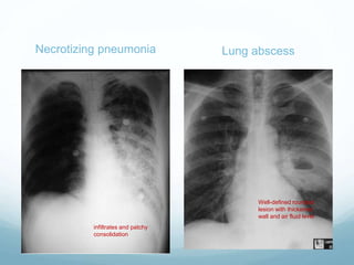
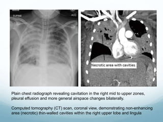
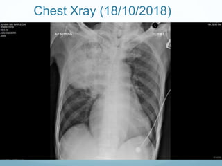
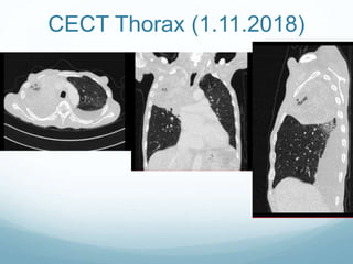
Necrotizing
Pneumonia
OVERVIEW
 Necrotizing lung infections constitute a
spectrum of disease severity ranging
from simple lung abscess to
necrotizing pneumonia to pulmonary
gangrene
 Necrotizing pneumonia is a rare and is
a severe complication of bacterial
community-acquired pneumonia (CAP)
 It is associated with high morbidity and
mortality.
 Patients who develop necrotising
pneumonia usually have concomitant
medical disease like diabetes mellitus
and alcohol abuse
 Is characterized by pulmonary
inflammation with consolidation,
peripheral necrosis and multiple small
cavities
Pathogen than cause necrotizing
pneumonia includes:
Staphylococcus aureus: particularly in
young immunocompetent patients
Klebsiella pneumoniae
Enterobacter spp.
Nocardia spp
Actinomyces spp
Pseudomonas spp
Pneumococcus spp.: especially type III
Pneumococcal sp.
Haemophilus influenzae
 Pathogenesis of necrotizing pneumonia is not clearly defined, but
most studies suggest that it is a result of inflammatory response
due to the toxins produced by the pathogen; or the associated
vasculitis and venous thrombosis.
 Is characterized by pulmonary inflammation with :
 consolidation
 peripheral necrosis and
 multiple small cavities
 It compromise of the bronchial and pulmonary vascular supply and
has the potential for devitalization of lung parenchyma.
 The lack of blood supply to the underperfused areas impedes
delivery of antibiotics, allowing for uncontrolled infection and
further destruction of lung tissue.
 Pulmonary gangrene is the “final stage in a continuum of
progressive devitalization of pulmonary parenchyma” and is
characterized by “sloughing of a pulmonary segment or lobe”
TYPICAL FINDINGS
Necrotizing Pneumonia Lung Abscess
Clinical signs /
symptoms
• severely ill patient with sepsis
• rapid clinical deterioration, may
required ventilator support and/or
• exhibiting signs of septic shock
within 72 h of presentation.
• patient presented weeks
after onset of initial
symptoms, with a
productive cough and
fevers that fail to resolve
despite usual courses of
outpatient antibiotics.
Pathologically Similar with lung abscess pneumonitis in the affected
lobe progresses to a region of
local tissue necrosis within 7
to 14 days, becoming walled-
off as a single cavity with a
fibrous capsule. Initial
radiographic examination
may reveal a cavity within a
pulmonary infiltrate
Necrotizing Pneumonia Lung Abscess
Radiographic
findings
necrotizing pneumonia present with
more extensive airspace disease often
involving multiple lobes.
CXRs : bulging fissures due to the
intense inflammatory exudation present.
CT Scan findings:
Multiple small cavities, <1 cm.
patchy inflammation with lack of
perfusion (lack of contrast uptake in the
parenchyma) and microabscesses .
tissue undergoes liquefactive necrosis
 multiple small abscesses may
coalesce to form larger cavities
Initial radiograph:
examination may reveal a
cavity within a pulmonary
infiltrate.
Serial x-rays may show a
single cavity with
thickened wall, often
containing an air-fluid
level, for several weeks
after the surrounding
airspace disease has
cleared
Necrotizing pneumonia Lung abscess
infiltrates and patchy
consolidation
Well-defined rounded
lesion with thickened
wall and air fluid level
Plain chest radiograph revealing cavitation in the right mid to upper zones,
pleural effusion and more general airspace changes bilaterally.
Computed tomography (CT) scan, coronal view, demonstrating non-enhancing
area (necrotic) thin-walled cavities within the right upper lobe and lingula
 Contrast-enhanced chest CT scans are more sensitive
than chest radiographs as
 standard imaging for making the diagnosis of
necrotizing pneumonia.
 The key diagnostic features are:
 poor or absent vascularity, loss of pulmonary
architecture, and finally cavity formation.
 Multiple small, gas- or fluid-filled, thin-walled cavities are
usually seen initially, often involving just a single lobe.
These are non-enhancing, indicating reduced perfusion
while loculated fluid or gas within the pleural cavity is also
generally present.
 As the lung undergoes further liquefactive necrosis, the
multiple small cavities may coalesce and form larger
cavities, including gas-filled pneumatoceles that develop
in the later stages of NP
CASE
 Mr A 62 years old with underlying
1. Smear negative PTB diagnosed in October 2018 – on
anti TB (Tab. Akurit 4)
2. Atrial tachycardia diagnosed in 2008. Holter done
normal. Discharged from cardio follow up.
3. Diabetes Mellitus (under GP follow up)

 First presentation to ED on 18/10/2018:
- Complaint of sudden onset of shortness of breath (SOB) associated
with cough prior.
- No fever
- General examinaton: GCS full, not tachypnoeic, normal vital signs.
- Lungs: Equal air entry, no crepitation / rhonchi.
- WCC: 13.9 (neutrophil predominant 86%)
- DDX: community acquired pneumonia with underlying negative smear
PTB.
- Patient was discharged from ED with Tab Augmentin 625mg
Chest Xray (16/10/2018)
 Second presentation to ED on 18/10/2018:
- Presented with complaint of fever x 3 days and generalised body weakness.
- General examination: GCS 11/15 (E4 V2 M5), septic looking, cachexic,
dehydrated, mild tachypneic and tachycardic.
- BP: 90/44mmHg, PR 150 (fast AF), spO2 95% under HFM
- ECG: fast AF (220bpm)
- Lung: reduce air entry over right upper zone with dullness on percussion.
- Blood investigation:
- FBC: WCC-7.5 (neutrophil predominant 83%), Hb-15.1, platlet 301.
Chest Xray (18/10/2018)
 Impression:
 1. Secondary bacterial infection; CAP with type 1 resp
failure with U/L smear negative PTB.
 2. AKI secondary to dehydration/ sepsis
 3. Fast AF secondary to sepsis.
Progress in ward (18/10/2018 –
11/11/2018)
- Patient was referred to respi team and investigated further TRO lung
malignancy.
- During admission, patient had episode of disorientation. NECT and
CECT Brain done.
- CT findings: Multiple intracranial rim enhancing lesions. DDx: cerebral
metastasis, tuberculoma.
- Flexible Bronchoscopy done on 26.10.2018:
- Findings: RUL broad based smooth surface mass obliterating RUL,
extending to RMB laterally. Hypervascularity seen on the surface of the
mass. Unable to see opening RUL. Able to pass through scope via RBI.
- Suggestive of malignancy, however unable to proceed with tissue biopsy
in view of hypervascularity of the mass and coagulopathy.
- Bronchial lavage cytology: No atypical cells.
CECT Thorax (1.11.2018)
consolidation and multiple, irregular, hypodense areas in the lung
parenchyma, indicating necrosis in the right upper lobe.
Bilateral pleural effusion
CECT Thorax (1.11.2018)

Findings:
The right upper lobe is consolidated, the segmental bronchi are filled with fluid. There are area of hypodensity with multiple air pockets within suggestive of
necrotic changes. Traversing vessels also seen within. The mass is seen infiltrating into the middle mediastinal region. The right upper lobe bronchi is
obstructed by the mass. There are multiple enlarged matted right hilar lymph nodes noted which is encasing the right main pulmonary artery. The ascending
branch of right main pulmonary artery is truncated. The pulmonary trunk and descending branch of right main pulmonary artery is opacified. The superior
vena cava is patent. No rib erosion seen. No extension of the mass seen to the cervical region. No invasion of the anterior chest wall noted. The trachea,
bronchus intermedius and left main bronchus are patent.
A small lung nodule seen at the left apical region measuring 0.5cm in diameter.
There are few calcified granuloma noted at the left upper lobe.
The rest of the lung fields are clear.
Bilateral pleural effusion noted.
Heart is enlarged. No pericardial effusion.
There is a lobulatd heterogenous enhancing mass seen at the left suprarenal region. Hypodense area seen in keeping with necrosis. The mass measures
approximately 9.2cm(ap) x5.5cm(w) x6.7cm(cc). The normal left adrenal gland is not visualised.
The rest of the visualised upper abdominal organs are unremarkable.
Presence of ascites.
No suspicious bony lesion.
Impression:
1. Right upper lobe finding is in consistent with necrotising pneumonia.
2. Bilateral pleural effusion.
3. Left upper lobe granuloma
4. Left adrenal cystic mass.
 The lung findings are suggestive of infective changes rather than malignancy. Should the left adrenal cystic mass pathophysiology is related to the right
upper lobe finding, tuberculosis needs to be considered.
 The lung findings are suggestive of infective changes rather than malignancy. Should the left adrenal cystic mass and the rim enhancing brain lesions from
previous CT are pathophysiology related to the right upper lobe finding, systemic infection such as nocardiosis and tuberculosis needs to be considered. A
proper CT Abdomen is useful to find other possible site of abscesses.
Another differential would be, the necrotising pneumonia is separate from the adrenal and brain findings in which the primary tumour is not visualised in this
study, either being superimposed by the infective changes or not in the lungs. Dx:1) TRO Lung malignancy with brain & Suprarenal mets

MDT Discussion 5.11.2018
 CT Thorax review:
- necrotic consolidative mass at right upper lobe, mass at right
hilar region obstructing right upper lobe bronchus with post
obstructive pneumonia and necrotizing pneumonia.
- mass encasing pulmonary artery and superior pulmonary
vein
- bilateral pleural effusion
- cardiomegaly
- mass seen at the left suprarenal region
- underlying emphysematous area with reticular ground glass
density
imp: central mass over right upper lobe with post obstructive
pneumonia and necrotizing pneumonia with left suprarenal
gland meastasis
consensus:
for CT guided biopsy of lung
 CT guided lung biopsy (RUL) on 9.11.2018
 Unfortunately, his condition worsens progressively and
he succumbed to the illness on day 24 of admission
(11.11.2018)


necrotizing pneumonia.ppt

  • 1.
  • 2.
    OVERVIEW  Necrotizing lunginfections constitute a spectrum of disease severity ranging from simple lung abscess to necrotizing pneumonia to pulmonary gangrene  Necrotizing pneumonia is a rare and is a severe complication of bacterial community-acquired pneumonia (CAP)  It is associated with high morbidity and mortality.  Patients who develop necrotising pneumonia usually have concomitant medical disease like diabetes mellitus and alcohol abuse  Is characterized by pulmonary inflammation with consolidation, peripheral necrosis and multiple small cavities Pathogen than cause necrotizing pneumonia includes: Staphylococcus aureus: particularly in young immunocompetent patients Klebsiella pneumoniae Enterobacter spp. Nocardia spp Actinomyces spp Pseudomonas spp Pneumococcus spp.: especially type III Pneumococcal sp. Haemophilus influenzae
  • 3.
     Pathogenesis ofnecrotizing pneumonia is not clearly defined, but most studies suggest that it is a result of inflammatory response due to the toxins produced by the pathogen; or the associated vasculitis and venous thrombosis.  Is characterized by pulmonary inflammation with :  consolidation  peripheral necrosis and  multiple small cavities  It compromise of the bronchial and pulmonary vascular supply and has the potential for devitalization of lung parenchyma.  The lack of blood supply to the underperfused areas impedes delivery of antibiotics, allowing for uncontrolled infection and further destruction of lung tissue.  Pulmonary gangrene is the “final stage in a continuum of progressive devitalization of pulmonary parenchyma” and is characterized by “sloughing of a pulmonary segment or lobe”
  • 4.
    TYPICAL FINDINGS Necrotizing PneumoniaLung Abscess Clinical signs / symptoms • severely ill patient with sepsis • rapid clinical deterioration, may required ventilator support and/or • exhibiting signs of septic shock within 72 h of presentation. • patient presented weeks after onset of initial symptoms, with a productive cough and fevers that fail to resolve despite usual courses of outpatient antibiotics. Pathologically Similar with lung abscess pneumonitis in the affected lobe progresses to a region of local tissue necrosis within 7 to 14 days, becoming walled- off as a single cavity with a fibrous capsule. Initial radiographic examination may reveal a cavity within a pulmonary infiltrate
  • 5.
    Necrotizing Pneumonia LungAbscess Radiographic findings necrotizing pneumonia present with more extensive airspace disease often involving multiple lobes. CXRs : bulging fissures due to the intense inflammatory exudation present. CT Scan findings: Multiple small cavities, <1 cm. patchy inflammation with lack of perfusion (lack of contrast uptake in the parenchyma) and microabscesses . tissue undergoes liquefactive necrosis  multiple small abscesses may coalesce to form larger cavities Initial radiograph: examination may reveal a cavity within a pulmonary infiltrate. Serial x-rays may show a single cavity with thickened wall, often containing an air-fluid level, for several weeks after the surrounding airspace disease has cleared
  • 6.
    Necrotizing pneumonia Lungabscess infiltrates and patchy consolidation Well-defined rounded lesion with thickened wall and air fluid level
  • 7.
    Plain chest radiographrevealing cavitation in the right mid to upper zones, pleural effusion and more general airspace changes bilaterally. Computed tomography (CT) scan, coronal view, demonstrating non-enhancing area (necrotic) thin-walled cavities within the right upper lobe and lingula
  • 8.
     Contrast-enhanced chestCT scans are more sensitive than chest radiographs as  standard imaging for making the diagnosis of necrotizing pneumonia.  The key diagnostic features are:  poor or absent vascularity, loss of pulmonary architecture, and finally cavity formation.  Multiple small, gas- or fluid-filled, thin-walled cavities are usually seen initially, often involving just a single lobe. These are non-enhancing, indicating reduced perfusion while loculated fluid or gas within the pleural cavity is also generally present.  As the lung undergoes further liquefactive necrosis, the multiple small cavities may coalesce and form larger cavities, including gas-filled pneumatoceles that develop in the later stages of NP
  • 9.
    CASE  Mr A62 years old with underlying 1. Smear negative PTB diagnosed in October 2018 – on anti TB (Tab. Akurit 4) 2. Atrial tachycardia diagnosed in 2008. Holter done normal. Discharged from cardio follow up. 3. Diabetes Mellitus (under GP follow up) 
  • 10.
     First presentationto ED on 18/10/2018: - Complaint of sudden onset of shortness of breath (SOB) associated with cough prior. - No fever - General examinaton: GCS full, not tachypnoeic, normal vital signs. - Lungs: Equal air entry, no crepitation / rhonchi. - WCC: 13.9 (neutrophil predominant 86%) - DDX: community acquired pneumonia with underlying negative smear PTB. - Patient was discharged from ED with Tab Augmentin 625mg
  • 11.
  • 12.
     Second presentationto ED on 18/10/2018: - Presented with complaint of fever x 3 days and generalised body weakness. - General examination: GCS 11/15 (E4 V2 M5), septic looking, cachexic, dehydrated, mild tachypneic and tachycardic. - BP: 90/44mmHg, PR 150 (fast AF), spO2 95% under HFM - ECG: fast AF (220bpm) - Lung: reduce air entry over right upper zone with dullness on percussion. - Blood investigation: - FBC: WCC-7.5 (neutrophil predominant 83%), Hb-15.1, platlet 301.
  • 13.
  • 14.
     Impression:  1.Secondary bacterial infection; CAP with type 1 resp failure with U/L smear negative PTB.  2. AKI secondary to dehydration/ sepsis  3. Fast AF secondary to sepsis.
  • 15.
    Progress in ward(18/10/2018 – 11/11/2018) - Patient was referred to respi team and investigated further TRO lung malignancy. - During admission, patient had episode of disorientation. NECT and CECT Brain done. - CT findings: Multiple intracranial rim enhancing lesions. DDx: cerebral metastasis, tuberculoma. - Flexible Bronchoscopy done on 26.10.2018: - Findings: RUL broad based smooth surface mass obliterating RUL, extending to RMB laterally. Hypervascularity seen on the surface of the mass. Unable to see opening RUL. Able to pass through scope via RBI. - Suggestive of malignancy, however unable to proceed with tissue biopsy in view of hypervascularity of the mass and coagulopathy. - Bronchial lavage cytology: No atypical cells.
  • 16.
  • 17.
    consolidation and multiple,irregular, hypodense areas in the lung parenchyma, indicating necrosis in the right upper lobe. Bilateral pleural effusion
  • 18.
    CECT Thorax (1.11.2018)  Findings: Theright upper lobe is consolidated, the segmental bronchi are filled with fluid. There are area of hypodensity with multiple air pockets within suggestive of necrotic changes. Traversing vessels also seen within. The mass is seen infiltrating into the middle mediastinal region. The right upper lobe bronchi is obstructed by the mass. There are multiple enlarged matted right hilar lymph nodes noted which is encasing the right main pulmonary artery. The ascending branch of right main pulmonary artery is truncated. The pulmonary trunk and descending branch of right main pulmonary artery is opacified. The superior vena cava is patent. No rib erosion seen. No extension of the mass seen to the cervical region. No invasion of the anterior chest wall noted. The trachea, bronchus intermedius and left main bronchus are patent. A small lung nodule seen at the left apical region measuring 0.5cm in diameter. There are few calcified granuloma noted at the left upper lobe. The rest of the lung fields are clear. Bilateral pleural effusion noted. Heart is enlarged. No pericardial effusion. There is a lobulatd heterogenous enhancing mass seen at the left suprarenal region. Hypodense area seen in keeping with necrosis. The mass measures approximately 9.2cm(ap) x5.5cm(w) x6.7cm(cc). The normal left adrenal gland is not visualised. The rest of the visualised upper abdominal organs are unremarkable. Presence of ascites. No suspicious bony lesion. Impression: 1. Right upper lobe finding is in consistent with necrotising pneumonia. 2. Bilateral pleural effusion. 3. Left upper lobe granuloma 4. Left adrenal cystic mass.  The lung findings are suggestive of infective changes rather than malignancy. Should the left adrenal cystic mass pathophysiology is related to the right upper lobe finding, tuberculosis needs to be considered.  The lung findings are suggestive of infective changes rather than malignancy. Should the left adrenal cystic mass and the rim enhancing brain lesions from previous CT are pathophysiology related to the right upper lobe finding, systemic infection such as nocardiosis and tuberculosis needs to be considered. A proper CT Abdomen is useful to find other possible site of abscesses. Another differential would be, the necrotising pneumonia is separate from the adrenal and brain findings in which the primary tumour is not visualised in this study, either being superimposed by the infective changes or not in the lungs. Dx:1) TRO Lung malignancy with brain & Suprarenal mets 
  • 19.
    MDT Discussion 5.11.2018 CT Thorax review: - necrotic consolidative mass at right upper lobe, mass at right hilar region obstructing right upper lobe bronchus with post obstructive pneumonia and necrotizing pneumonia. - mass encasing pulmonary artery and superior pulmonary vein - bilateral pleural effusion - cardiomegaly - mass seen at the left suprarenal region - underlying emphysematous area with reticular ground glass density imp: central mass over right upper lobe with post obstructive pneumonia and necrotizing pneumonia with left suprarenal gland meastasis consensus: for CT guided biopsy of lung
  • 20.
     CT guidedlung biopsy (RUL) on 9.11.2018  Unfortunately, his condition worsens progressively and he succumbed to the illness on day 24 of admission (11.11.2018) 

Editor's Notes

  • #12 Patchy area of consolidation at right upper zone with thickened oblique fissure
  • #14 Plain chest radiograph Consolidation in the right mid to upper zones with area of lucency (cavitation)   Bulging of fissure