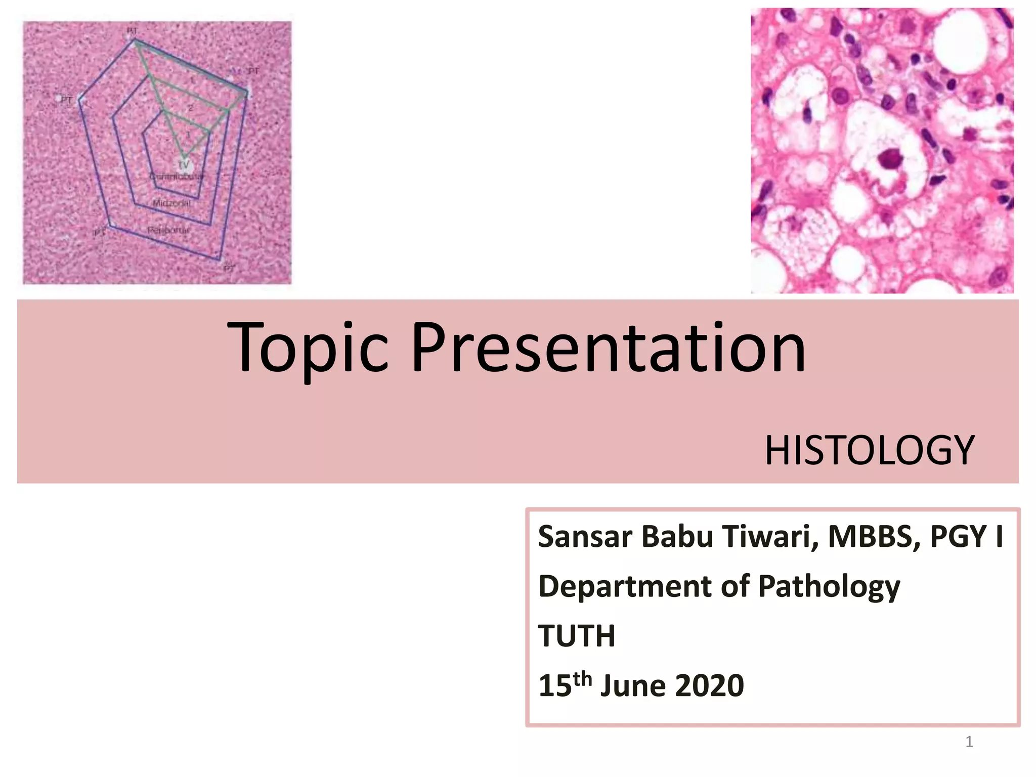
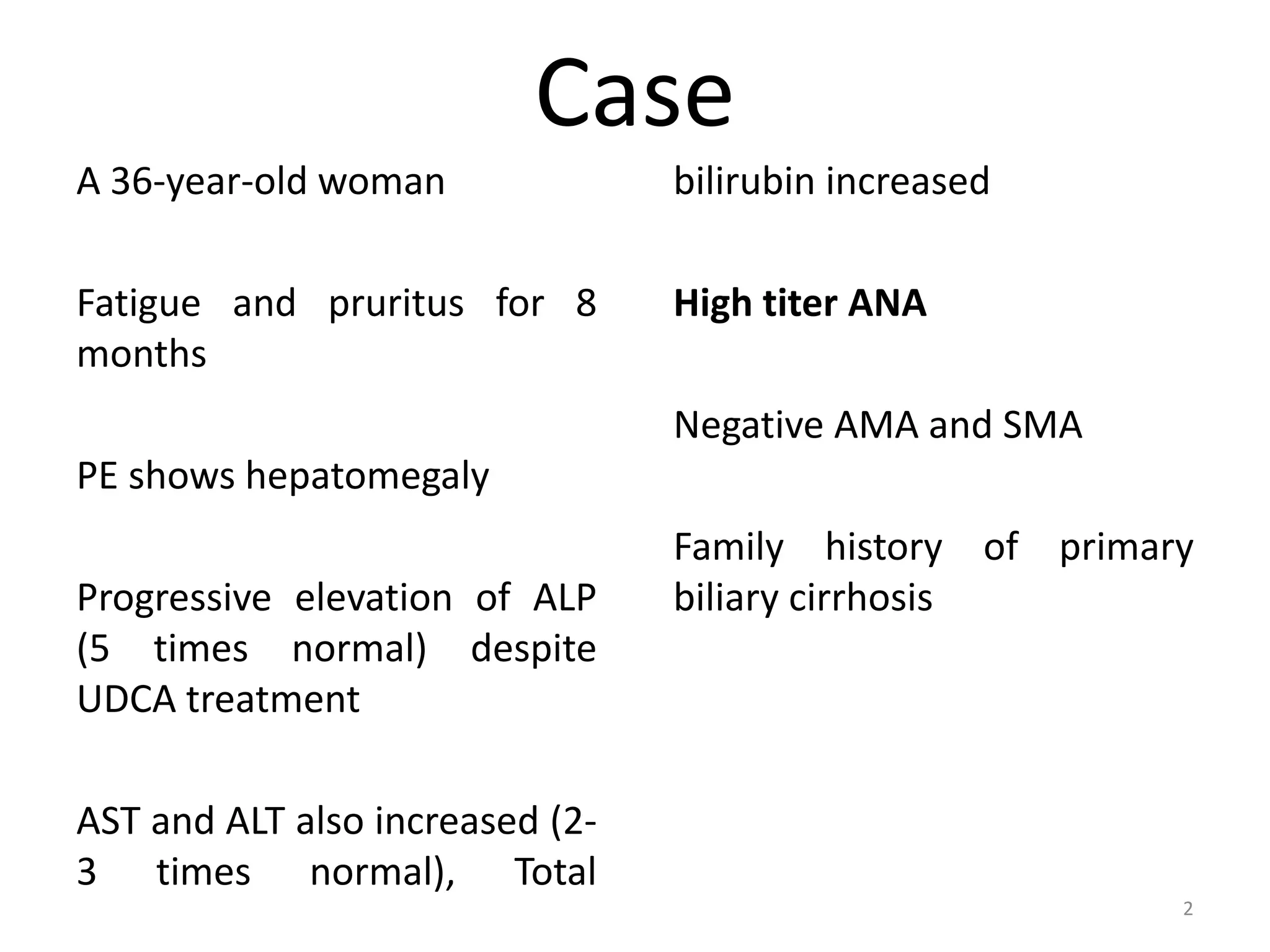
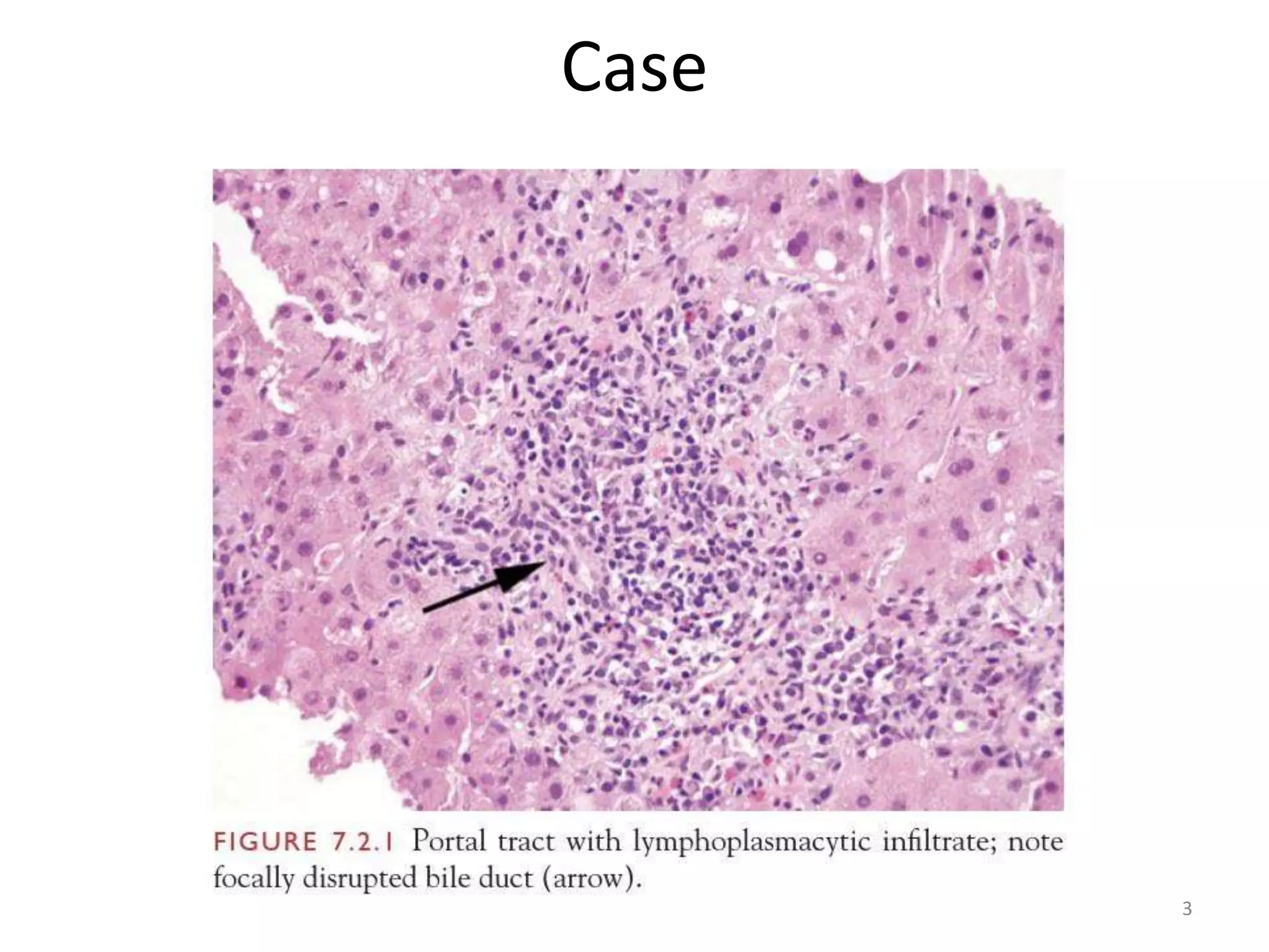
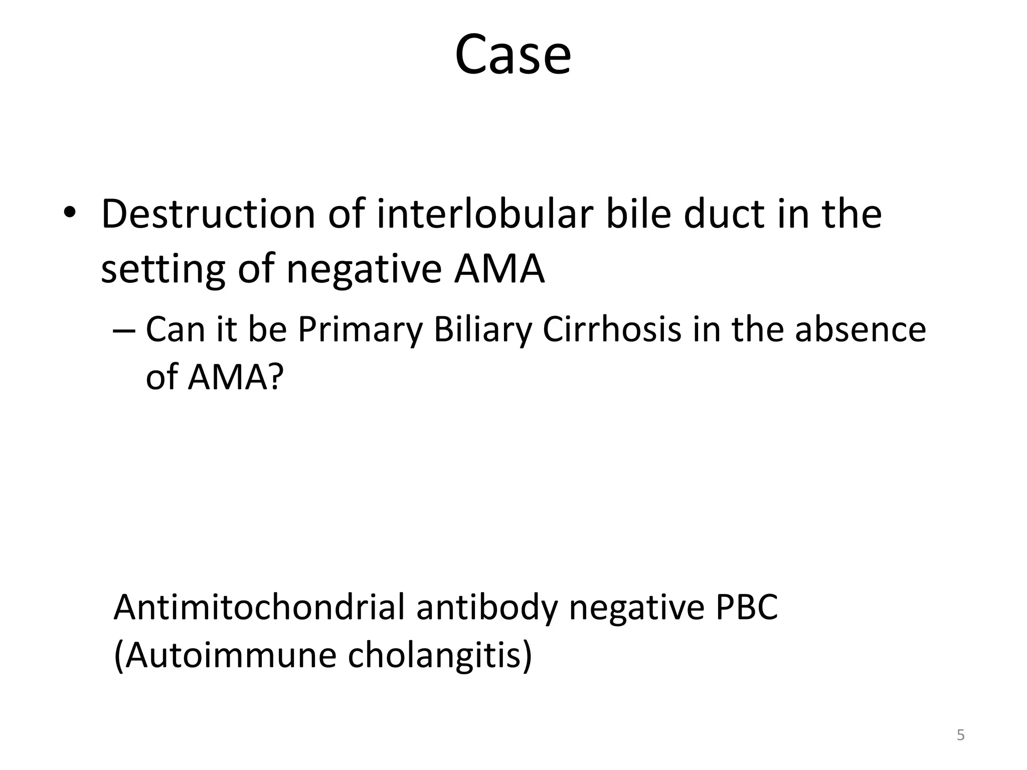
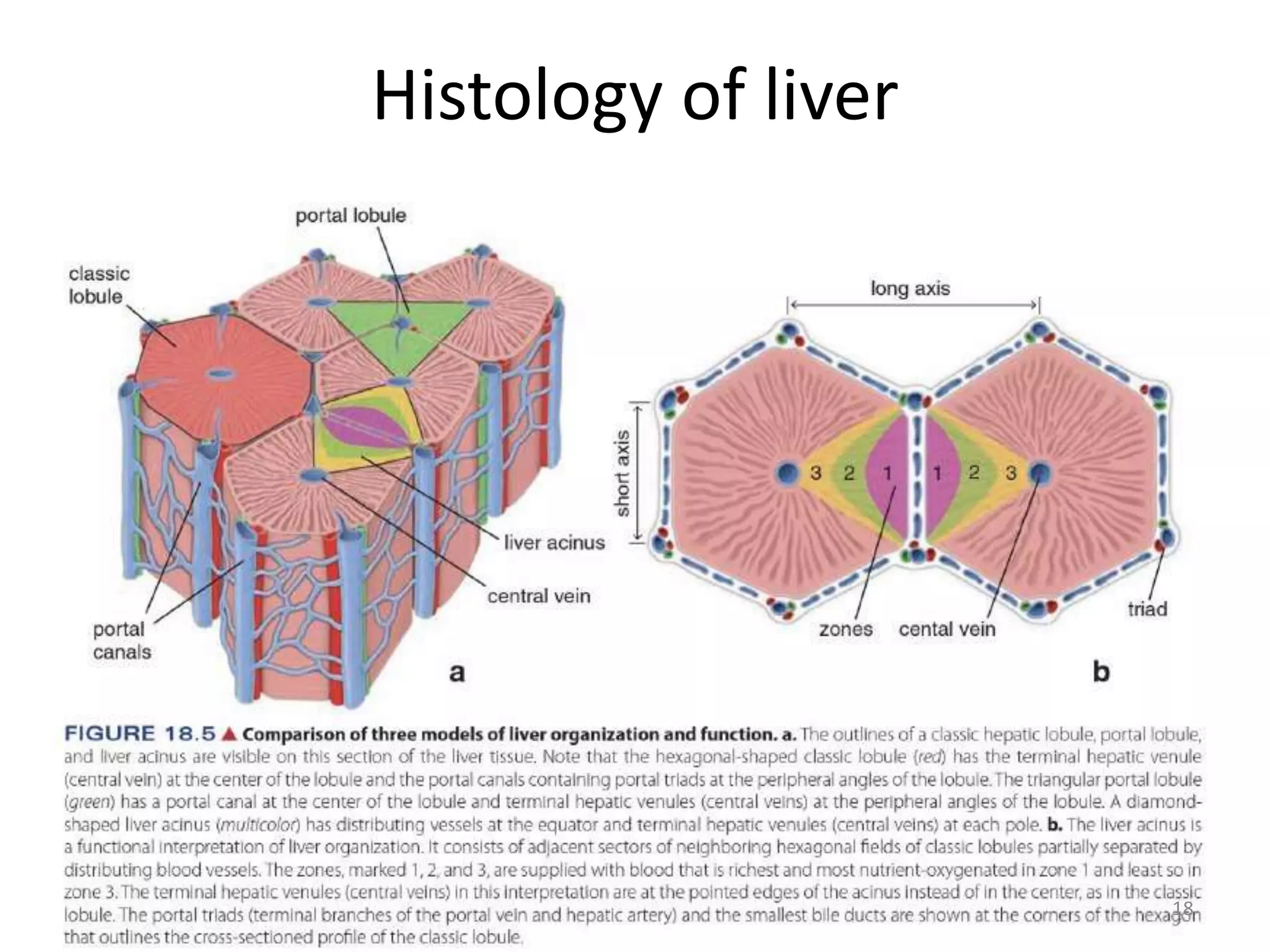
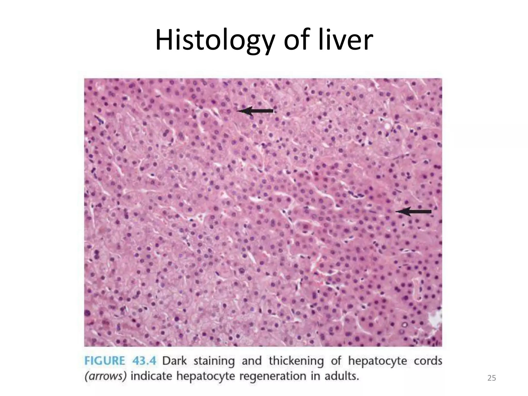
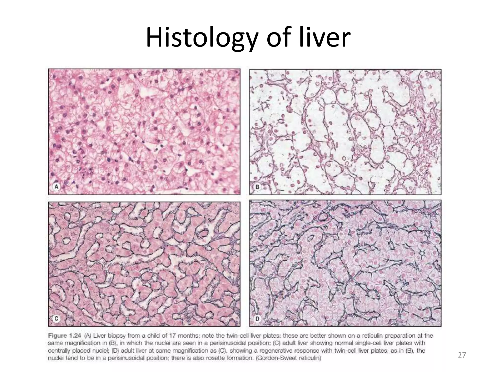
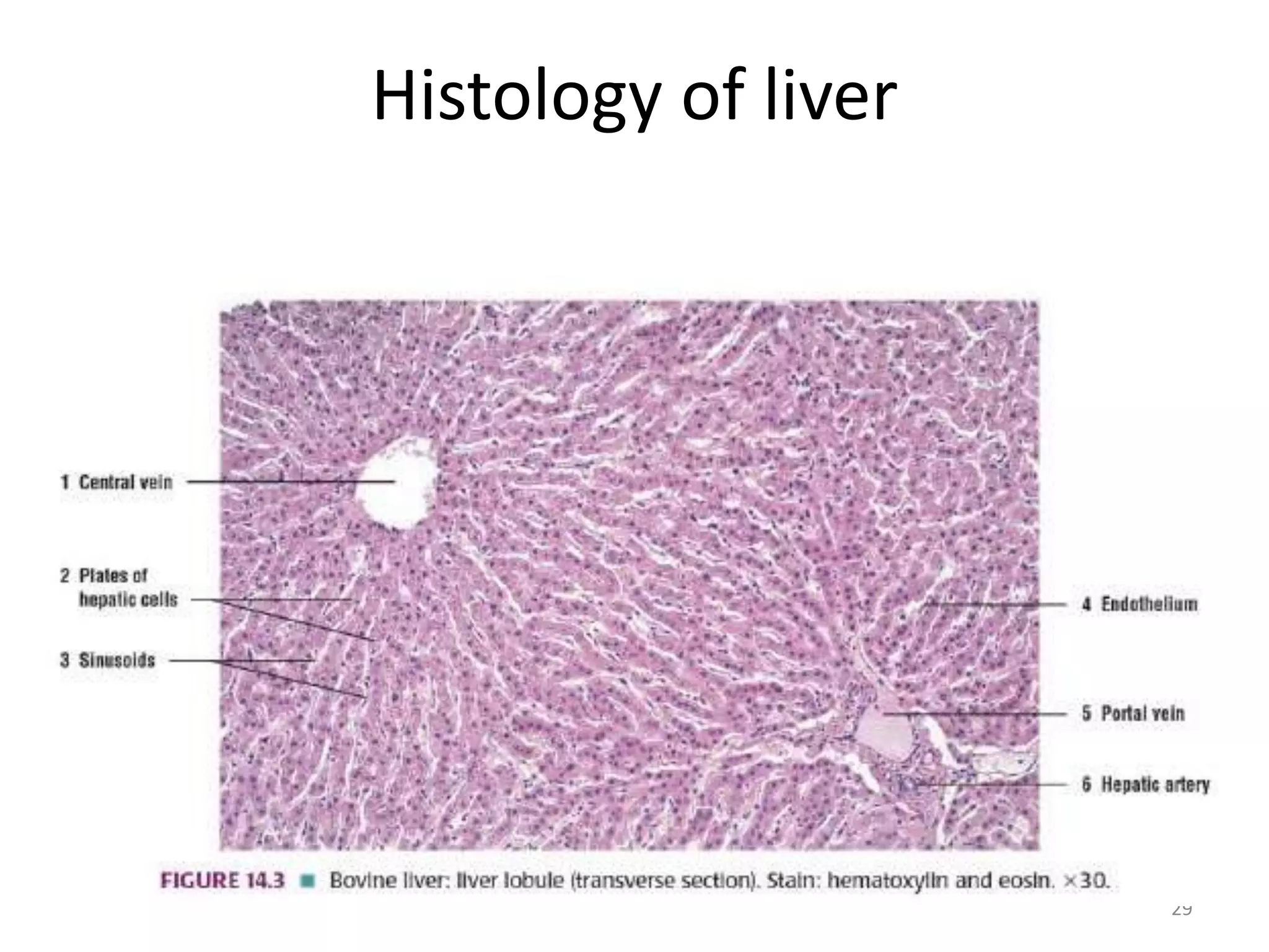
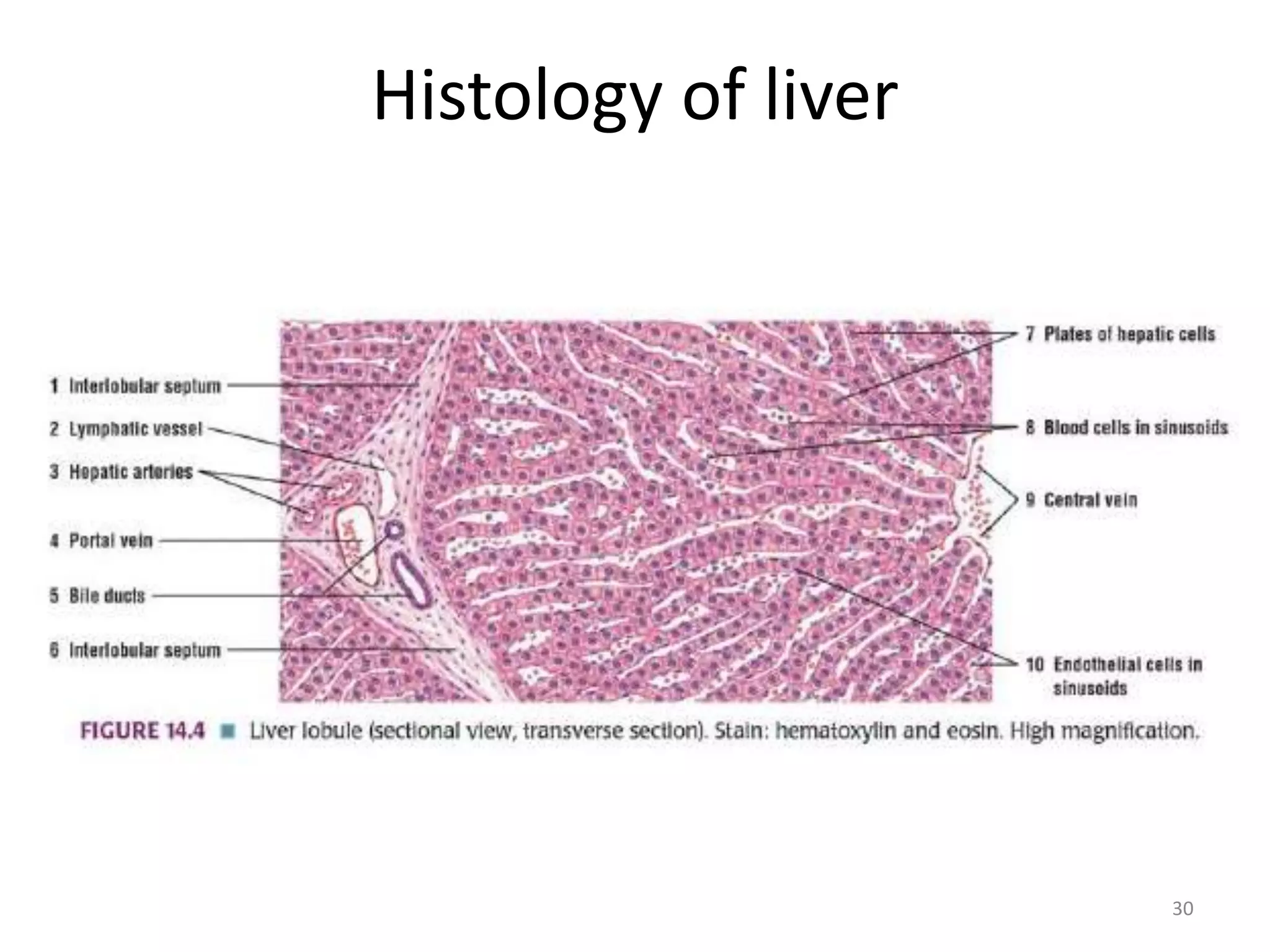
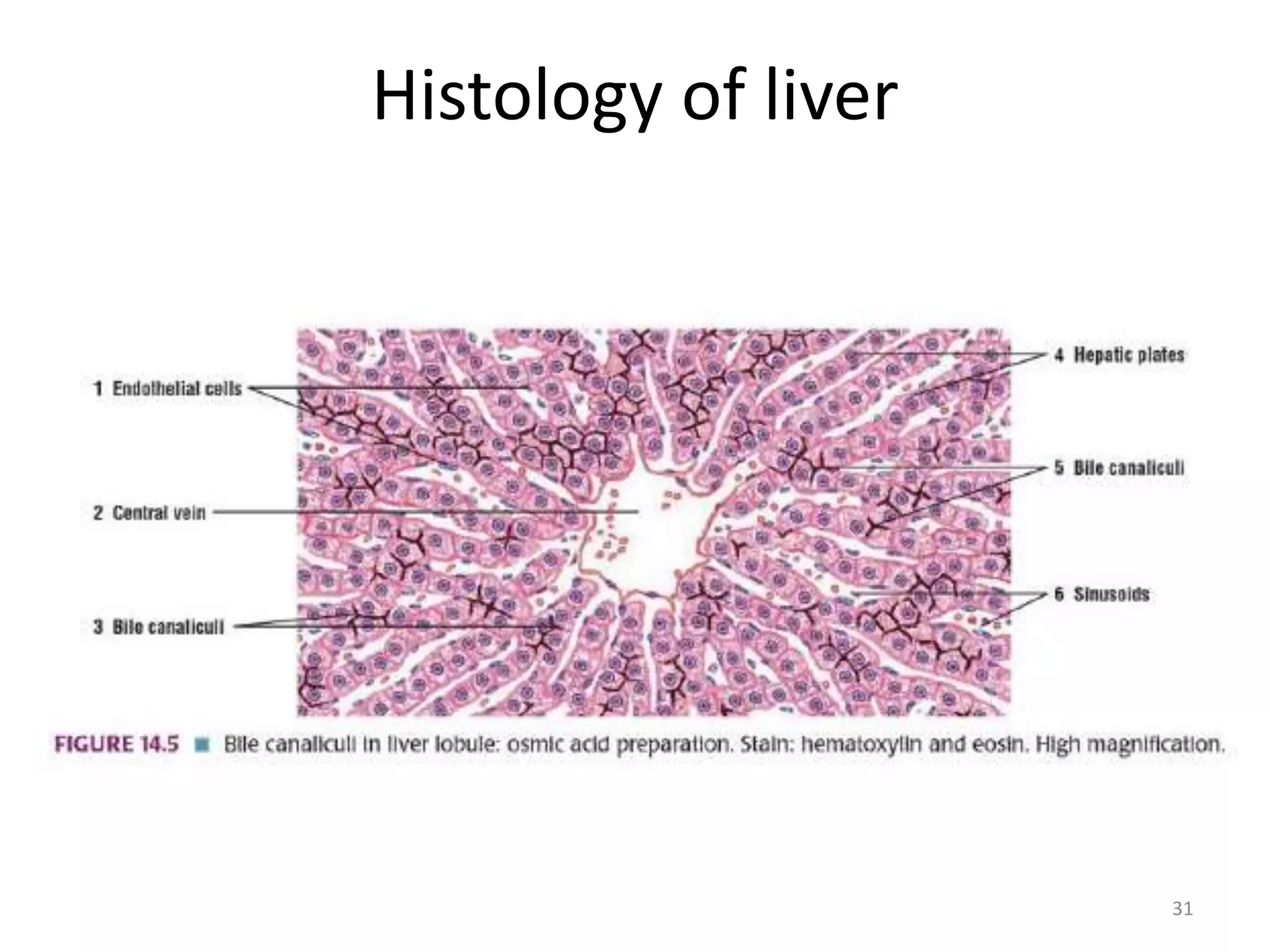
Topic Presentation
HISTOLOGY
Sansar Babu Tiwari, MBBS, PGY I
Department of Pathology
TUTH
15th June 2020
1
Case
A 36-year-old woman
Fatigue and pruritus for 8
months
PE shows hepatomegaly
Progressive elevation of ALP
(5 times normal) despite
UDCA treatment
AST and ALT also increased (2-
3 times normal), Total
bilirubin increased
High titer ANA
Negative AMA and SMA
Family history of primary
biliary cirrhosis
2
3
Case
4
Case
5
Case
• Destruction of interlobular bile duct in the
setting of negative AMA
– Can it be Primary Biliary Cirrhosis in the absence
of AMA?
Antimitochondrial antibody negative PBC
(Autoimmune cholangitis)
6
Case
• 5 years later liver
transplant was
performed and
explant biopsy
showed
– Jigsaw pattern
of biliary
cirrhosis
Liver Biopsy
• Liver biopsy methods
– Percutaneous (blind)
– Transjugular or transvenous
– Laparoscopic or open
– CT or USG-guided
With the advent of modern non-invasive tests like
elastography (fibroscan) the diagnostic importance
of liver biopsy as a gold standard has now been
decreased.
7
Liver Biopsy
8
Liver Biopsy
• Specimen handling:
– Should be at least 1.5 cm long, if not another pass
is recommended
– If tumor is suspected touch preparation can be
made immediately
– Should not be placed on a dry gauge, which tends
to dehydrate cells, resulting in a prominent
nuclear artifact
9
Liver Biopsy
• Specimen handling:
– Color, consistency and tendency to float or
fragment is documented by the person
performing biopsy
– Tumor or granulomas: white areas in a
background of reddish brown tissue
– Yellow in fatty liver, dark red in congested liver,
rusty brown in hemochromatosis
– Fragmentation suggests advanced fibrosis or
cirrhosis
10
Liver Biopsy
• Overview:
– Low scan magnification to appreciate
• The lobular architecture
• Presence and quantity of normal structures
• Focal changes if any
• Type and location of inflammation and steatosis
• Careful scrutiny of zone 3 acinus where congestion,
steatosis, necrosis, cholestasis, pigment are often found
• It is often the topographic and functional relationships
of the structures of the liver that contribute to a
clinically meaningful diagnosis
11
Liver Biopsy
• Overview:
– Usually started without taking into account the
clinical history and laboratory information
– As soon as the morphological changes and
generalized pattern on injury are appreciated,
differentials are made combining the clinical and
laboratory information
– Clinical history and laboratory information should
always be reviewed before submitting final
diagnosis
12
Liver Biopsy
• Overview:
– Often times the sample provided may be missing
the pathology concerned
• Eg. in PSC, the characteristic bile duct injury and
periductal concentric fibrosis may not be present at all.
So reviewing radiological findings may help the
diagnosis.
13
Liver Biopsy
• Overview:
– Crawford showed that the number of portal tracts in a
biopsy specimen was proportional to the total length
of the specimen submitted
– Portal vein is not present most of the time (38%),
followed by hepatic artery in 9% and bile duct in 7%.
(All three structures may not be present)
– Paired bile duct and a hepatic artery are of equal
diameters
– During processing fragmentation is a common
phenomenon, so the original record of fragmentation
is important
14
15
Liver Biopsy
• Overview:
– If the biopsy is taken
from 2 mm of the
subcapsular area, it
may mimic cirrhosis,
but increased blood
vessels and density and
maturity of fibrous
tissue gives us some
hint.
16
Liver Biopsy
• Overview:
– If a biopsy is taken at the
end of a long surgical
procedure, small tight
clusters of neutrophils
within or under hepatic
capsule, sinusoids
around central venules,
portal tract and hepatic
plate is seen, and that
they resemble
microabscess.
17
Histology of liver
18
Histology of liver
19
Histology of liver
20
Zonation
21
Histology of liver
22
Histology of liver
23
Histology of liver
24
Histology of liver
25
Histology of liver
26
Histology of liver
27
Histology of liver
28
29
Histology of liver
30
Histology of liver
31
Histology of liver
Histology of liver
32
Fatty liver disease
• What is the threshold?
– 5% lipid is normal
• Micro Vs Macro vesicles?
– Nuclei central Vs Peripheral
– Encephalopathy and hepatic failure Vs Chronicity and
fibrosis
• Acute fatty liver of pregnancy
• Reye Syndrome
33
Micro Vs Macrovesicles
34
ALD
• Spectrum of liver disease
• Mixed large and small droplet macro-vesicular
steatosis with or without lobular and portal
inflammation
• Steato-hepatitis with or without fibrosis
• Alcoholic hepatitis
• Alcoholic cirrhosis with or without steatosis
• Alcoholic foamy degeneration
35
ALD Vs NAFLD for Pathologists?
• ALD to NAFLD Index (ANI):
-58.5 +0.637(MCV) + 3.91 (AST/ALT) -0.406 (BMI) + 6.35
for men
ANI > 0: ALD
ANI< 0: NAFLD
Probability of patient having ALD than NAFLD: eANI /
(1+eANI)
• Steatosis or Steato-hepatitis  Enough?
• Let the clinician decide based on clinical
situations
• We can give some clue however
36
ALD Vs NAFLD for Pathologists?
• Canalicular cholestasis
• Marked ductular reaction
• Acute inflammation in the portal region
• Periportal fibrosis
• Steatosis is not always seen in ALD, whereas
NAFLD is generally associated with a greater
degree of steatosis and nuclear vacuolization
37
Alcohol-Induced Liver Disease
38
ALT and AST
• ToAST the alcohol
– Usually AST and ALT are not more than 500IU
– Ratio AST/ALT > 1 suggests ALD, 90% alcoholic liver
disease proved on biopsy shows AST/ALT > 2, and 96%
show AST/ALT > 3.
– Why?
• Due to underproduction of ALT, in which pyridoxal 5
phosphate (deficient in alcoholics) is used as a cofactor.
– GGT 8-10 times the upper limit:
• Not specific can be increased in biliary and pancreatic
diseases and with phenytoin and barbiturates
39
Alcohol-Induced Liver Disease
40
• Family, twins and ethnicity
• Gender
• Genes
• CYP(450)2E1
• TNF-α
• IL-10
Do all alcoholics land up into ALD?
41
• Even among heavy drinkers, less than 10-15%
end up into ALD.
– Men: >60g/day
– Women: >30g/day
– Female > Male
– Non-meal time > Meal time
– Daily > Weekend
– Use of acetaminophen and coffee increases risk
Alcohol threshold
• Significant alcohol consumption as:
– > 210 g of alcohol per week in men
– > 140 g of alcohol per week in women
– Over at least 2 year period
• Quantification: 14 gm alcohol = 1 standard drink
– 360 mL beer
– 150 mL wine
– 45 mL 80-proof spirit
– >15 drinks per week in men and > 10 drinks per week
in women
42
ALD
43
• Pathogenesis:
– Hepatocellular oxidative stress
– Dehydrogenases and MEOS produces acetate that
causes steatosis
– Direct hepatotoxic effect by acetaldehyde and
free radicals  collagen synthesis by stellate cells
– Accumulation of intracellular proteins, lipids,
water and electrolytes with loss of the hepatocyte
structural keratins K8 and K18.
ALD
44
• Pathogenesis:
– Excess alcohol leads to increased gut permeability
and increased portal venous exposure to gut-
derived endotoxins (CD14/TLR4 complex
activation  innate immunity), Estrogen
– Unique to ALD is the additional involvement of
hepatocytes and sinusoidal endothelial cells in
fibrogenesis through TGF-B and fibronectin
– Hypoxia induced by ethanol metabolism in zone 3
upregulates VEGF.
DO all Alcoholics need liver biopsy?
• NO
• One can make a sure-shot diagnosis with
history of alcohol use, laboratory features and
in absence of risk of other liver diseases and
testing for other causes of hepatitis is
negative.
• When diagnosis is uncertain or one need
severity of the disease.
45
Indications
• Persistent elevation of aminotrasnferases for 6
months without a clear explanation even if
the patient is asymptomatic
• Elevated aminos and clinical evidence of
hepatic dysfuntion (hypoalbuminemia and
abnormal PT)  if coagulopathy transjugular
is safer than percutaneous
• Diagnosis of ALD is difficult based on Hx and
PE
46
Indications
• When there is more than one type of liver
disease, liver biopsy may help determine the
relative contribution of these factors
• When more detailed understanding of
prognosis is desired
47
ALD
48
• Pathology:
– Grossly, steatosis causes greasy enlargement of
liver
– Advanced fibrosis and cirrhosis: Firm and contains
micronodules (≤3mm diameter), may be small or
enlarged  progressively coalesce to become a
larger nodule
– HCC stand out on background cirrhosis as raised,
green-tinged or darker nodules that usually are
larger than 8 mm in diameter.
ALD
49
• Pathology:
– Acinar zone 3 is involved initially
I. Steatosis: Typically macrovesicular
II. Steatohepatitis: Steatosis + Hepatocyte injury
Hepatocyte injury: Ballooning, apoptotic or lytic
Bridging necrosis is rare
Lipogranulomas
Satellitosis
The presence of neutrophil is the hallmark of
steatohepatitis and is unusual in chronic viral hepatitis
ALD
50
51
ALD
52
ALD
53
• Pathology:
– Acinar zone 3 is involved initially
I. Steatosis: Typically macrovesicular
II. Steatohepatitis: Steatosis + Hepatocyte injury
III. Alcoholic Hepatitis:
+ canalicular cholestasis, dense perisinusoidal fibrosis and
perivenular lesion (sclerosing hyaline necrosis)
MDB + satellitosis + obliterative fibrosis of outflow vein
The absence of steatosis doesnot rule out alcohol-induced
hepatitis
ALD
54
• Pathology:
III. Alcoholic Hepatitis:
perivenular lesion ZONE 3 fibrosis (sclerosing hyaline
necrosis)
Since patients who abuse alcohol often deny having a
drinking problem, the presence of zone 3 fibrosis can
sometimes be used to convince a patient who drinks
heavily that his or her drinking is a problem and that
eventual progression to ESLD is virtually certain with
continued alcohol abuse
ALD
55
ALD
56
• Pathology:
– Acinar zone 3 is involved initially
I. Steatosis: Typically macrovesicular
II. Steatohepatitis: Steatosis + Hepatocyte injury
III. Alcoholic Hepatitis
IV. Alcoholic Foamy degeneration: Reversible with
abstinence
Diffuse, microvesicular steatosis without inflammation or
ballooning or marked fibrosis with or without canalicular
cholestasis
MDBs are uncommon
Clinically, indistinguisable from extra-hepatic biliary obstruction,
liver biopsy can help differentiate
ALD
57
ALD
58
• Early fibrosis: Pericellular or perisinusoidal
pattern
– Collagen deposition in the space of Disse
– Chicken wire fibrosis
– Starts from zone 3 acini
– When it is present in the absence of steatohepatitis, it
suggests the patient has at least one prior episode of
steatohepatitis
– When perisinusoidal fibrosis is seen, patient is already
developing portal hypertension even if cirrhosis is
absent
ALD
59
• Late fibrosis: Perivenular pattern
– Prognostic indicator of progression and
continuation of alchoholism
– Peri-portal, bridging fibrosis and ductular
(proliferative) reaction in late stage
– Holly leaf
– Ductular reaction, characterized by proliferation of
K7- and/or K19- positive ovoid cells in an
inflammatory matrix due to impaired regeneration
of hepatocytes ( the effect of alcohol)
ALD
60
ALD
61
• Late fibrosis:
– Micronodular, mixed or macronodular
– Macronodular cirrhosis can be difficult to
diagnose in biopsy specimen, clues to abnormal
architecture can be sought by reticulin stain
– Clusters of oncocytic hepstocytes suggest
macronodular cirrhosis.
62
63
64
65
Micronodular cirrhosis
66
Other features
• Megamitochondria:
– Identified by light microscopy as intracytoplasmic,
round or cigar-shaped, eosinophilic structures,
mostly in hepatocytes that contain microvesicular
steatosis.
– Other causes:
• Normal pregnancy
• Acute fatty liver of pregnancy
• Wilson disease
67
Megamitochondria
68
Other features
• Megamitochondria
• Iron deposition: usually non zonal
– Mildly increased in common
– Rarely moderate or severe increase in iron
deposition
– Possible reasons
• Dysregulation of hepcidin production
• Intestinal iron absorption
• Iron in beverages
• Upregulation of transferrin receptor
69
Iron deposition in nodular cirrhosis
70
Other features
• Megamitochondria
• Iron deposition: usually non zonal
• Acute and chronic cholestasis
– Canalicular bile thrombi seen in 15-32 % of ALD
– Severe fatty liver with portal tract features of
cholangitis and cholestasis is a specific
clinicopathological cholestatic syndrome in ALD
– Advanced stage, cytoplasm maybe ground glass (due
to SER) or oxyphilic. Previously they are indicative of
ongoing alcohol consumption, but now research
shows these are found after abstinence
71
Cholestasis
72
Other features
• Megamitochondria
• Iron deposition: usually non zonal
• Acute and chronic cholestasis
• Treatment effects:
– Alcoholic steatosis may resolve completely within 4
weeks of alcohol abstinence
– Alcoholic hepatitis and steatohepatitis may persist for
as long as 6 months
– With abstinence, there is usually an increase in the
degree of portal lymphocytic infiltration
73
ALD VS NAFLD
74
Primary Biliary Cirrhosis
• Middle aged to elderly women (F:M >10)
• Increased expression of estrogen receptor in bile
duct and hepatocytes
• Male: less pruritus and skin pigmentation and
increased risk of HCC
• Symptoms
– Intense pruritus
– Skin pigmentation
– Cholestatic jaundice
– Xanthema/xanthelesma in 30% patients
75
Primary Biliary Cirrhosis
• Associations
– Sjogren like symptoms in 50%
– CREST
– Celiac disease
– SLE
– Renal tubular acidosis
• Serum AMA in 95% ( E2 component of PDC)
76
Primary Biliary Cirrhosis
77
Primary Biliary Cirrhosis
• Pathological features:
– Bile duct injury: The smaller one are the first to
disappear
– Swollen bile duct epithelium with vacuolated
cytoplasm and irregular luminal border
– Proliferative epithelium with stratification
– Rupture of basement membrane
– Majority of lymphocytes are CD4 and CD8 + and few B
cells forming follicles
– Non-caseating epithelioid granulomas with bile duct
injury  characteristic findings of PBC.
78
Primary Biliary Cirrhosis
• Pathological features:
– Later the bile duct disappear completely.
– The presence of arteries unaccompanied by ducts
is used a s a useful, yet rough, marker of bile duct
loss or ductopenia
– Septal and large intrahepatic ducts are preserved
even in the late stage of the course
79
Primary Biliary Cirrhosis
80
Primary Biliary Cirrhosis
81
Primary Biliary Cirrhosis
82
Primary Biliary Cirrhosis
83
Primary Biliary Cirrhosis
84
Primary Sclerosing cholangitis
• PSC is a chronic cholestatic disorder
characterized by nonspecific inflammatory
fibrosis in the wall of the biliary tree leading to
unevenly distributed stenosis and ectasia
(“beading” in imaging)
• Usually affects extrahepatic ducts and large
intrahepatic bile ducts
• Small bile ducts involvement rare: Small-duct
PSC
85
Primary Sclerosing cholangitis
• Diagnostic criteria:
– Absence of previous biliary tract surgery
– Absence of choledocholithiasis
– Diffuse involvement of the extrahepatic biliary
tract
– Exclusion of cholangiocarcinoma
86
Primary Sclerosing cholangitis
• Any age group
• Male preponderance 2-3:1
• Associated with Chronic IBD usually UC (70%).
• Fatigue, vague upper abdominal pain,
intermittent or progressive jaundice
• Rare associations with celiac disease and
hypereosinophilic syndrome
87
Primary Sclerosing cholangitis
• Pathologenesis:
– It has been proposed that T cells activated in the
damaged mucosa of patients with ulcerative colitis
migrate to the liver where they recognize a cross
reacting bile duct antigen.
– 65% of patients have atypical perinuclear
antineutrophil cytoplasmic antibodies (pANCA)
targeting a nuclear envelope protein.
88
Primary Sclerosing cholangitis
89
Primary Sclerosing cholangitis
• Pathology:
– Large duct lesion: Acute neutrophilic infiltration of
the epithelium superimposed on a chronic
inflammatory background
– Inflamed areas develop strictures because edema
and inflammation narrows the lumen or because
of subsequent scarring.
– Small duct lesion: Little inflammation with “onion
skin” fibrosis around an atrophic duct lumen 
tombstone scar
90
Primary Sclerosing cholangitis
• Pathology:
– May ultimately land up into biliary cirrhosis
(secondary biliary cirrhosis)
– Sometimes BIN may develop and cholangio-
carcinoma appears usually with a fatal outcomes
(20% life time risk).
91
Primary Sclerosing cholangitis
92
PBC Vs PSC
93
• Thankyou!!!
94

ALD, Cirrhosis, PBC and PSC

  • 1.
    Topic Presentation HISTOLOGY Sansar BabuTiwari, MBBS, PGY I Department of Pathology TUTH 15th June 2020 1
  • 2.
    Case A 36-year-old woman Fatigueand pruritus for 8 months PE shows hepatomegaly Progressive elevation of ALP (5 times normal) despite UDCA treatment AST and ALT also increased (2- 3 times normal), Total bilirubin increased High titer ANA Negative AMA and SMA Family history of primary biliary cirrhosis 2
  • 3.
  • 4.
  • 5.
    5 Case • Destruction ofinterlobular bile duct in the setting of negative AMA – Can it be Primary Biliary Cirrhosis in the absence of AMA? Antimitochondrial antibody negative PBC (Autoimmune cholangitis)
  • 6.
    6 Case • 5 yearslater liver transplant was performed and explant biopsy showed – Jigsaw pattern of biliary cirrhosis
  • 7.
    Liver Biopsy • Liverbiopsy methods – Percutaneous (blind) – Transjugular or transvenous – Laparoscopic or open – CT or USG-guided With the advent of modern non-invasive tests like elastography (fibroscan) the diagnostic importance of liver biopsy as a gold standard has now been decreased. 7
  • 8.
  • 9.
    Liver Biopsy • Specimenhandling: – Should be at least 1.5 cm long, if not another pass is recommended – If tumor is suspected touch preparation can be made immediately – Should not be placed on a dry gauge, which tends to dehydrate cells, resulting in a prominent nuclear artifact 9
  • 10.
    Liver Biopsy • Specimenhandling: – Color, consistency and tendency to float or fragment is documented by the person performing biopsy – Tumor or granulomas: white areas in a background of reddish brown tissue – Yellow in fatty liver, dark red in congested liver, rusty brown in hemochromatosis – Fragmentation suggests advanced fibrosis or cirrhosis 10
  • 11.
    Liver Biopsy • Overview: –Low scan magnification to appreciate • The lobular architecture • Presence and quantity of normal structures • Focal changes if any • Type and location of inflammation and steatosis • Careful scrutiny of zone 3 acinus where congestion, steatosis, necrosis, cholestasis, pigment are often found • It is often the topographic and functional relationships of the structures of the liver that contribute to a clinically meaningful diagnosis 11
  • 12.
    Liver Biopsy • Overview: –Usually started without taking into account the clinical history and laboratory information – As soon as the morphological changes and generalized pattern on injury are appreciated, differentials are made combining the clinical and laboratory information – Clinical history and laboratory information should always be reviewed before submitting final diagnosis 12
  • 13.
    Liver Biopsy • Overview: –Often times the sample provided may be missing the pathology concerned • Eg. in PSC, the characteristic bile duct injury and periductal concentric fibrosis may not be present at all. So reviewing radiological findings may help the diagnosis. 13
  • 14.
    Liver Biopsy • Overview: –Crawford showed that the number of portal tracts in a biopsy specimen was proportional to the total length of the specimen submitted – Portal vein is not present most of the time (38%), followed by hepatic artery in 9% and bile duct in 7%. (All three structures may not be present) – Paired bile duct and a hepatic artery are of equal diameters – During processing fragmentation is a common phenomenon, so the original record of fragmentation is important 14
  • 15.
  • 16.
    Liver Biopsy • Overview: –If the biopsy is taken from 2 mm of the subcapsular area, it may mimic cirrhosis, but increased blood vessels and density and maturity of fibrous tissue gives us some hint. 16
  • 17.
    Liver Biopsy • Overview: –If a biopsy is taken at the end of a long surgical procedure, small tight clusters of neutrophils within or under hepatic capsule, sinusoids around central venules, portal tract and hepatic plate is seen, and that they resemble microabscess. 17
  • 18.
  • 19.
  • 20.
  • 21.
  • 22.
  • 23.
  • 24.
  • 25.
  • 26.
  • 27.
  • 28.
  • 29.
  • 30.
  • 31.
  • 32.
  • 33.
    Fatty liver disease •What is the threshold? – 5% lipid is normal • Micro Vs Macro vesicles? – Nuclei central Vs Peripheral – Encephalopathy and hepatic failure Vs Chronicity and fibrosis • Acute fatty liver of pregnancy • Reye Syndrome 33
  • 34.
  • 35.
    ALD • Spectrum ofliver disease • Mixed large and small droplet macro-vesicular steatosis with or without lobular and portal inflammation • Steato-hepatitis with or without fibrosis • Alcoholic hepatitis • Alcoholic cirrhosis with or without steatosis • Alcoholic foamy degeneration 35
  • 36.
    ALD Vs NAFLDfor Pathologists? • ALD to NAFLD Index (ANI): -58.5 +0.637(MCV) + 3.91 (AST/ALT) -0.406 (BMI) + 6.35 for men ANI > 0: ALD ANI< 0: NAFLD Probability of patient having ALD than NAFLD: eANI / (1+eANI) • Steatosis or Steato-hepatitis  Enough? • Let the clinician decide based on clinical situations • We can give some clue however 36
  • 37.
    ALD Vs NAFLDfor Pathologists? • Canalicular cholestasis • Marked ductular reaction • Acute inflammation in the portal region • Periportal fibrosis • Steatosis is not always seen in ALD, whereas NAFLD is generally associated with a greater degree of steatosis and nuclear vacuolization 37
  • 38.
  • 39.
    ALT and AST •ToAST the alcohol – Usually AST and ALT are not more than 500IU – Ratio AST/ALT > 1 suggests ALD, 90% alcoholic liver disease proved on biopsy shows AST/ALT > 2, and 96% show AST/ALT > 3. – Why? • Due to underproduction of ALT, in which pyridoxal 5 phosphate (deficient in alcoholics) is used as a cofactor. – GGT 8-10 times the upper limit: • Not specific can be increased in biliary and pancreatic diseases and with phenytoin and barbiturates 39
  • 40.
    Alcohol-Induced Liver Disease 40 •Family, twins and ethnicity • Gender • Genes • CYP(450)2E1 • TNF-α • IL-10
  • 41.
    Do all alcoholicsland up into ALD? 41 • Even among heavy drinkers, less than 10-15% end up into ALD. – Men: >60g/day – Women: >30g/day – Female > Male – Non-meal time > Meal time – Daily > Weekend – Use of acetaminophen and coffee increases risk
  • 42.
    Alcohol threshold • Significantalcohol consumption as: – > 210 g of alcohol per week in men – > 140 g of alcohol per week in women – Over at least 2 year period • Quantification: 14 gm alcohol = 1 standard drink – 360 mL beer – 150 mL wine – 45 mL 80-proof spirit – >15 drinks per week in men and > 10 drinks per week in women 42
  • 43.
    ALD 43 • Pathogenesis: – Hepatocellularoxidative stress – Dehydrogenases and MEOS produces acetate that causes steatosis – Direct hepatotoxic effect by acetaldehyde and free radicals  collagen synthesis by stellate cells – Accumulation of intracellular proteins, lipids, water and electrolytes with loss of the hepatocyte structural keratins K8 and K18.
  • 44.
    ALD 44 • Pathogenesis: – Excessalcohol leads to increased gut permeability and increased portal venous exposure to gut- derived endotoxins (CD14/TLR4 complex activation  innate immunity), Estrogen – Unique to ALD is the additional involvement of hepatocytes and sinusoidal endothelial cells in fibrogenesis through TGF-B and fibronectin – Hypoxia induced by ethanol metabolism in zone 3 upregulates VEGF.
  • 45.
    DO all Alcoholicsneed liver biopsy? • NO • One can make a sure-shot diagnosis with history of alcohol use, laboratory features and in absence of risk of other liver diseases and testing for other causes of hepatitis is negative. • When diagnosis is uncertain or one need severity of the disease. 45
  • 46.
    Indications • Persistent elevationof aminotrasnferases for 6 months without a clear explanation even if the patient is asymptomatic • Elevated aminos and clinical evidence of hepatic dysfuntion (hypoalbuminemia and abnormal PT)  if coagulopathy transjugular is safer than percutaneous • Diagnosis of ALD is difficult based on Hx and PE 46
  • 47.
    Indications • When thereis more than one type of liver disease, liver biopsy may help determine the relative contribution of these factors • When more detailed understanding of prognosis is desired 47
  • 48.
    ALD 48 • Pathology: – Grossly,steatosis causes greasy enlargement of liver – Advanced fibrosis and cirrhosis: Firm and contains micronodules (≤3mm diameter), may be small or enlarged  progressively coalesce to become a larger nodule – HCC stand out on background cirrhosis as raised, green-tinged or darker nodules that usually are larger than 8 mm in diameter.
  • 49.
    ALD 49 • Pathology: – Acinarzone 3 is involved initially I. Steatosis: Typically macrovesicular II. Steatohepatitis: Steatosis + Hepatocyte injury Hepatocyte injury: Ballooning, apoptotic or lytic Bridging necrosis is rare Lipogranulomas Satellitosis The presence of neutrophil is the hallmark of steatohepatitis and is unusual in chronic viral hepatitis
  • 50.
  • 51.
  • 52.
  • 53.
    ALD 53 • Pathology: – Acinarzone 3 is involved initially I. Steatosis: Typically macrovesicular II. Steatohepatitis: Steatosis + Hepatocyte injury III. Alcoholic Hepatitis: + canalicular cholestasis, dense perisinusoidal fibrosis and perivenular lesion (sclerosing hyaline necrosis) MDB + satellitosis + obliterative fibrosis of outflow vein The absence of steatosis doesnot rule out alcohol-induced hepatitis
  • 54.
    ALD 54 • Pathology: III. AlcoholicHepatitis: perivenular lesion ZONE 3 fibrosis (sclerosing hyaline necrosis) Since patients who abuse alcohol often deny having a drinking problem, the presence of zone 3 fibrosis can sometimes be used to convince a patient who drinks heavily that his or her drinking is a problem and that eventual progression to ESLD is virtually certain with continued alcohol abuse
  • 55.
  • 56.
    ALD 56 • Pathology: – Acinarzone 3 is involved initially I. Steatosis: Typically macrovesicular II. Steatohepatitis: Steatosis + Hepatocyte injury III. Alcoholic Hepatitis IV. Alcoholic Foamy degeneration: Reversible with abstinence Diffuse, microvesicular steatosis without inflammation or ballooning or marked fibrosis with or without canalicular cholestasis MDBs are uncommon Clinically, indistinguisable from extra-hepatic biliary obstruction, liver biopsy can help differentiate
  • 57.
  • 58.
    ALD 58 • Early fibrosis:Pericellular or perisinusoidal pattern – Collagen deposition in the space of Disse – Chicken wire fibrosis – Starts from zone 3 acini – When it is present in the absence of steatohepatitis, it suggests the patient has at least one prior episode of steatohepatitis – When perisinusoidal fibrosis is seen, patient is already developing portal hypertension even if cirrhosis is absent
  • 59.
    ALD 59 • Late fibrosis:Perivenular pattern – Prognostic indicator of progression and continuation of alchoholism – Peri-portal, bridging fibrosis and ductular (proliferative) reaction in late stage – Holly leaf – Ductular reaction, characterized by proliferation of K7- and/or K19- positive ovoid cells in an inflammatory matrix due to impaired regeneration of hepatocytes ( the effect of alcohol)
  • 60.
  • 61.
    ALD 61 • Late fibrosis: –Micronodular, mixed or macronodular – Macronodular cirrhosis can be difficult to diagnose in biopsy specimen, clues to abnormal architecture can be sought by reticulin stain – Clusters of oncocytic hepstocytes suggest macronodular cirrhosis.
  • 62.
  • 63.
  • 64.
  • 65.
  • 66.
  • 67.
    Other features • Megamitochondria: –Identified by light microscopy as intracytoplasmic, round or cigar-shaped, eosinophilic structures, mostly in hepatocytes that contain microvesicular steatosis. – Other causes: • Normal pregnancy • Acute fatty liver of pregnancy • Wilson disease 67
  • 68.
  • 69.
    Other features • Megamitochondria •Iron deposition: usually non zonal – Mildly increased in common – Rarely moderate or severe increase in iron deposition – Possible reasons • Dysregulation of hepcidin production • Intestinal iron absorption • Iron in beverages • Upregulation of transferrin receptor 69
  • 70.
    Iron deposition innodular cirrhosis 70
  • 71.
    Other features • Megamitochondria •Iron deposition: usually non zonal • Acute and chronic cholestasis – Canalicular bile thrombi seen in 15-32 % of ALD – Severe fatty liver with portal tract features of cholangitis and cholestasis is a specific clinicopathological cholestatic syndrome in ALD – Advanced stage, cytoplasm maybe ground glass (due to SER) or oxyphilic. Previously they are indicative of ongoing alcohol consumption, but now research shows these are found after abstinence 71
  • 72.
  • 73.
    Other features • Megamitochondria •Iron deposition: usually non zonal • Acute and chronic cholestasis • Treatment effects: – Alcoholic steatosis may resolve completely within 4 weeks of alcohol abstinence – Alcoholic hepatitis and steatohepatitis may persist for as long as 6 months – With abstinence, there is usually an increase in the degree of portal lymphocytic infiltration 73
  • 74.
  • 75.
    Primary Biliary Cirrhosis •Middle aged to elderly women (F:M >10) • Increased expression of estrogen receptor in bile duct and hepatocytes • Male: less pruritus and skin pigmentation and increased risk of HCC • Symptoms – Intense pruritus – Skin pigmentation – Cholestatic jaundice – Xanthema/xanthelesma in 30% patients 75
  • 76.
    Primary Biliary Cirrhosis •Associations – Sjogren like symptoms in 50% – CREST – Celiac disease – SLE – Renal tubular acidosis • Serum AMA in 95% ( E2 component of PDC) 76
  • 77.
  • 78.
    Primary Biliary Cirrhosis •Pathological features: – Bile duct injury: The smaller one are the first to disappear – Swollen bile duct epithelium with vacuolated cytoplasm and irregular luminal border – Proliferative epithelium with stratification – Rupture of basement membrane – Majority of lymphocytes are CD4 and CD8 + and few B cells forming follicles – Non-caseating epithelioid granulomas with bile duct injury  characteristic findings of PBC. 78
  • 79.
    Primary Biliary Cirrhosis •Pathological features: – Later the bile duct disappear completely. – The presence of arteries unaccompanied by ducts is used a s a useful, yet rough, marker of bile duct loss or ductopenia – Septal and large intrahepatic ducts are preserved even in the late stage of the course 79
  • 80.
  • 81.
  • 82.
  • 83.
  • 84.
  • 85.
    Primary Sclerosing cholangitis •PSC is a chronic cholestatic disorder characterized by nonspecific inflammatory fibrosis in the wall of the biliary tree leading to unevenly distributed stenosis and ectasia (“beading” in imaging) • Usually affects extrahepatic ducts and large intrahepatic bile ducts • Small bile ducts involvement rare: Small-duct PSC 85
  • 86.
    Primary Sclerosing cholangitis •Diagnostic criteria: – Absence of previous biliary tract surgery – Absence of choledocholithiasis – Diffuse involvement of the extrahepatic biliary tract – Exclusion of cholangiocarcinoma 86
  • 87.
    Primary Sclerosing cholangitis •Any age group • Male preponderance 2-3:1 • Associated with Chronic IBD usually UC (70%). • Fatigue, vague upper abdominal pain, intermittent or progressive jaundice • Rare associations with celiac disease and hypereosinophilic syndrome 87
  • 88.
    Primary Sclerosing cholangitis •Pathologenesis: – It has been proposed that T cells activated in the damaged mucosa of patients with ulcerative colitis migrate to the liver where they recognize a cross reacting bile duct antigen. – 65% of patients have atypical perinuclear antineutrophil cytoplasmic antibodies (pANCA) targeting a nuclear envelope protein. 88
  • 89.
  • 90.
    Primary Sclerosing cholangitis •Pathology: – Large duct lesion: Acute neutrophilic infiltration of the epithelium superimposed on a chronic inflammatory background – Inflamed areas develop strictures because edema and inflammation narrows the lumen or because of subsequent scarring. – Small duct lesion: Little inflammation with “onion skin” fibrosis around an atrophic duct lumen  tombstone scar 90
  • 91.
    Primary Sclerosing cholangitis •Pathology: – May ultimately land up into biliary cirrhosis (secondary biliary cirrhosis) – Sometimes BIN may develop and cholangio- carcinoma appears usually with a fatal outcomes (20% life time risk). 91
  • 92.
  • 93.
  • 94.