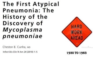
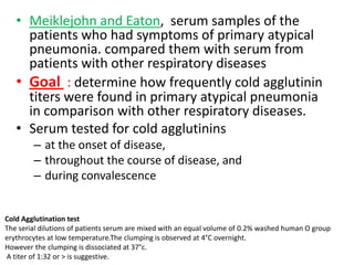
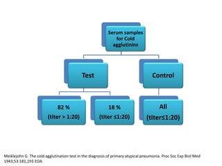
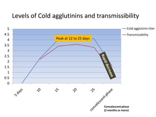
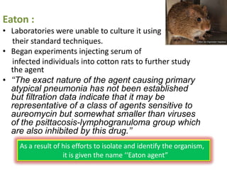
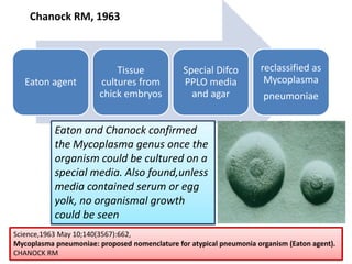
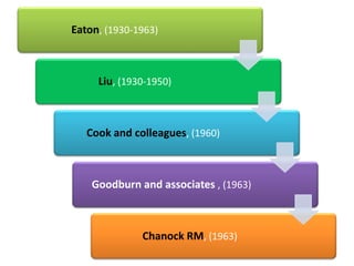
ATYPICAL PNEUMONIA

      A series
Pneumonia
                               An inflammatory
                               condition of the lung -
                                especially affecting the
                                  microscopic air sacs (alveoli)
                                  associated with
                                   •fever,
                                   •chest symptoms, and
                                   •lack of air space (consolidation) on a chest X-ray

Fishman’s Pulmonary Diseases and Disorders, vol 2, 3rd edn, McGraw Hill, 1996
What is atypical pneumonia ?
• In 1930’s , the term ‘atypical pneumonia’ was first applied
  to viral community-acquired pneumonias (CAP) that were
  clinically and radiologically distinct from bacterial CAPs
• Over the past decades, atypical pneumonia has come to
  mean lower respiratory tract infections due to specific
  respiratory pathogens
  Chlamydia psittaci (psittacosis)
  Francisella tularensis (tularemia)
  Coxiella burnetii (Q fever)
  Chlamydia pneumoniae
  Mycoplasma pneumoniae or
  Legionella species

Fishman’s Pulmonary Diseases and Disorders, vol 2, 3rd edn, McGraw Hill, 1996
(Typical)Pneumonia                                        Atypical Pneumonia
    • Typical CAPs with clinical and                          • Systemic infectious disease
      laboratory findings limited to                            with a pulmonary component
      the lungs
    • Typical bacterial pathogens                             • In contrast, most of the
      have classically responded to                             atypical pathogens do not
      b-lactam antimicrobial therapy                            have a bacterial cell wall and
                                                                some are
      because they have a cell wall                             intracellular, e.g., Legionella, a
      amenable to b-lactam                                      nd still others are
      disruption.                                               paracellular, e.g., M.
    • Chest radiograph will show                                pneumoniae
      lobar or segmental                                      • This finding can also be seen in
      homogeneous opacity in over                               nearly half the cases of
      80% of typical bacterial                                  atypical infection, but diffuse
      pneumonias.                                               patchy or ground glass
                                                                shadows are more commonly
                                                                observed.
*Fishman’s Pulmonary Diseases and Disorders, vol 2, 3rd edn, McGraw Hill, 1996
*The atypical pneumonias: clinical diagnosis and importance,B.A.Cunha,Clin Microbiol Infect 2006; 12 (Suppl. 3): 12–24
Clinical
                                                      pneumonia


   No extrapulmonary features                                     Extrapulmonary features
                                                                  (loose stools,abdominal
  (typical bacterial pneumonia)                                  pain,meningism,facial rash)


            Streptococcus
             pneumoniae                        No zoonotic contact history                 Zoonotic contact history
      Haemophilus influenzae                            Mycoplasma,                               Psittacosis,
                                                     Chlamydia (no RB),                         Q fever (RB+) ,
        Group A streptococci
                                                Legionnaires’ disease(RB +)                    Tularemia (no RB)
    Klebsiella pneumoniae,etc.

RB : Relative Bradycardia, + : present
The atypical pneumonias: clinical diagnosis and importance,B.A.Cunha,Clin Microbiol Infect 2006; 12 (Suppl. 3): 12–24
J Infect. 1996 Nov;33(3):185-91.Relative bradycardia in infectious diseases.Ostergaard L, Huniche B, Andersen PL
• Atypical CAPs represent approximately 15% of
  all CAPs.
• From India, atypical organisms have rarely
  been isolated from patients with CAP, mainly
  due to lack of facilities for culture/serology of
  these organisms.


ǂ Porath A, Schaeffer F, Lieberman D. The epidemiology of community acquired pneumonia among
  hospitalized adults. J Infect 1997 ; 34 : 41 - 48.
1930 to 1963
•   Nonfiltrable viruses,1938
     •   Serum used to infect animals, 1942
     •   No growth on standard medium or agar
     •   1943,Peterson and colleagues - suggested the
         use of the cold agglutination study as a
         diagnostic test.


Filterable virus
A virus particle small enough to pass through a filter of diatomaceous earth or porcelain, which will
not pass bacteria:chiefly historical or an informal indicator of size.
• Meiklejohn and Eaton, serum samples of the
     patients who had symptoms of primary atypical
     pneumonia. compared them with serum from
     patients with other respiratory diseases
   • Goal : determine how frequently cold agglutinin
     titers were found in primary atypical pneumonia
     in comparison with other respiratory diseases.
   • Serum tested for cold agglutinins
        – at the onset of disease,
        – throughout the course of disease, and
        – during convalescence


Cold Agglutination test
The serial dilutions of patients serum are mixed with an equal volume of 0.2% washed human O group
erythrocytes at low temperature.The clumping is observed at 4°C overnight.
However the clumping is dissociated at 37°c.
A titer of 1:32 or > is suggestive.
Serum samples
                                                             for Cold
                                                            agglutinins


                                            Test                                   Control


                              82 %                         18 %                        All
                        (titer > 1:20)               (titer ≤1:20)              (titer≤1:20)


Meiklejohn G. The cold agglutination test in the diagnosis of primary atypical pneumonia. Proc Soc Exp Biol Med
1943;53:181,193 EOA.
Levels of Cold agglutinins and transmissibility
  5                                                           Cold agglutinin titer
4.5                                                           Transmissibility
  4                   Peak at 12 to 25 days
3.5
  3
2.5
  2
1.5
  1
0.5
  0


                                              Convalescent phase
                                              (2 months or more)
• Confounding factor : storage of samples
 • Despite that fact , this was the first time that a
   specific test was identified as directly aiding in
   the diagnosis of this clinical syndrome.
 • Meiklejohn himself said that this was a
   “ROUNDABOUT WAY“ of testing for the
   disease.


Roundabout way - one that is used temporarily while a main route is blocked
Eaton :
• Laboratories were unable to culture it using
   their standard techniques.
• Began experiments injecting serum of
   infected individuals into cotton rats to further study
  the agent
• ‘‘The exact nature of the agent causing primary
  atypical pneumonia has not been established
  but filtration data indicate that it may be
  representative of a class of agents sensitive to
  aureomycin but somewhat smaller than viruses
  of the psittacosis-lymphogranuloma group which
  are also inhibited by this drug.’’
    As a result of his efforts to isolate and identify the organism,
                   it is given the name ‘‘Eaton agent”
• 1954 : Watson developed Indirect Fluorescent
  Antibody(IFA) test for the Eaton agent.
• 1957 : Liu tested original and new samples.(Liu
  antibody test)
• Revealed that among cases of clinically diagnosed
  primary atypical pneumonia with positive cold
  agglutinin titers, IFA test was posotive in 67-90%
  patients.
• Confirming Eaton agent as the causative
  organism.
By 1960, Cook and colleagues
 • Prove the link between cold agglutinin and the
   Liu antibody test.
 • Inoculate cotton rats intranasally with isolates
   from these samples
 • The serum samples that were obtained from
   these atypical pneumonia patients were retested
   and found that most had either a positive cold
   agglutinin test or a positive streptococcus MG
   test, while in control group both these tests were
   negative.
  All the efforts of Cook and colleagues was directed towards proving Eaton agent
  as the cause of this atypical pneumonia syndrome

Streptococcus MG test :
The test is performed by mixing serial dilutions of patients serum with heat killed suspension of
Streptococcus MG. The sample is incubated at 37°c.
The agglutination titer of 1:20 or > is suggestive.
Cook and colleagues
• Inoculated all strains available to them in chick embryos and
  subsequently into an animal model.
• Cold agglutinin,streptococcus MG test and indirect
  fluorescent antibody test to confirm the presence of Eaton
  agent.
• Analyzed lung tissue from the infected animals.
• Performed complement fixation studies on their
  samples, searching for
   –   Q fever,
   –   psittacosis,
   –   lymphogranuloma venereum,
   –   adenovirus,
   –   coxsackie B, and
   –   respiratory syncytial virus
• Performed hemaglutination inhibition study on all samples
Inferences from the work of Cook and
              colleagues
• Indirect antibody testing for Eaton agent had a
  relatively high specificity for primary atypical
  pneumonia.
• The disease principally affects younger
  individuals, and rather than occurring in large
  outbreaks, could occur sporadically at any
  time of the year.
• Excluded other differential diagnoses
• 1960, Chanock and colleagues studied atypical
  pneumonia in children using the same
  technique as Cook and colleagues.Study
  showed that the indirect antibody test may
  have been even more specific than the adult
  study had proven.
In 1961, Chanock and colleagues
• studied a population of 238 Marine Corps with
  atypical pneumonia
• All demonstrated an elevated cold agglutinin titer
•
• Eaton and Liu had never confirmed their thoughts on
  the precise nature of the organism
• 1963, Goodburn and his associates had been
  experimenting with
   – Giemsa staining
   – Tissue cultures
   – Chick embryos (which had been the only method of growing the
     organism)
      for evidence of Eaton agent.
• Finally able to visualize coccobacillary bodies on
  staining.
• Owing to the
  –   Organism’s size,
  –   Clinical manifestations,
  –   Giemsa staining evidence, and
  –   Sensitivity of the organism to antibiotics and some organic
      gold compounds,
    they felt that rather than being a virus, it
 was a pleuropneumonia-like organism (PPLO) of
 the mycoplasma group.
Chanock RM, 1963


                            Tissue              Special Difco        reclassified as
   Eaton agent          cultures from           PPLO media            Mycoplasma
                        chick embryos             and agar            pneumoniae


           Eaton and Chanock confirmed
           the Mycoplasma genus once the
           organism could be cultured on a
           special media. Also found,unless
           media contained serum or egg
           yolk, no organismal growth
           could be seen
Science,1963 May 10;140(3567):662,
Mycoplasma pneumoniae: proposed nomenclature for atypical pneumonia organism (Eaton agent).
CHANOCK RM
Eaton, (1930-1963)


     Liu, (1930-1950)


   Cook and colleagues, (1960)


    Goodburn and associates , (1963)


              Chanock RM, (1963)
Thank

you
Powerpoint Templates

Atypical pneumonia

  • 1.
  • 2.
    Pneumonia An inflammatory condition of the lung - especially affecting the microscopic air sacs (alveoli) associated with •fever, •chest symptoms, and •lack of air space (consolidation) on a chest X-ray Fishman’s Pulmonary Diseases and Disorders, vol 2, 3rd edn, McGraw Hill, 1996
  • 3.
    What is atypicalpneumonia ? • In 1930’s , the term ‘atypical pneumonia’ was first applied to viral community-acquired pneumonias (CAP) that were clinically and radiologically distinct from bacterial CAPs • Over the past decades, atypical pneumonia has come to mean lower respiratory tract infections due to specific respiratory pathogens Chlamydia psittaci (psittacosis) Francisella tularensis (tularemia) Coxiella burnetii (Q fever) Chlamydia pneumoniae Mycoplasma pneumoniae or Legionella species Fishman’s Pulmonary Diseases and Disorders, vol 2, 3rd edn, McGraw Hill, 1996
  • 4.
    (Typical)Pneumonia Atypical Pneumonia • Typical CAPs with clinical and • Systemic infectious disease laboratory findings limited to with a pulmonary component the lungs • Typical bacterial pathogens • In contrast, most of the have classically responded to atypical pathogens do not b-lactam antimicrobial therapy have a bacterial cell wall and some are because they have a cell wall intracellular, e.g., Legionella, a amenable to b-lactam nd still others are disruption. paracellular, e.g., M. • Chest radiograph will show pneumoniae lobar or segmental • This finding can also be seen in homogeneous opacity in over nearly half the cases of 80% of typical bacterial atypical infection, but diffuse pneumonias. patchy or ground glass shadows are more commonly observed. *Fishman’s Pulmonary Diseases and Disorders, vol 2, 3rd edn, McGraw Hill, 1996 *The atypical pneumonias: clinical diagnosis and importance,B.A.Cunha,Clin Microbiol Infect 2006; 12 (Suppl. 3): 12–24
  • 5.
    Clinical pneumonia No extrapulmonary features Extrapulmonary features (loose stools,abdominal (typical bacterial pneumonia) pain,meningism,facial rash) Streptococcus pneumoniae No zoonotic contact history Zoonotic contact history Haemophilus influenzae Mycoplasma, Psittacosis, Chlamydia (no RB), Q fever (RB+) , Group A streptococci Legionnaires’ disease(RB +) Tularemia (no RB) Klebsiella pneumoniae,etc. RB : Relative Bradycardia, + : present The atypical pneumonias: clinical diagnosis and importance,B.A.Cunha,Clin Microbiol Infect 2006; 12 (Suppl. 3): 12–24 J Infect. 1996 Nov;33(3):185-91.Relative bradycardia in infectious diseases.Ostergaard L, Huniche B, Andersen PL
  • 6.
    • Atypical CAPsrepresent approximately 15% of all CAPs. • From India, atypical organisms have rarely been isolated from patients with CAP, mainly due to lack of facilities for culture/serology of these organisms. ǂ Porath A, Schaeffer F, Lieberman D. The epidemiology of community acquired pneumonia among hospitalized adults. J Infect 1997 ; 34 : 41 - 48.
  • 7.
  • 8.
    Nonfiltrable viruses,1938 • Serum used to infect animals, 1942 • No growth on standard medium or agar • 1943,Peterson and colleagues - suggested the use of the cold agglutination study as a diagnostic test. Filterable virus A virus particle small enough to pass through a filter of diatomaceous earth or porcelain, which will not pass bacteria:chiefly historical or an informal indicator of size.
  • 9.
    • Meiklejohn andEaton, serum samples of the patients who had symptoms of primary atypical pneumonia. compared them with serum from patients with other respiratory diseases • Goal : determine how frequently cold agglutinin titers were found in primary atypical pneumonia in comparison with other respiratory diseases. • Serum tested for cold agglutinins – at the onset of disease, – throughout the course of disease, and – during convalescence Cold Agglutination test The serial dilutions of patients serum are mixed with an equal volume of 0.2% washed human O group erythrocytes at low temperature.The clumping is observed at 4°C overnight. However the clumping is dissociated at 37°c. A titer of 1:32 or > is suggestive.
  • 10.
    Serum samples for Cold agglutinins Test Control 82 % 18 % All (titer > 1:20) (titer ≤1:20) (titer≤1:20) Meiklejohn G. The cold agglutination test in the diagnosis of primary atypical pneumonia. Proc Soc Exp Biol Med 1943;53:181,193 EOA.
  • 11.
    Levels of Coldagglutinins and transmissibility 5 Cold agglutinin titer 4.5 Transmissibility 4 Peak at 12 to 25 days 3.5 3 2.5 2 1.5 1 0.5 0 Convalescent phase (2 months or more)
  • 12.
    • Confounding factor: storage of samples • Despite that fact , this was the first time that a specific test was identified as directly aiding in the diagnosis of this clinical syndrome. • Meiklejohn himself said that this was a “ROUNDABOUT WAY“ of testing for the disease. Roundabout way - one that is used temporarily while a main route is blocked
  • 13.
    Eaton : • Laboratorieswere unable to culture it using their standard techniques. • Began experiments injecting serum of infected individuals into cotton rats to further study the agent • ‘‘The exact nature of the agent causing primary atypical pneumonia has not been established but filtration data indicate that it may be representative of a class of agents sensitive to aureomycin but somewhat smaller than viruses of the psittacosis-lymphogranuloma group which are also inhibited by this drug.’’ As a result of his efforts to isolate and identify the organism, it is given the name ‘‘Eaton agent”
  • 14.
    • 1954 :Watson developed Indirect Fluorescent Antibody(IFA) test for the Eaton agent. • 1957 : Liu tested original and new samples.(Liu antibody test) • Revealed that among cases of clinically diagnosed primary atypical pneumonia with positive cold agglutinin titers, IFA test was posotive in 67-90% patients. • Confirming Eaton agent as the causative organism.
  • 15.
    By 1960, Cookand colleagues • Prove the link between cold agglutinin and the Liu antibody test. • Inoculate cotton rats intranasally with isolates from these samples • The serum samples that were obtained from these atypical pneumonia patients were retested and found that most had either a positive cold agglutinin test or a positive streptococcus MG test, while in control group both these tests were negative. All the efforts of Cook and colleagues was directed towards proving Eaton agent as the cause of this atypical pneumonia syndrome Streptococcus MG test : The test is performed by mixing serial dilutions of patients serum with heat killed suspension of Streptococcus MG. The sample is incubated at 37°c. The agglutination titer of 1:20 or > is suggestive.
  • 16.
    Cook and colleagues •Inoculated all strains available to them in chick embryos and subsequently into an animal model. • Cold agglutinin,streptococcus MG test and indirect fluorescent antibody test to confirm the presence of Eaton agent. • Analyzed lung tissue from the infected animals. • Performed complement fixation studies on their samples, searching for – Q fever, – psittacosis, – lymphogranuloma venereum, – adenovirus, – coxsackie B, and – respiratory syncytial virus • Performed hemaglutination inhibition study on all samples
  • 17.
    Inferences from thework of Cook and colleagues • Indirect antibody testing for Eaton agent had a relatively high specificity for primary atypical pneumonia. • The disease principally affects younger individuals, and rather than occurring in large outbreaks, could occur sporadically at any time of the year. • Excluded other differential diagnoses
  • 18.
    • 1960, Chanockand colleagues studied atypical pneumonia in children using the same technique as Cook and colleagues.Study showed that the indirect antibody test may have been even more specific than the adult study had proven.
  • 19.
    In 1961, Chanockand colleagues • studied a population of 238 Marine Corps with atypical pneumonia • All demonstrated an elevated cold agglutinin titer •
  • 20.
    • Eaton andLiu had never confirmed their thoughts on the precise nature of the organism • 1963, Goodburn and his associates had been experimenting with – Giemsa staining – Tissue cultures – Chick embryos (which had been the only method of growing the organism) for evidence of Eaton agent. • Finally able to visualize coccobacillary bodies on staining.
  • 21.
    • Owing tothe – Organism’s size, – Clinical manifestations, – Giemsa staining evidence, and – Sensitivity of the organism to antibiotics and some organic gold compounds, they felt that rather than being a virus, it was a pleuropneumonia-like organism (PPLO) of the mycoplasma group.
  • 22.
    Chanock RM, 1963 Tissue Special Difco reclassified as Eaton agent cultures from PPLO media Mycoplasma chick embryos and agar pneumoniae Eaton and Chanock confirmed the Mycoplasma genus once the organism could be cultured on a special media. Also found,unless media contained serum or egg yolk, no organismal growth could be seen Science,1963 May 10;140(3567):662, Mycoplasma pneumoniae: proposed nomenclature for atypical pneumonia organism (Eaton agent). CHANOCK RM
  • 23.
    Eaton, (1930-1963) Liu, (1930-1950) Cook and colleagues, (1960) Goodburn and associates , (1963) Chanock RM, (1963)
  • 25.

Editor's Notes

  • #24 Liu, worked to strengthen disease agent relationship(1930-1950)Cook and colleagues, Excluded all other differential diagnoses and proved Eaton agent as the Causative agent(1960)Goodburn and associates , directed the identification towards right direction by proposing that it cannot be a virus.(1963)Chanock RM, grew the organism on special Difco PPLO media and agar.(1963)