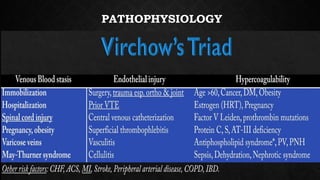
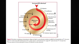
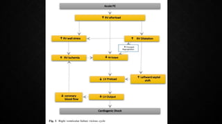
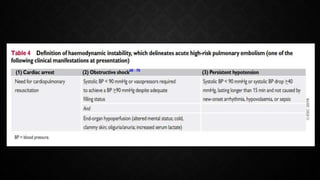
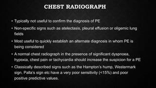
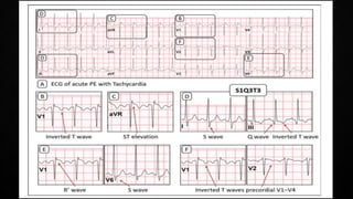
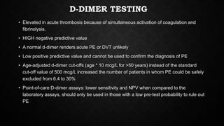
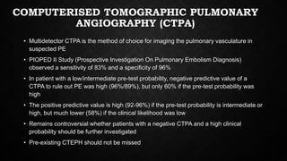
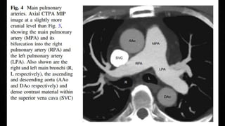
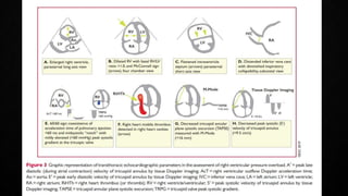
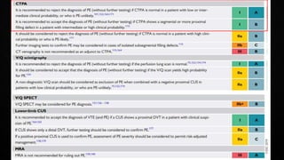
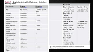
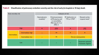
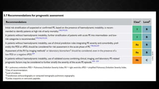
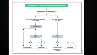
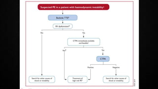
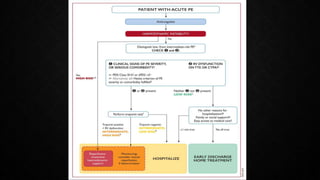
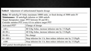
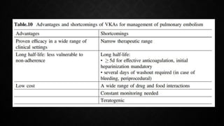
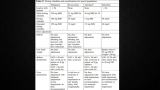
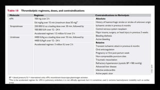
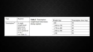
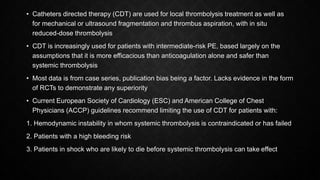
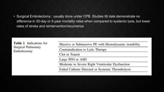
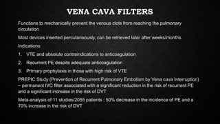
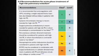
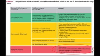
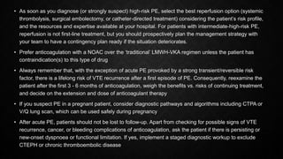
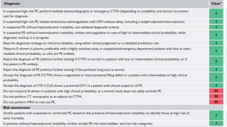
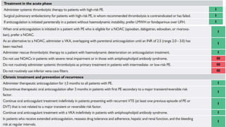
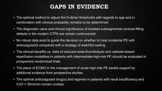
PULMONARY EMBOLISM
AND OBSTRUCTIVE SHOCK
PRESENTER: DR DILEEP N
MODERATOR: DR VINOD SINGH
ESC 2019 Guidelines for the diagnosis and management of acute
pulmonary embolism developed in collaboration with the European
Respiratory Society (ERS)
Textbook of Critical Care 8th Edition (2023) by Jean-Louis Vincent et
al (previously known as Fink’s)
OVERVIEW
• Introduction
• Epidemiology
• Predisposing Factors
• Pathophysiology
• Clinical presentation
• Diagnosis – investigations and testing strategies
• Assessment of severity and prognosis
• Treatment – medical and surgical management
• Summary of Guidelines
• Key messages and Gaps in evidence
INTRODUCTION
• Venous thromboembolism (VTE) encompasses the continuum of the disease that
includes deep venous thrombosis (DVT) and pulmonary embolism (PE)
• Prototypical VTE event occurs when a clot forms in the deep veins of the legs, detaches
and then embolises through the vasculature to lodge in the pulmonary arteries, where it
is referred to as a PE
• Wide variety of presentation
• Diagnostic and therapeutic challenge in the ICU scenario
EPIDEMIOLOGY
• Globally the third most frequent acute cardiovascular syndrome, behind only myocardial
infarction and stroke.
• Annual incidence rates for PE range from 39-115 per 100000 population
• Incidence 8 times higher in individuals >80 years
• Longitudinal studies have revealed a rising tendency in annual PE incidence rates over
the years
• 34% died suddenly or within a few hours of presentation before therapy could be
initiated or take effect.
• Though the exact incidence is not known in the Indian subcontinent, the clinical
relevance and incidence is not expected to be any different from the Western population
• Patients with symptomatic DVT in India in the PROVE registry had similar baseline
characteristics and medical histories to patients enrolled in other countries
PATHOPHYSIOLOGY
DIAGNOSIS
• Increasing awareness of VTE and universal availability of CTPA has generated a
tendency for clinicians to suspect and initiate workup for PE more frequently than in the
past
• Rates of PE confirmation were as low as 5% in the recent North American diagnostic
studies, in contrast to 50% in the 1980s
• Safely excluding PE is of paramount importance to conserve resources and reduce the
financial burden
• Assessment of pre-test probability
• Clinical judgement
• Prediction scores- Geneva rule, Well’s criteria
AVOIDING OVERUSE OF DIAGNOSTIC TESTS
CHEST RADIOGRAPH
• Typically not useful to confirm the diagnosis of PE
• Non-specific signs such as atelectasis, pleural effusion or oligemic lung
fields
• Most useful to quickly establish an alternate diagnosis in whom PE is
being considered
• A normal chest radiograph in the presence of significant dyspnoea,
hypoxia, chest pain or tachycardia should increase the suspicion for a PE
• Classically described signs such as the Hampton’s hump, Westermark
sign, Palla’s sign etc have a very poor sensitivity (<15%) and poor
positive predictive values.
HAMPTON’S HUMP
WESTERMARK SIGN
PALLA’S SIGN
ECG
• Non-specific findings
• Most common ECG in a PE: Sinus tachycardia
• Many patients have a normal ECG!
• Classically described triad of S1Q3T3 occurs only in a small minority of
patients
• Atrial tachyarrhythmias are common
D-DIMER TESTING
• Elevated in acute thrombosis because of simultaneous activation of coagulation and
fibrinolysis.
• HIGH negative predictive value
• A normal d-dimer renders acute PE or DVT unlikely
• Low positive predictive value and cannot be used to confirm the diagnosis of PE
• Age-adjusted d-dimer cut-offs (age * 10 mcg/L for >50 years) instead of the standard
cut-off value of 500 mcg/L increased the number of patients in whom PE could be safely
excluded from 6.4 to 30%
• Point-of-care D-dimer assays: lower sensitivity and NPV when compared to the
laboratory assays, should only be used in those with a low pre-test probability to rule out
PE
COMPUTERISED TOMOGRAPHIC PULMONARY
ANGIOGRAPHY (CTPA)
• Multidetector CTPA is the method of choice for imaging the pulmonary vasculature in
suspected PE
• PIOPED II Study (Prospective Investigation On Pulmonary Embolism Diagnosis)
observed a sensitivity of 83% and a specificity of 96%
• In patient with a low/intermediate pre-test probability, negative predictive value of a
CTPA to rule out PE was high (96%/89%), but only 60% if the pre-test probability was
high
• The positive predictive value is high (92-96%) if the pre-test probability is intermediate or
high, but much lower (58%) if the clinical likelihood was low
• Remains controversial whether patients with a negative CTPA and a high clinical
probability should be further investigated
• Pre-existing CTEPH should not be missed
RADIONUCLIDE LUNG SCANNING
• Planar ventilation/perfusion (V/Q lung scintigraphy) scan to increase the specificity
• Tracers: Xenon-133, Krypton-81, Technetium-99
• In acute PE, ventilation is expected to be normal in hypo-perfused segments
(mismatched)
• Applied in those with a low clinical probability and normal chest X-ray particularly in
1. Female patients
2. Pregnant women
3. History of contrast medium allergy/anaphylaxis
4. Renal failure
• PIOPED study three-tier classification: normal scan (excluding PE), high-probability
scan (diagnostic of PE in most) and non-diagnostic scan
• High frequency of non-diagnostic scans is a limitation
OTHER DIAGNOSTIC MODALITIES
• SPECT (single photon emission CT ) : V/Q SPECT or preferably hybrid
SPECT/CT allows a more accurate exploration of all lung segments and better
identification of segmental and subsegmental perfusion defects typical of PE
Reduces the frequency of non-diagnostic studies
Better sensitivity and specificity than planar CTPA, needs more evidence
• PULMONARY ANGIOGRAPHY: Was the gold standard for decades
previously, but rarely performed now with the advent of CTPA with similar
diagnostic accuracy
• MAGNETIC RESONANCE ANGIOGRAPHY: promising, needs more
evidence
ECHOCARDIOGRAPHY IN PE
• Manifestations of RV pressure overload and dysfunction
• RV dilatation seen in >25% of patients with PE on TTE and is useful for risk stratification
of the disease
• 60/60 Sign : combination of a pulmonary ejection acceleration time (measured in the RV
outflow tract) of <60ms AND a peak systolic tricuspid valve gradient of <60mmHg
• McConnell’s sign : depressed contractility of the RV free wall when compared to the RV
apex
• Finding present only in 12-20% of unselected PE patients
• Decreased tricuspid annular systolic plane excursion (TAPSE) may also be seen in PE
• Mobile right heart thrombi (in up to 18% of PE in the ICU) essentially confirm the
diagnosis of PE and associated with high mortality rates
• Echo is not mandatory as a part of workup in hemodynamically stable patients with PE
• BUT in suspected high-risk PE, absence of echocardiographic signs of RV overload
practically excludes PE as the cause of hemodynamic instability. Can give a pointer
towards the differential diagnosis of the actual cause of shock
• Conversely, in a hemodynamically compromised patient with suspected PE, unequivocal
signs of RV pressure overload such as the McConnell’s sign or the 60/60 sign justify
emergency reperfusion treatment for PE if immediate CTPA is not feasible in a patient
with high clinical probability and no other obvious causes for RV pressure overload.
COMPRESSION ULTRASONOGRAPHY (CUS)
• PE originates from a lower limb DVT in majority of the cases
• Lower limb CUS has replaced traditional venography for diagnosis of DVT
• Sensitivity of >90% AND Specificity ~95%
• CUS shows a DVT in 30-50% of those with PE and finding a proximal DVT in a
suspected PE in sufficient to warrant anti-coagulation treatment without further testing
• In the setting of a suspected PE, CUS can be limited to a simple 4-point examination
(bilateral groins and bilateral popliteal fossae)
• Only validated diagnostic criterion is incomplete compressibility of the vein which
indicates the presence of a clot
• Flow measurements are unreliable
• Specificity improves when combined with a cardiac ultrasound
LABORATORY BIOMARKERS
• Markers of myocardial injury – elevated cardiac troponins associated with an increased
mortality and worse prognosis
• Age-adjusted high-sensitivity troponin assays have a high negative predictive value
(98%) for excluding an adverse in-hospital clinical outcome
• Heart-type fatty acid-binding protein (H-FABP) >6ng/ml associated with adverse short
term outcomes and all-cause mortality
• Markers of RV dysfunction – BNP/NTProBNP : low sensitivity and positive predictive
value but low levels are capable of excluding an unfavourable clinical outcome
• Copeptin (surrogate marker of Vasopressin) elevated due to endogenous stress,
hypotension and low cardiac output
BLOOD GAS ANALYSIS
• Hypoxemia
• Hypocapnia
• Respiratory alkalosis
• Mixed acidosis in the setting of hemodynamic compromise
• Lactate is a marker of imbalance between tissue oxygen supply and demand, and
consequently of severe PE with overt or imminent hemodynamic compromise
• Elevated arterial plasma levels >2 mmol/L predict PE-related complications
• Utility of ABG in the diagnosis of PE is minimal
SEVERITY ASSESSMENT AND RISK
STRATIFICATION
• Pulmonary Embolism Severity Index (PESI)/simplified PESI
• Extensively validated
• Reliable identification of patients at low risk for 30-day mortality
INTEGRATED RISK-ADAPTED DIAGNOSIS AND
MANAGEMENT
• Hemodynamic and respiratory support – oxygen therapy/ventilation, pharmacological
treatment of acute right ventricular failure, mechanical circulatory support and
oxygenation, advanced life support in cardiac arrest
• Initial anticoagulation – parenteral, NOACs, VKAs
• Reperfusion therapies – systemic thrombolysis, percutaneous catheter-directed
thrombolysis, surgical embolectomy
• Vena cava filters
ANTICOAGULATION
• Should be initiated in patients with intermediate/high clinical probability
• Subcutaneous weight-adjusted LMWH or Fondaparinux preferred over UFH (lower
risk of major bleeding/HIT)
• NOACs – Apixaban, Rivaroxaban : Phase 3 clinical trials have demonstrated non-
inferiority. Conclusive evidence in PE lacking
• Use of UFH restricted to patients with overt or imminent hemodynamic
decompensation in whom reperfusion strategies may be necessary. Also
recommended for serious renal impairment with a CrCl <30ml/min
• VKAs : anticoagulation with UFH/LMWH/Fondaparinux should be continued in
parallel for 5-7 days until a target INR of 2-3 is achieved.
REPERFUSION
• Greatest benefit when started within 48 hours of symptom onset, but can still be useful
up to 6-14 days
• Meta-analysis of thrombolysis trials in patients with high-risk PE indicated a significant
reduction in the combined outcome of mortality and recurrent PE, with a 9.9% rate of
severe bleeding and 1.9% of intracranial haemorrhage
• PEITHO trial (Pulmonary Embolism Thrombolysis) in normotensive patients with
intermediate-risk PE : significant reduction in the risk of hemodynamic decompensation
or collapse, BUT an increased risk of severe bleeding and intracranial haemorrhage
• CDT or surgical embolectomy are reserved for patients who are not candidates for
systemic thrombolysis or for patients who have a higher risk of bleeding with systemic
thrombolytic therapy and who have access to the expertise and resources required to
perform CDT
• Catheters directed therapy (CDT) are used for local thrombolysis treatment as well as
for mechanical or ultrasound fragmentation and thrombus aspiration, with in situ
reduced-dose thrombolysis
• CDT is increasingly used for patients with intermediate-risk PE, based largely on the
assumptions that it is more efficacious than anticoagulation alone and safer than
systemic thrombolysis
• Most data is from case series, publication bias being a factor. Lacks evidence in the form
of RCTs to demonstrate any superiority
• Current European Society of Cardiology (ESC) and American College of Chest
Physicians (ACCP) guidelines recommend limiting the use of CDT for patients with:
1. Hemodynamic instability in whom systemic thrombolysis is contraindicated or has failed
2. Patients with a high bleeding risk
3. Patients in shock who are likely to die before systemic thrombolysis can take effect
• Surgical Embolectomy : usually done under CPB. Studies till date demonstrate no
difference in 30-day or 5-year mortality rates when compared to systemic lysis, but lower
rates of stroke and reintervention/recurrence
VENA CAVA FILTERS
Functions to mechanically prevent the venous clots from reaching the pulmonary
circulation
Most devices inserted percutaneously, can be retrieved later after weeks/months
Indications:
1. VTE and absolute contraindications to anticoagulation
2. Recurrent PE despite adequate anticoagulation
3. Primary prophylaxis in those with high risk of VTE
PREPIC Study (Prevention of Recurrent Pulmonary Embolism by Vena cava Interruption)
– permanent IVC filter associated with a significant reduction in the risk of recurrent PE
and a significant increase in the risk of DVT
Meta-analysis of 11 studies/2055 patients : 50% decrease in the incidence of PE and a
70% increase in the risk of DVT
PE IN SPECIAL SITUATIONS
• Recurrent PE
• PE in the immediate post-operative period
• PE in Pregnancy
• Amniotic Fluid Embolism
• PE in Cancer
• PE in Covid-19
• Chronic Thromboembolic Pulmonary Hypertension (CTEPH)
• Non-thrombotic Pulmonary Embolism : septic emboli, fat embolism, foreign material
emboli, air embolism, tumour emboli
SUMMARY
• In patients presenting with hemodynamic instability, perform bedside TTE as a fast, immediate step to
differentiate suspected high-risk PE from other acute life-threatening situations
• If you suspect acute PE, institute anticoagulation therapy as soon as possible, while the diagnostic workup
is ongoing, unless the patient is bleeding or has absolute contraindications to this therapy
• Use recommended, validated diagnostic algorithms for PE, including standardized assessment of (pre-test)
clinical probability and D-dimer testing. They help to avoid unnecessary, expensive, and potentially harmful
imaging tests and exposure to ionizing radiation
• If the CTPA report suggests single subsegmental PE, consider the possibility of a false-positive finding.
Discuss the findings again with the radiologist and/or seek a second opinion to avoid misdiagnosis, and
unnecessary, potentially harmful anticoagulation treatment
• Confirmation of PE in a patient, without hemodynamic instability, must be followed by further risk
assessment involving clinical findings, evaluation of the size and/or function of the RV, and laboratory
biomarkers as appropriate. This information will help you to decide on the need for reperfusion treatment or
monitoring for patients at elevated risk.
• As soon as you diagnose (or strongly suspect) high-risk PE, select the best reperfusion option (systemic
thrombolysis, surgical embolectomy, or catheter-directed treatment) considering the patient’s risk profile,
and the resources and expertise available at your hospital. For patients with intermediate-high-risk PE,
reperfusion is not first-line treatment, but you should prospectively plan the management strategy with
your team to have a contingency plan ready if the situation deteriorates.
• Prefer anticoagulation with a NOAC over the ‘traditional’ LMWH-VKA regimen unless the patient has
contraindication(s) to this type of drug
• Always remember that, with the exception of acute PE provoked by a strong transient/reversible risk
factor, there is a lifelong risk of VTE recurrence after a first episode of PE. Consequently, reexamine the
patient after the first 3 - 6 months of anticoagulation, weigh the benefits vs. risks of continuing treatment,
and decide on the extension and dose of anticoagulant therapy
• If you suspect PE in a pregnant patient, consider diagnostic pathways and algorithms including CTPA or
V/Q lung scan, which can be used safely during pregnancy
• After acute PE, patients should not be lost to follow-up. Apart from checking for possible signs of VTE
recurrence, cancer, or bleeding complications of anticoagulation, ask the patient if there is persisting or
new-onset dyspnoea or functional limitation. If yes, implement a staged diagnostic workup to exclude
CTEPH or chronic thromboembolic disease
GAPS IN EVIDENCE
• The optimal method to adjust the D-dimer thresholds with regards to age and in
combination with clinical probability remains to be determined
• The diagnostic value and clinical significance of isolated subsegmental contrast-filling
defects in the modern CTPA era remain controversial
• No robust data exist to guide the decision on whether to treat incidental PE with
anticoagulants compared with a strategy of watchful waiting
• The clinical benefits vs. risks of reduced-dose thrombolysis and catheter-based
reperfusion modalities in patients with intermediate-high-risk PE should be evaluated in
prospective randomized trials
• The place of ECMO in the management of acute high-risk PE awaits support by
additional evidence from prospective studies
• The optimal anticoagulant drug(s) and regimen in patients with renal insufficiency and
CrCl < 30ml/min remain unclear
THANK YOU

Pulmonary Embolism.pptx

  • 1.
    PULMONARY EMBOLISM AND OBSTRUCTIVESHOCK PRESENTER: DR DILEEP N MODERATOR: DR VINOD SINGH
  • 2.
    ESC 2019 Guidelinesfor the diagnosis and management of acute pulmonary embolism developed in collaboration with the European Respiratory Society (ERS) Textbook of Critical Care 8th Edition (2023) by Jean-Louis Vincent et al (previously known as Fink’s)
  • 3.
    OVERVIEW • Introduction • Epidemiology •Predisposing Factors • Pathophysiology • Clinical presentation • Diagnosis – investigations and testing strategies • Assessment of severity and prognosis • Treatment – medical and surgical management • Summary of Guidelines • Key messages and Gaps in evidence
  • 4.
    INTRODUCTION • Venous thromboembolism(VTE) encompasses the continuum of the disease that includes deep venous thrombosis (DVT) and pulmonary embolism (PE) • Prototypical VTE event occurs when a clot forms in the deep veins of the legs, detaches and then embolises through the vasculature to lodge in the pulmonary arteries, where it is referred to as a PE • Wide variety of presentation • Diagnostic and therapeutic challenge in the ICU scenario
  • 5.
    EPIDEMIOLOGY • Globally thethird most frequent acute cardiovascular syndrome, behind only myocardial infarction and stroke. • Annual incidence rates for PE range from 39-115 per 100000 population • Incidence 8 times higher in individuals >80 years • Longitudinal studies have revealed a rising tendency in annual PE incidence rates over the years • 34% died suddenly or within a few hours of presentation before therapy could be initiated or take effect. • Though the exact incidence is not known in the Indian subcontinent, the clinical relevance and incidence is not expected to be any different from the Western population • Patients with symptomatic DVT in India in the PROVE registry had similar baseline characteristics and medical histories to patients enrolled in other countries
  • 8.
  • 13.
    DIAGNOSIS • Increasing awarenessof VTE and universal availability of CTPA has generated a tendency for clinicians to suspect and initiate workup for PE more frequently than in the past • Rates of PE confirmation were as low as 5% in the recent North American diagnostic studies, in contrast to 50% in the 1980s • Safely excluding PE is of paramount importance to conserve resources and reduce the financial burden • Assessment of pre-test probability • Clinical judgement • Prediction scores- Geneva rule, Well’s criteria
  • 16.
    AVOIDING OVERUSE OFDIAGNOSTIC TESTS
  • 17.
    CHEST RADIOGRAPH • Typicallynot useful to confirm the diagnosis of PE • Non-specific signs such as atelectasis, pleural effusion or oligemic lung fields • Most useful to quickly establish an alternate diagnosis in whom PE is being considered • A normal chest radiograph in the presence of significant dyspnoea, hypoxia, chest pain or tachycardia should increase the suspicion for a PE • Classically described signs such as the Hampton’s hump, Westermark sign, Palla’s sign etc have a very poor sensitivity (<15%) and poor positive predictive values.
  • 18.
  • 19.
  • 20.
  • 21.
    ECG • Non-specific findings •Most common ECG in a PE: Sinus tachycardia • Many patients have a normal ECG! • Classically described triad of S1Q3T3 occurs only in a small minority of patients • Atrial tachyarrhythmias are common
  • 24.
    D-DIMER TESTING • Elevatedin acute thrombosis because of simultaneous activation of coagulation and fibrinolysis. • HIGH negative predictive value • A normal d-dimer renders acute PE or DVT unlikely • Low positive predictive value and cannot be used to confirm the diagnosis of PE • Age-adjusted d-dimer cut-offs (age * 10 mcg/L for >50 years) instead of the standard cut-off value of 500 mcg/L increased the number of patients in whom PE could be safely excluded from 6.4 to 30% • Point-of-care D-dimer assays: lower sensitivity and NPV when compared to the laboratory assays, should only be used in those with a low pre-test probability to rule out PE
  • 25.
    COMPUTERISED TOMOGRAPHIC PULMONARY ANGIOGRAPHY(CTPA) • Multidetector CTPA is the method of choice for imaging the pulmonary vasculature in suspected PE • PIOPED II Study (Prospective Investigation On Pulmonary Embolism Diagnosis) observed a sensitivity of 83% and a specificity of 96% • In patient with a low/intermediate pre-test probability, negative predictive value of a CTPA to rule out PE was high (96%/89%), but only 60% if the pre-test probability was high • The positive predictive value is high (92-96%) if the pre-test probability is intermediate or high, but much lower (58%) if the clinical likelihood was low • Remains controversial whether patients with a negative CTPA and a high clinical probability should be further investigated • Pre-existing CTEPH should not be missed
  • 31.
    RADIONUCLIDE LUNG SCANNING •Planar ventilation/perfusion (V/Q lung scintigraphy) scan to increase the specificity • Tracers: Xenon-133, Krypton-81, Technetium-99 • In acute PE, ventilation is expected to be normal in hypo-perfused segments (mismatched) • Applied in those with a low clinical probability and normal chest X-ray particularly in 1. Female patients 2. Pregnant women 3. History of contrast medium allergy/anaphylaxis 4. Renal failure • PIOPED study three-tier classification: normal scan (excluding PE), high-probability scan (diagnostic of PE in most) and non-diagnostic scan • High frequency of non-diagnostic scans is a limitation
  • 35.
    OTHER DIAGNOSTIC MODALITIES •SPECT (single photon emission CT ) : V/Q SPECT or preferably hybrid SPECT/CT allows a more accurate exploration of all lung segments and better identification of segmental and subsegmental perfusion defects typical of PE Reduces the frequency of non-diagnostic studies Better sensitivity and specificity than planar CTPA, needs more evidence • PULMONARY ANGIOGRAPHY: Was the gold standard for decades previously, but rarely performed now with the advent of CTPA with similar diagnostic accuracy • MAGNETIC RESONANCE ANGIOGRAPHY: promising, needs more evidence
  • 39.
    ECHOCARDIOGRAPHY IN PE •Manifestations of RV pressure overload and dysfunction • RV dilatation seen in >25% of patients with PE on TTE and is useful for risk stratification of the disease • 60/60 Sign : combination of a pulmonary ejection acceleration time (measured in the RV outflow tract) of <60ms AND a peak systolic tricuspid valve gradient of <60mmHg • McConnell’s sign : depressed contractility of the RV free wall when compared to the RV apex • Finding present only in 12-20% of unselected PE patients • Decreased tricuspid annular systolic plane excursion (TAPSE) may also be seen in PE • Mobile right heart thrombi (in up to 18% of PE in the ICU) essentially confirm the diagnosis of PE and associated with high mortality rates
  • 46.
    • Echo isnot mandatory as a part of workup in hemodynamically stable patients with PE • BUT in suspected high-risk PE, absence of echocardiographic signs of RV overload practically excludes PE as the cause of hemodynamic instability. Can give a pointer towards the differential diagnosis of the actual cause of shock • Conversely, in a hemodynamically compromised patient with suspected PE, unequivocal signs of RV pressure overload such as the McConnell’s sign or the 60/60 sign justify emergency reperfusion treatment for PE if immediate CTPA is not feasible in a patient with high clinical probability and no other obvious causes for RV pressure overload.
  • 47.
    COMPRESSION ULTRASONOGRAPHY (CUS) •PE originates from a lower limb DVT in majority of the cases • Lower limb CUS has replaced traditional venography for diagnosis of DVT • Sensitivity of >90% AND Specificity ~95% • CUS shows a DVT in 30-50% of those with PE and finding a proximal DVT in a suspected PE in sufficient to warrant anti-coagulation treatment without further testing • In the setting of a suspected PE, CUS can be limited to a simple 4-point examination (bilateral groins and bilateral popliteal fossae) • Only validated diagnostic criterion is incomplete compressibility of the vein which indicates the presence of a clot • Flow measurements are unreliable • Specificity improves when combined with a cardiac ultrasound
  • 49.
    LABORATORY BIOMARKERS • Markersof myocardial injury – elevated cardiac troponins associated with an increased mortality and worse prognosis • Age-adjusted high-sensitivity troponin assays have a high negative predictive value (98%) for excluding an adverse in-hospital clinical outcome • Heart-type fatty acid-binding protein (H-FABP) >6ng/ml associated with adverse short term outcomes and all-cause mortality • Markers of RV dysfunction – BNP/NTProBNP : low sensitivity and positive predictive value but low levels are capable of excluding an unfavourable clinical outcome • Copeptin (surrogate marker of Vasopressin) elevated due to endogenous stress, hypotension and low cardiac output
  • 50.
    BLOOD GAS ANALYSIS •Hypoxemia • Hypocapnia • Respiratory alkalosis • Mixed acidosis in the setting of hemodynamic compromise • Lactate is a marker of imbalance between tissue oxygen supply and demand, and consequently of severe PE with overt or imminent hemodynamic compromise • Elevated arterial plasma levels >2 mmol/L predict PE-related complications • Utility of ABG in the diagnosis of PE is minimal
  • 53.
    SEVERITY ASSESSMENT ANDRISK STRATIFICATION • Pulmonary Embolism Severity Index (PESI)/simplified PESI • Extensively validated • Reliable identification of patients at low risk for 30-day mortality
  • 60.
    INTEGRATED RISK-ADAPTED DIAGNOSISAND MANAGEMENT • Hemodynamic and respiratory support – oxygen therapy/ventilation, pharmacological treatment of acute right ventricular failure, mechanical circulatory support and oxygenation, advanced life support in cardiac arrest • Initial anticoagulation – parenteral, NOACs, VKAs • Reperfusion therapies – systemic thrombolysis, percutaneous catheter-directed thrombolysis, surgical embolectomy • Vena cava filters
  • 62.
    ANTICOAGULATION • Should beinitiated in patients with intermediate/high clinical probability • Subcutaneous weight-adjusted LMWH or Fondaparinux preferred over UFH (lower risk of major bleeding/HIT) • NOACs – Apixaban, Rivaroxaban : Phase 3 clinical trials have demonstrated non- inferiority. Conclusive evidence in PE lacking • Use of UFH restricted to patients with overt or imminent hemodynamic decompensation in whom reperfusion strategies may be necessary. Also recommended for serious renal impairment with a CrCl <30ml/min • VKAs : anticoagulation with UFH/LMWH/Fondaparinux should be continued in parallel for 5-7 days until a target INR of 2-3 is achieved.
  • 67.
    REPERFUSION • Greatest benefitwhen started within 48 hours of symptom onset, but can still be useful up to 6-14 days • Meta-analysis of thrombolysis trials in patients with high-risk PE indicated a significant reduction in the combined outcome of mortality and recurrent PE, with a 9.9% rate of severe bleeding and 1.9% of intracranial haemorrhage • PEITHO trial (Pulmonary Embolism Thrombolysis) in normotensive patients with intermediate-risk PE : significant reduction in the risk of hemodynamic decompensation or collapse, BUT an increased risk of severe bleeding and intracranial haemorrhage • CDT or surgical embolectomy are reserved for patients who are not candidates for systemic thrombolysis or for patients who have a higher risk of bleeding with systemic thrombolytic therapy and who have access to the expertise and resources required to perform CDT
  • 70.
    • Catheters directedtherapy (CDT) are used for local thrombolysis treatment as well as for mechanical or ultrasound fragmentation and thrombus aspiration, with in situ reduced-dose thrombolysis • CDT is increasingly used for patients with intermediate-risk PE, based largely on the assumptions that it is more efficacious than anticoagulation alone and safer than systemic thrombolysis • Most data is from case series, publication bias being a factor. Lacks evidence in the form of RCTs to demonstrate any superiority • Current European Society of Cardiology (ESC) and American College of Chest Physicians (ACCP) guidelines recommend limiting the use of CDT for patients with: 1. Hemodynamic instability in whom systemic thrombolysis is contraindicated or has failed 2. Patients with a high bleeding risk 3. Patients in shock who are likely to die before systemic thrombolysis can take effect
  • 71.
    • Surgical Embolectomy: usually done under CPB. Studies till date demonstrate no difference in 30-day or 5-year mortality rates when compared to systemic lysis, but lower rates of stroke and reintervention/recurrence
  • 72.
    VENA CAVA FILTERS Functionsto mechanically prevent the venous clots from reaching the pulmonary circulation Most devices inserted percutaneously, can be retrieved later after weeks/months Indications: 1. VTE and absolute contraindications to anticoagulation 2. Recurrent PE despite adequate anticoagulation 3. Primary prophylaxis in those with high risk of VTE PREPIC Study (Prevention of Recurrent Pulmonary Embolism by Vena cava Interruption) – permanent IVC filter associated with a significant reduction in the risk of recurrent PE and a significant increase in the risk of DVT Meta-analysis of 11 studies/2055 patients : 50% decrease in the incidence of PE and a 70% increase in the risk of DVT
  • 77.
    PE IN SPECIALSITUATIONS • Recurrent PE • PE in the immediate post-operative period • PE in Pregnancy • Amniotic Fluid Embolism • PE in Cancer • PE in Covid-19 • Chronic Thromboembolic Pulmonary Hypertension (CTEPH) • Non-thrombotic Pulmonary Embolism : septic emboli, fat embolism, foreign material emboli, air embolism, tumour emboli
  • 78.
    SUMMARY • In patientspresenting with hemodynamic instability, perform bedside TTE as a fast, immediate step to differentiate suspected high-risk PE from other acute life-threatening situations • If you suspect acute PE, institute anticoagulation therapy as soon as possible, while the diagnostic workup is ongoing, unless the patient is bleeding or has absolute contraindications to this therapy • Use recommended, validated diagnostic algorithms for PE, including standardized assessment of (pre-test) clinical probability and D-dimer testing. They help to avoid unnecessary, expensive, and potentially harmful imaging tests and exposure to ionizing radiation • If the CTPA report suggests single subsegmental PE, consider the possibility of a false-positive finding. Discuss the findings again with the radiologist and/or seek a second opinion to avoid misdiagnosis, and unnecessary, potentially harmful anticoagulation treatment • Confirmation of PE in a patient, without hemodynamic instability, must be followed by further risk assessment involving clinical findings, evaluation of the size and/or function of the RV, and laboratory biomarkers as appropriate. This information will help you to decide on the need for reperfusion treatment or monitoring for patients at elevated risk.
  • 79.
    • As soonas you diagnose (or strongly suspect) high-risk PE, select the best reperfusion option (systemic thrombolysis, surgical embolectomy, or catheter-directed treatment) considering the patient’s risk profile, and the resources and expertise available at your hospital. For patients with intermediate-high-risk PE, reperfusion is not first-line treatment, but you should prospectively plan the management strategy with your team to have a contingency plan ready if the situation deteriorates. • Prefer anticoagulation with a NOAC over the ‘traditional’ LMWH-VKA regimen unless the patient has contraindication(s) to this type of drug • Always remember that, with the exception of acute PE provoked by a strong transient/reversible risk factor, there is a lifelong risk of VTE recurrence after a first episode of PE. Consequently, reexamine the patient after the first 3 - 6 months of anticoagulation, weigh the benefits vs. risks of continuing treatment, and decide on the extension and dose of anticoagulant therapy • If you suspect PE in a pregnant patient, consider diagnostic pathways and algorithms including CTPA or V/Q lung scan, which can be used safely during pregnancy • After acute PE, patients should not be lost to follow-up. Apart from checking for possible signs of VTE recurrence, cancer, or bleeding complications of anticoagulation, ask the patient if there is persisting or new-onset dyspnoea or functional limitation. If yes, implement a staged diagnostic workup to exclude CTEPH or chronic thromboembolic disease
  • 82.
    GAPS IN EVIDENCE •The optimal method to adjust the D-dimer thresholds with regards to age and in combination with clinical probability remains to be determined • The diagnostic value and clinical significance of isolated subsegmental contrast-filling defects in the modern CTPA era remain controversial • No robust data exist to guide the decision on whether to treat incidental PE with anticoagulants compared with a strategy of watchful waiting • The clinical benefits vs. risks of reduced-dose thrombolysis and catheter-based reperfusion modalities in patients with intermediate-high-risk PE should be evaluated in prospective randomized trials • The place of ECMO in the management of acute high-risk PE awaits support by additional evidence from prospective studies • The optimal anticoagulant drug(s) and regimen in patients with renal insufficiency and CrCl < 30ml/min remain unclear
  • 83.