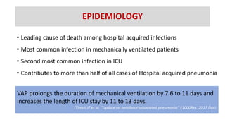
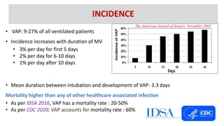
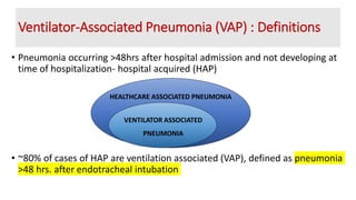
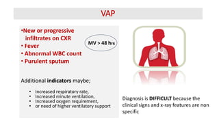
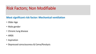
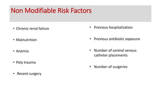
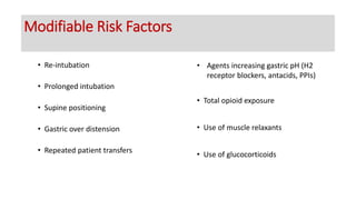
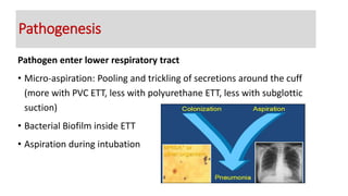
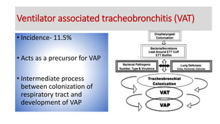
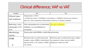
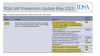
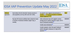
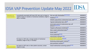
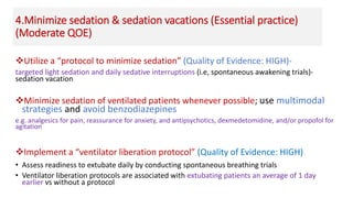
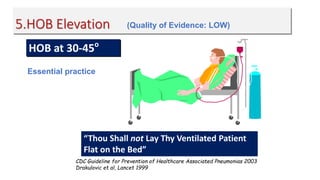
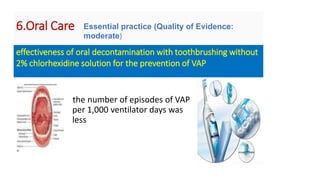
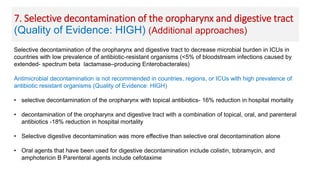
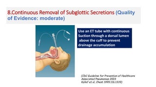
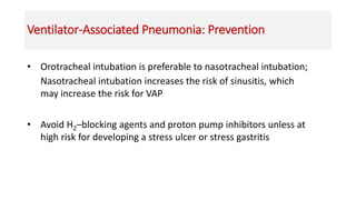
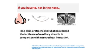
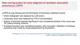
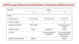
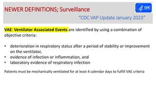
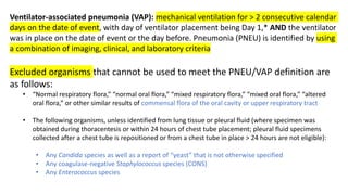
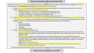
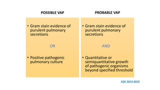
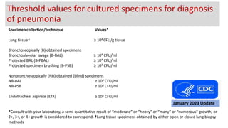
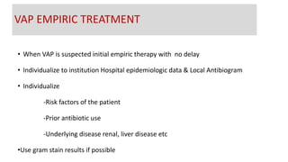
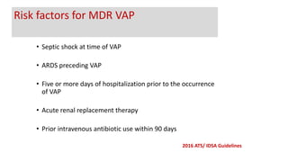
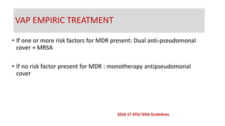
2. EPIDEMIOLOGY
• Leading cause of death among hospital acquired infections
• Most common infection in mechanically ventilated patients
• Second most common infection in ICU
• Contributes to more than half of all cases of Hospital acquired pneumonia
VAP prolongs the duration of mechanical ventilation by 7.6 to 11 days and
increases the length of ICU stay by 11 to 13 days.
(Timsit JF et al. “Update on ventilator-associated pneumonia” F1000Res. 2017 Nov)
3. • VAP: 9-27% of all ventilated patients
• Incidence increases with duration of MV
• 3% per day for first 5 days
• 2% per day for 6-10 days
• 1% per day after 10 days
• Mean duration between intubation and development of VAP- 3.3 days
Mortality higher than any of other healthcare associated infection
• As per IDSA 2016, VAP has a mortality rate : 20-50%
• As per CDC 2020, VAP accounts for mortality rate : 60%
INCIDENCE
The American Journal of Surgery. November 2003
4. VAP Rate
• VAP RATE
• Incidence increases with increase in duration of
ventilation
Total no. of VAP cases in a time period
1000
Total no. of days pts. were on ventilator
for same time period
5. Ventilator-Associated Pneumonia (VAP) : Definitions
• Pneumonia occurring >48hrs after hospital admission and not developing at
time of hospitalization- hospital acquired (HAP)
• ~80% of cases of HAP are ventilation associated (VAP), defined as pneumonia
>48 hrs. after endotracheal intubation
HEALTHCARE ASSOCIATED PNEUMONIA
VENTILATOR ASSOCIATED
PNEUMONIA
6. VAP
•New or progressive
infiltrates on CXR
• Fever
• Abnormal WBC count
• Purulent sputum
Additional indicators maybe;
• Increased respiratory rate,
• Increased minute ventilation,
• Increased oxygen requirement,
• or need of higher ventilatory support
MV > 48 hrs
Diagnosis is DIFFICULT because the
clinical signs and x-ray features are non
specific
7. Most significant risk factor: Mechanical ventilation
• Older Age
• Male gender
• Chronic lung disease
• ARDS
• Aspiration
• Depressed consciousness & Coma/Paralysis
Risk Factors; Non Modifiable
8. • Chronic renal failure
• Malnutrition
• Anemia
• Poly trauma
• Recent surgery
• Previous hospitalization
• Previous antibiotic exposure
• Number of central venous
catheter placements
• Number of surgeries
Non Modifiable Risk Factors
9. • Re-intubation
• Prolonged intubation
• Supine positioning
• Gastric over distension
• Repeated patient transfers
• Agents increasing gastric pH (H2
receptor blockers, antacids, PPIs)
• Total opioid exposure
• Use of muscle relaxants
• Use of glucocorticoids
Modifiable Risk Factors
10. Pathogen enter lower respiratory tract
• Micro-aspiration: Pooling and trickling of secretions around the cuff
(more with PVC ETT, less with polyurethane ETT, less with subglottic
suction)
• Bacterial Biofilm inside ETT
• Aspiration during intubation
Pathogenesis
11. • Incidence- 11.5%
• Acts as a precursor for VAP
• Intermediate process
between colonization of
respiratory tract and
development of VAP
Ventilator associated tracheobronchitis (VAT)
18. 1.Avoid intubation and reintubation (Quality of Evidence:
HIGH)
Intubation is the single most powerful predictor of nosocomial pneumonia & is a
continuous risk for development of pneumonia- Use high-flow nasal oxygen or
non-invasive positive pressure ventilation (NIPPV)
19. NIPPV
lower rates of intubation,
reintubation, VAP and overall
mortality
Consider Non Invasive Ventilation
20. 2. Provide early enteral rather than parenteral
nutrition
• Provide early enteral rather than parenteral nutrition (Quality of Evidence:
HIGH)
• Early enteral nutrition is associated with a lower risk of nosocomial
pneumonia, shorter ICU length of stay compared to early parenteral
nutrition
21. 3. Maintain ventilator circuits and strategies to reduce use
of contaminated equipment
• DO NOT routinely change the ventilator breathing circuit. Change ONLY if visibly
soiled or mechanically malfunctioning (Quality of Evidence: HIGH)
• Periodically drain and discard any condensate that collects in the tubing of a
mechanical ventilator, taking precautions not to allow condensate to drain
toward the patient
• Wear personnel protective equipment to perform the above procedure or
handle the fluid/secretions
22. 4.Minimize sedation & sedation vacations (Essential practice)
(Moderate QOE)
Utilize a “protocol to minimize sedation” (Quality of Evidence: HIGH)-
targeted light sedation and daily sedative interruptions (i.e, spontaneous awakening trials)-
sedation vacation
Minimize sedation of ventilated patients whenever possible; use multimodal
strategies and avoid benzodiazepines
e.g. analgesics for pain, reassurance for anxiety, and antipsychotics, dexmedetomidine, and/or propofol for
agitation
Implement a “ventilator liberation protocol” (Quality of Evidence: HIGH)
• Assess readiness to extubate daily by conducting spontaneous breathing trials
• Ventilator liberation protocols are associated with extubating patients an average of 1 day
earlier vs without a protocol
23. 5.HOB Elevation
HOB at 30-45o
CDC Guideline for Prevention of Healthcare Associated Pneumonias 2003
Drakulovic et al, Lancet 1999
“Thou Shall not Lay Thy Ventilated Patient
Flat on the Bed”
(Quality of Evidence: LOW)
Essential practice
24. 6.Oral Care
the number of episodes of VAP
per 1,000 ventilator days was
less
effectiveness of oral decontamination with toothbrushing without
2% chlorhexidine solution for the prevention of VAP
Essential practice (Quality of Evidence:
moderate)
25. Selective decontamination of the oropharynx and digestive tract to decrease microbial burden in ICUs in
countries with low prevalence of antibiotic-resistant organisms (<5% of bloodstream infections caused by
extended- spectrum beta lactamase–producing Enterobacterales)
Antimicrobial decontamination is not recommended in countries, regions, or ICUs with high prevalence of
antibiotic resistant organisms (Quality of Evidence: HIGH)
• selective decontamination of the oropharynx with topical antibiotics- 16% reduction in hospital mortality
• decontamination of the oropharynx and digestive tract with a combination of topical, oral, and parenteral
antibiotics -18% reduction in hospital mortality
• Selective digestive decontamination was more effective than selective oral decontamination alone
• Oral agents that have been used for digestive decontamination include colistin, tobramycin, and
amphotericin B Parenteral agents include cefotaxime
7. Selective decontamination of the oropharynx and digestive tract
(Quality of Evidence: HIGH) (Additional approaches)
26. 8.Continuous Removal of Subglottic Secretions (Quality
of Evidence: moderate)
(CDC Guideline for Prevention of Healthcare
Associated Pneumonias 2003
Kollef et al, Chest 1999;116;1339)
Use an ET tube with continuous
Suction through a dorsal lumen
above the cuff to prevent
drainage accumulation
27. 9. Consider early tracheostomy In Patient Who Shall
Need Prolonged Support
(Quality of Evidence: moderate)
28. Ventilator-Associated Pneumonia: Prevention
• Orotracheal intubation is preferable to nasotracheal intubation;
Nasotracheal intubation increases the risk of sinusitis, which
may increase the risk for VAP
• Avoid H2–blocking agents and proton pump inhibitors unless at
high risk for developing a stress ulcer or stress gastritis
29. If you have to, not in the nose…
+ =
Salord F et al. Nosocomial maxillary sinusitis during mechanical ventilation: a prospective
comparison of orotracheal versus the nasotracheal route for intubation. Intensive Care Med.
1990;16(6):390-3.
long-term orotracheal intubation reduced
the incidence of maxillary sinusitis in
comparison with nasotracheal intubation.
30. Naso vs. Orogastric Tubes
• NG tubes do cause nosocomial sinusitis
• NG tubes are a risk factor for nosocomial pneumonia
• Patients with nosocomial sinusitis are more likely to get nosocomial
pneumonia
(AJRCCM 94; 150:776-783)
31. Use HME Devices”
• It recycle the heat and humidification of exhaled air of the patient in the ventilator circuit-
prevent condensation in the ventilator circuits and their bacterial colonization; decrease in
the incidence of VAP, advantageous; can be used for periods longer than 48 hours
• HME is associated with increased viscosity of bronchial secretions, which facilitate
atelectasis, increasing dead space and reducing CO2 clearance. Therefore, it is NOT
recommended for preventing VAP
(CoelhoL et. al. Airway and Respiratory Devices in the Prevention of Ventilator-Associated Pneumonia. Medicina.
2023)
32. Quantitative prediction model using clinical criteria
• May improve clinical diagnosis of VAP
• 72%-85% sensitive, 85%-91% specific
• CPIS on diagnosis with 5 criteria; tracheal secretions, Chest X-ray infiltrates, temp.,
TLC, P/F ratio
• CPIS on day 3 with 7 criteria: add x-ray progression, microbiology culture data.
(modified CPIS)*
• Score >6 is considered suggestive of VAP
• But recent studies have consistently shown low specificity and sensitivity of CPIS
Pugin J, Auckenthaler R, Mili N, et al. Am Rev Respir Dis 1991;143:1121-1129,
Hubmayr RD et al ATS Consensus Statement Intensive Care Med 2002;28:1521-1536,
*Singh N et al. AJRCCM 2000;162:505-511
CLINICAL PULMONARY INFECTION SCORE: (CPIS)
34. New scoring system for early diagnosis of ventilator-associated
pneumonia: LUPPIS
LUPPIS (Lung Ultrasound and Pentraxin-3 Pulmonary Infection Score)
• Chest radiograph was replaced by LUS score;
• Leukocyte count was replaced by PTX-3 concentration;
• Culture of tracheal aspirate significance was considered positive if the count was
104 colony-forming units/ml;
• Tracheal secretion was considered positive only if purulent. Definition of tracheal
purulence was made by visual assessment by physicians.
(Haliloglu M, et al. A new scoring system for early diagnosis of ventilator-associated pneumonia:
LUPPIS. Archives of Medical Science. 2020)
36. VAE: Ventilator Associated Events are identified by using a combination of
objective criteria:
• deterioration in respiratory status after a period of stability or improvement
on the ventilator,
• evidence of infection or inflammation, and
• laboratory evidence of respiratory infection
Patients must be mechanically ventilated for at least 4 calendar days to fulfill VAE criteria
NEWER DEFINITIONS; Surveillance
“CDC VAP Update January 2023”
37. Ventilator-associated pneumonia (VAP): mechanical ventilation for > 2 consecutive calendar
days on the date of event, with day of ventilator placement being Day 1,* AND the ventilator
was in place on the date of event or the day before. Pneumonia (PNEU) is identified by using
a combination of imaging, clinical, and laboratory criteria
Excluded organisms that cannot be used to meet the PNEU/VAP definition are
as follows:
• “Normal respiratory flora,” “normal oral flora,” “mixed respiratory flora,” “mixed oral flora,” “altered
oral flora,” or other similar results of commensal flora of the oral cavity or upper respiratory tract
• The following organisms, unless identified from lung tissue or pleural fluid (where specimen was
obtained during thoracentesis or within 24 hours of chest tube placement; pleural fluid specimens
collected after a chest tube is repositioned or from a chest tube in place > 24 hours are not eligible):
• Any Candida species as well as a report of “yeast” that is not otherwise specified
• Any coagulase-negative Staphylococcus species (CONS)
• Any Enterococcus species
38. Ventilator Associated Events(VAE); Surveillance Algorithm
• VAE algorithm is for use in surveillance only
• Not intended for use in clinical management
• Detects all ventilator associated
complications in a 3 tier system; VAC, IVAC
and possible/probable VAP
January 2023 Update
39.
40.
41.
42.
43. • Gram stain evidence of
purulent pulmonary
secretions
OR
• Positive pathogenic
pulmonary culture
• Gram stain evidence of
purulent pulmonary
secretions
AND
• Quantitative or
semiquantitative growth
of pathogenic organisms
beyond specified threshold
POSSIBLE VAP PROBABLE VAP
CDC 2013-2015
44. Specimen collection/technique Values*
Lung tissue† ≥ 104 CFU/g tissue
Bronchoscopically (B) obtained specimens
Bronchoalveolar lavage (B-BAL) ≥ 104 CFU/ml
Protected BAL (B-PBAL) ≥ 104 CFU/ml
Protected specimen brushing (B-PSB) ≥ 103 CFU/ml
Nonbronchoscopically (NB) obtained (blind) specimens
NB-BAL ≥ 104 CFU/ml
NB-PSB ≥ 103 CFU/ml
Endotracheal aspirate (ETA) ≥ 105 CFU/ml
*Consult with your laboratory, a semi-quantitative result of “moderate” or “heavy” or “many” or “numerous” growth, or
2+, 3+, or 4+ growth is considered to correspond. †Lung tissue specimens obtained by either open or closed lung biopsy
methods
Threshold values for cultured specimens for diagnosis
of pneumonia
January 2023 Update
45. Initiation of antibiotic therapy
• Recommends Clinical criteria alone , rather than using serum PCT , CRP
plus clinical criteria
• Suggest clinical criteria alone, rather than using CPIS plus clinical criteria
46. • When VAP is suspected initial empiric therapy with no delay
• Individualize to institution Hospital epidemiologic data & Local Antibiogram
• Individualize
-Risk factors of the patient
-Prior antibiotic use
-Underlying disease renal, liver disease etc
•Use gram stain results if possible
VAP EMPIRIC TREATMENT
47. • Septic shock at time of VAP
• ARDS preceding VAP
• Five or more days of hospitalization prior to the occurrence
of VAP
• Acute renal replacement therapy
• Prior intravenous antibiotic use within 90 days
2016 ATS/ IDSA Guidelines
Risk factors for MDR VAP
48. • If one or more risk factors for MDR present: Dual anti-pseudomonal
cover + MRSA
• If no risk factor present for MDR : monotherapy antipseudomonal
cover
VAP EMPIRIC TREATMENT
2016-17 ATS/ IDSA Guidelines
50. • Intravenous antibiotic treatment during the prior 90 day
• Treatment in a unit where prevalence of MRSA is not known or is > 20%
• Prior detection of MRSA by culture or non-culture screening may also
increase the risk of MRSA
2016-17 ATS/ IDSA Guidelines
Indications for MRSA coverage
51. • If MRSA coverage is not used, cover MSSA
• Piperacillin-tazobactam
• cefepime
• levofloxacin
• Imipenem, meropenem
• Proven MSSA prefer
• Oxacillin, nafcillin, and cefazolin
• not used in an empiric regimen
MRSA
2016-17 ATS/ IDSA Guidelines
52. Pseudomonas aureginosa
Dual antipseudomonal antibiotics from 2 different classes;
• >10% of gram-negative isolates are resistant to monotherapy
• Local antimicrobial susceptibility rates are not available
• Septic shock
• Risk of mortality > 25%
• Factors increasing gram-negative infection
• Structural lung disease increasing the risk of gram negative infection
(bronchiectasis or cystic fibrosis)
GRAM NEGATIVE ORGANISMS
2016-17 ATS/ IDSA Guidelines
53. Acinetobacter
• Treatment with either a carbapenem or ampicillin/sulbactam
• If sensitive only to polymyxins- Intravenous polymyxin adjunctive inhaled
colistin
• VAP caused by Acinetobacter species that is sensitive only to colistin, not
using adjunctive rifampicin (weak recommendation)
• Against the use of tigecycline (strong recommendation)
GRAM NEGATIVE ORGANISMS
2016-17 ATS/ IDSA Guidelines
54. DURATION OF TREATMENT
• 7-day course of antimicrobial therapy
• Antibiotic therapy: de-escalate rather than fixed
• Suggest using PCT levels plus clinical criteria to guide the discontinuation of antibiotic
• Suggest not using the CPIS to guide the discontinuation of antibiotic therapy
2016-17 ATS/ IDSA Guidelines