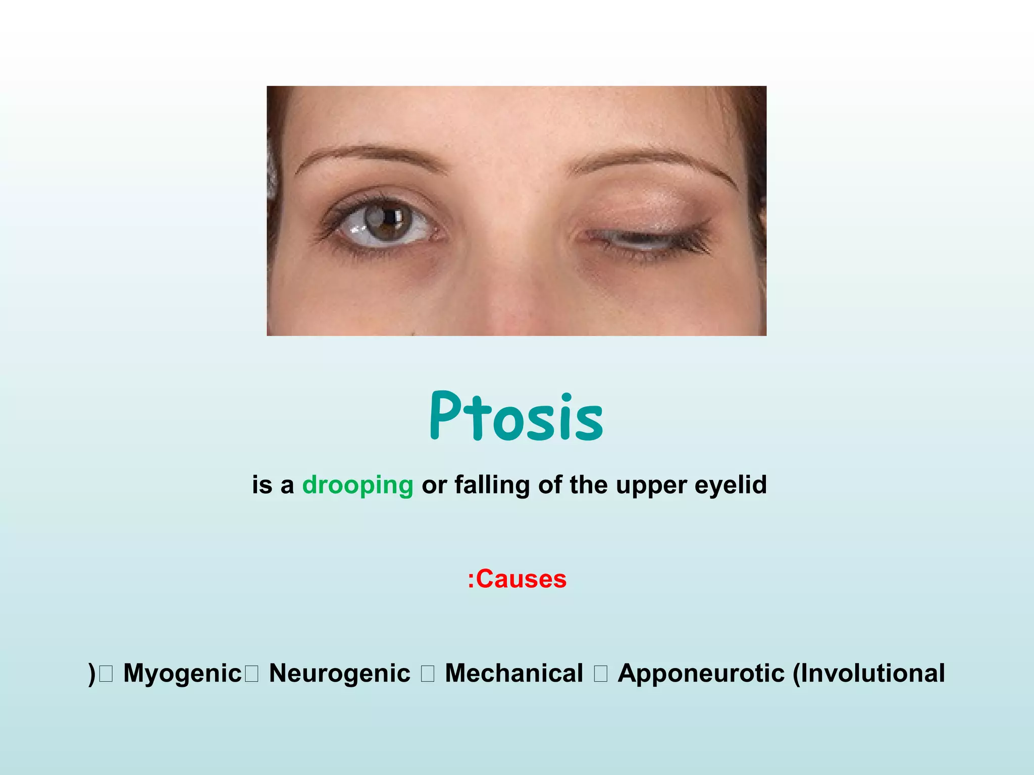
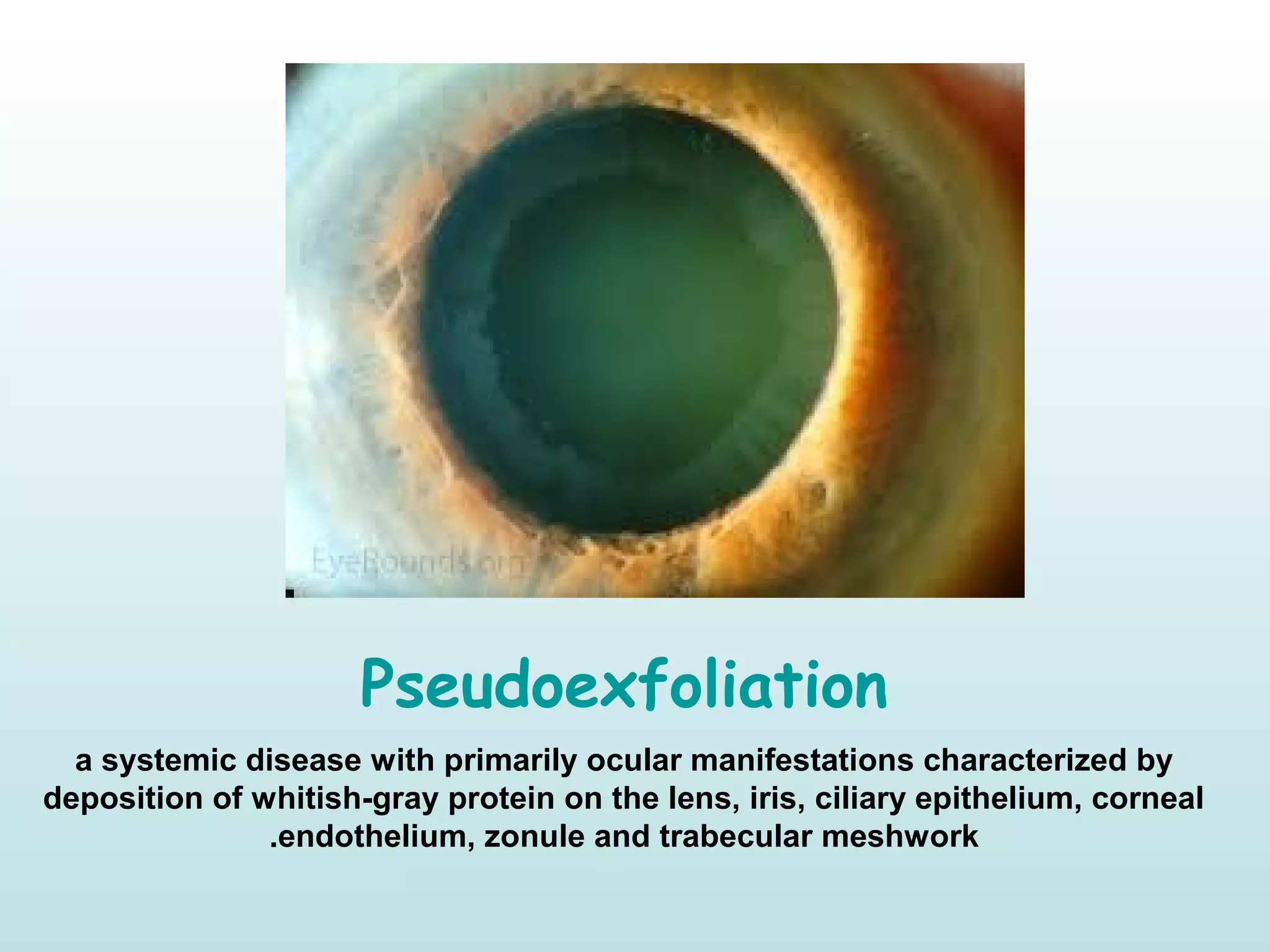
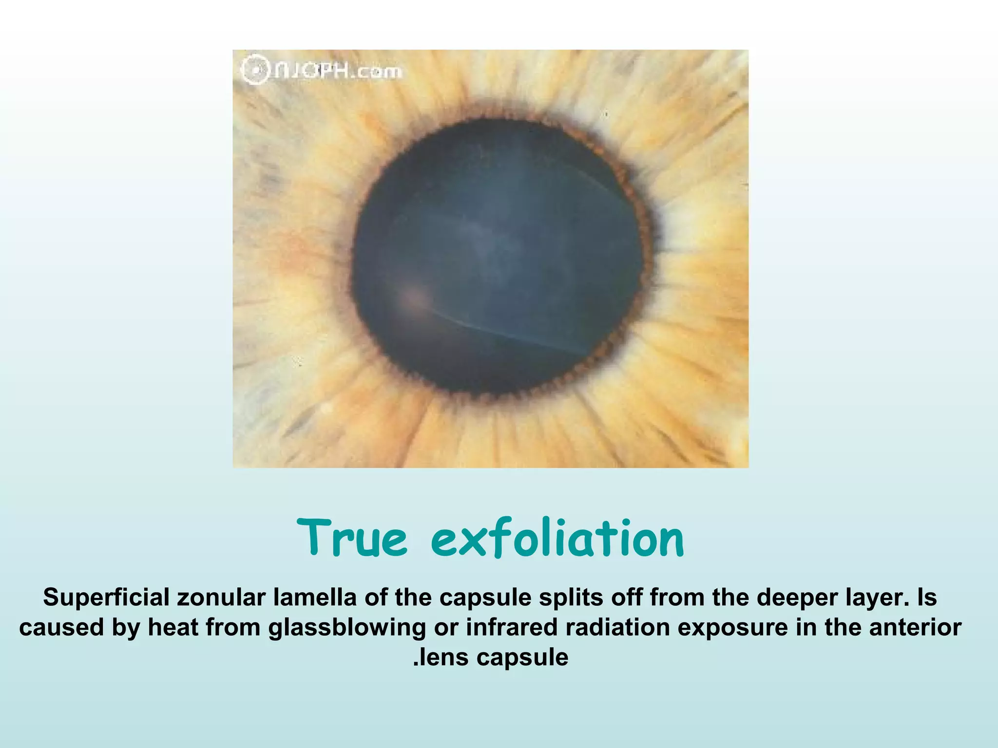
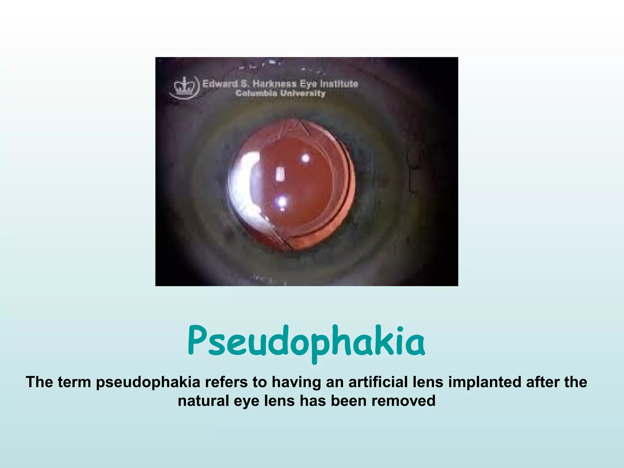
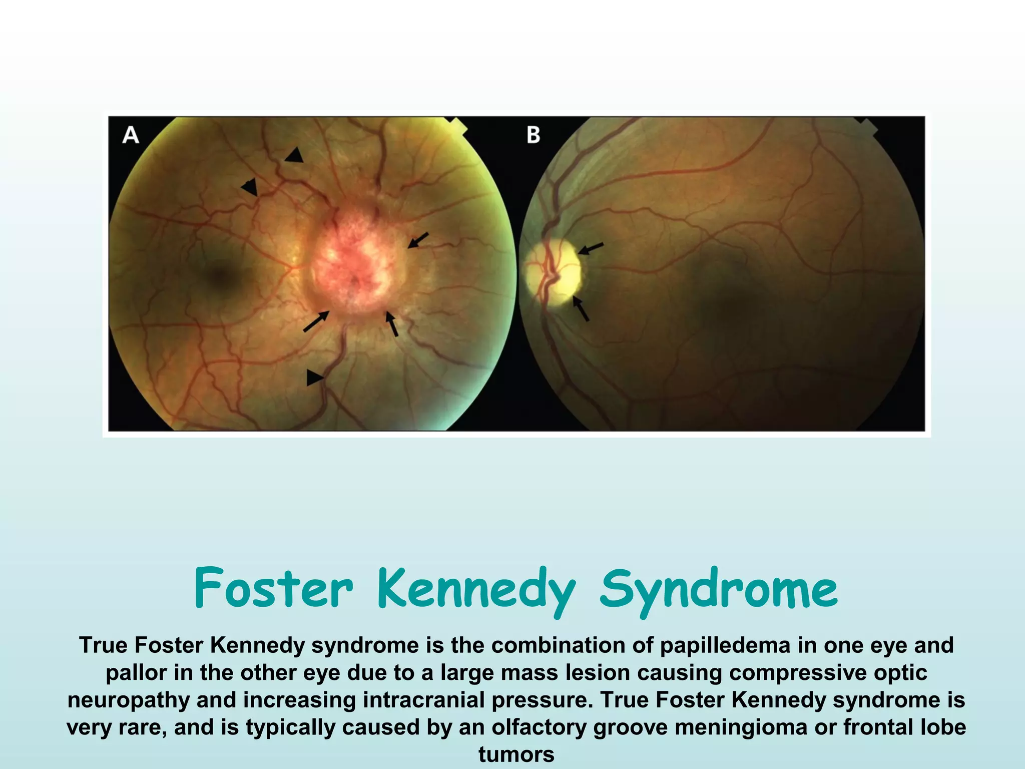
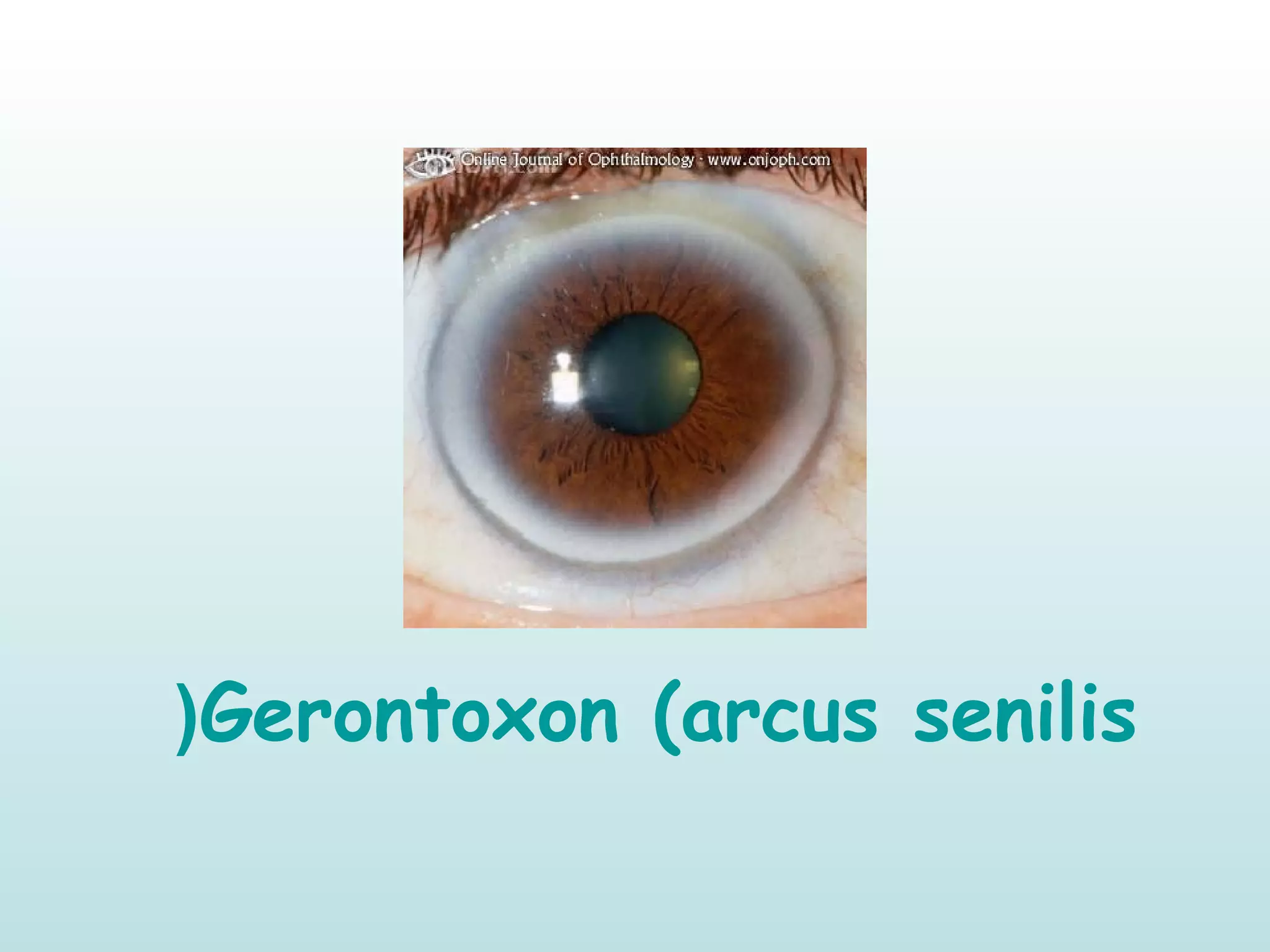
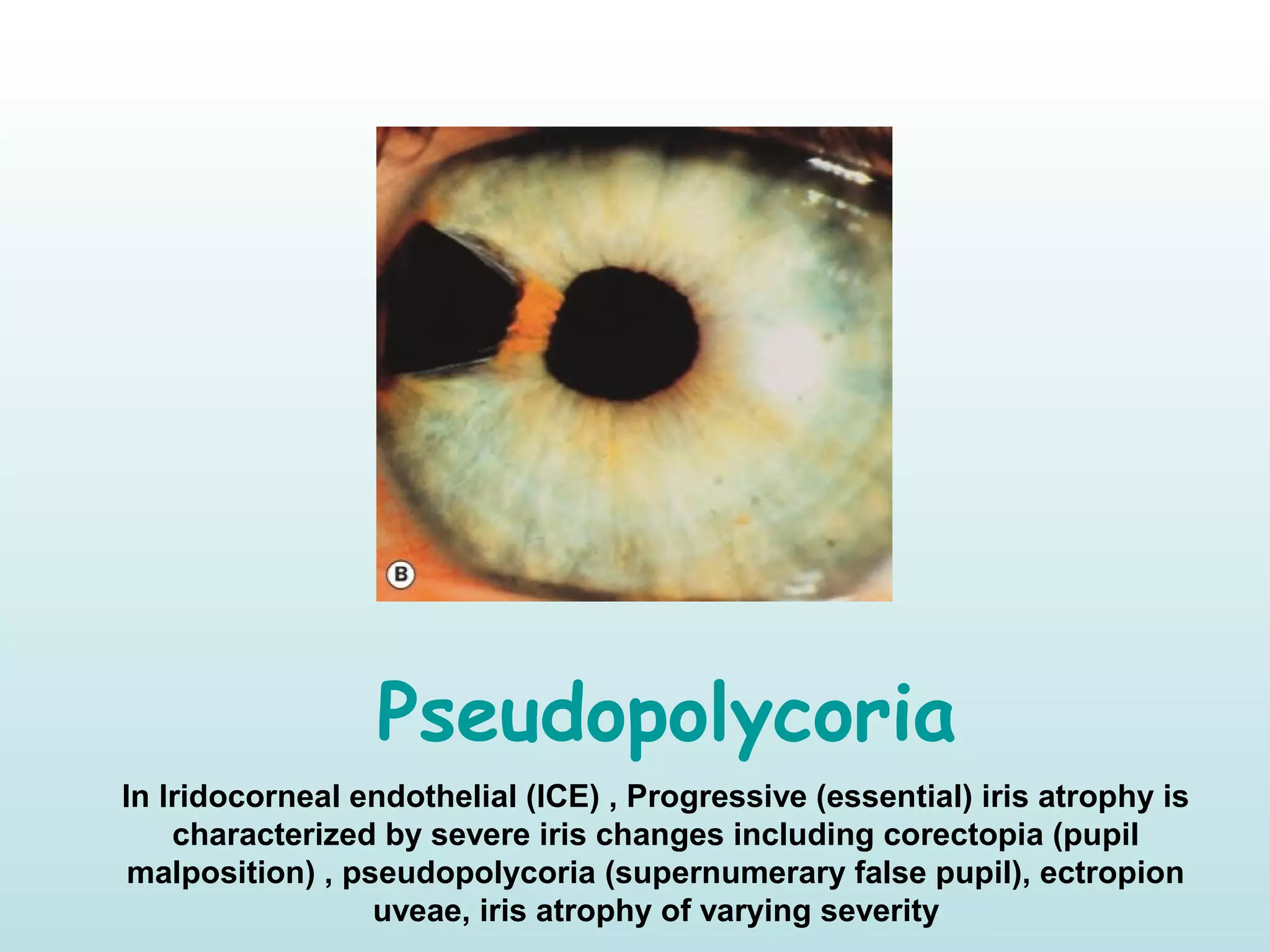
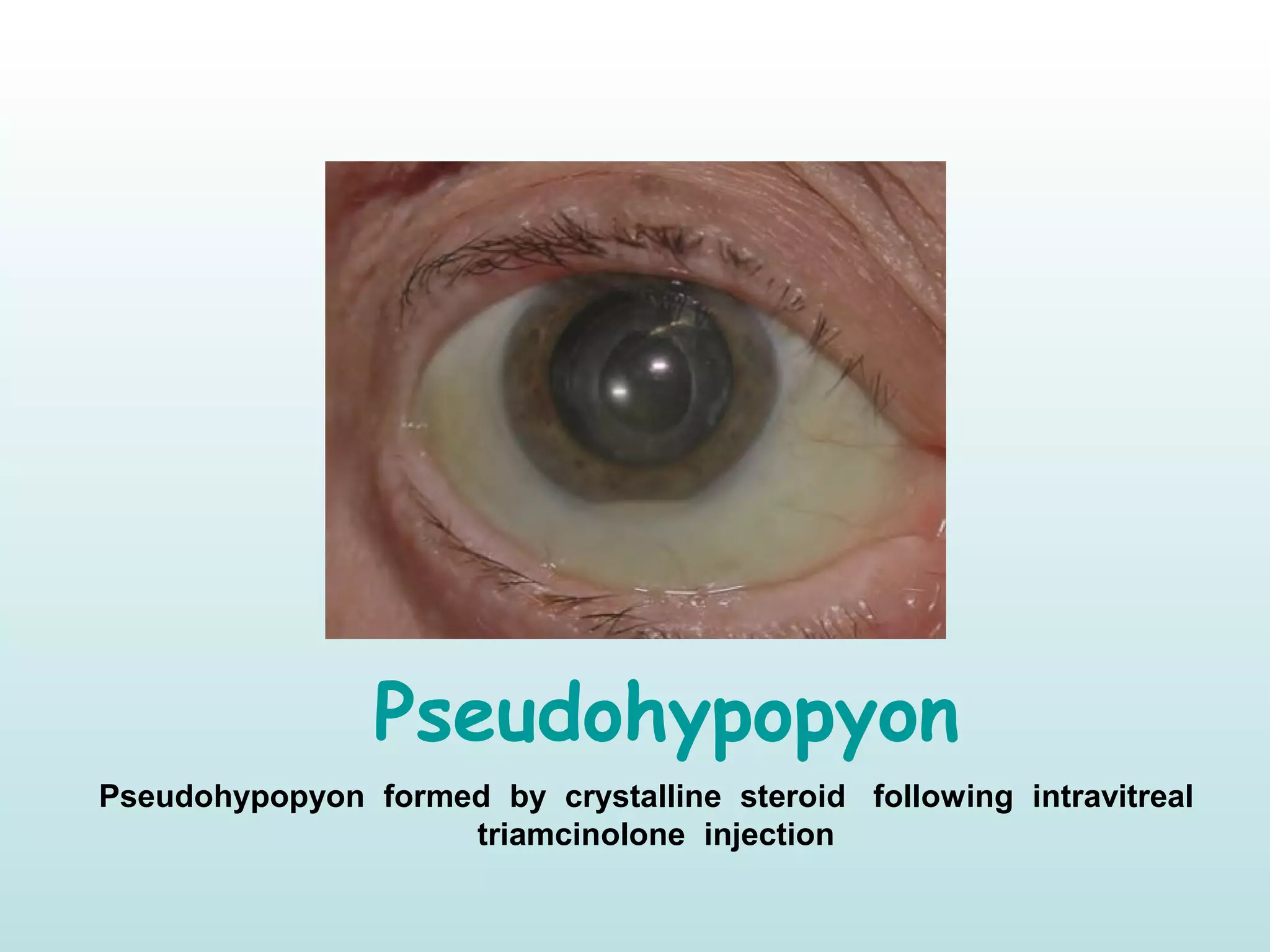
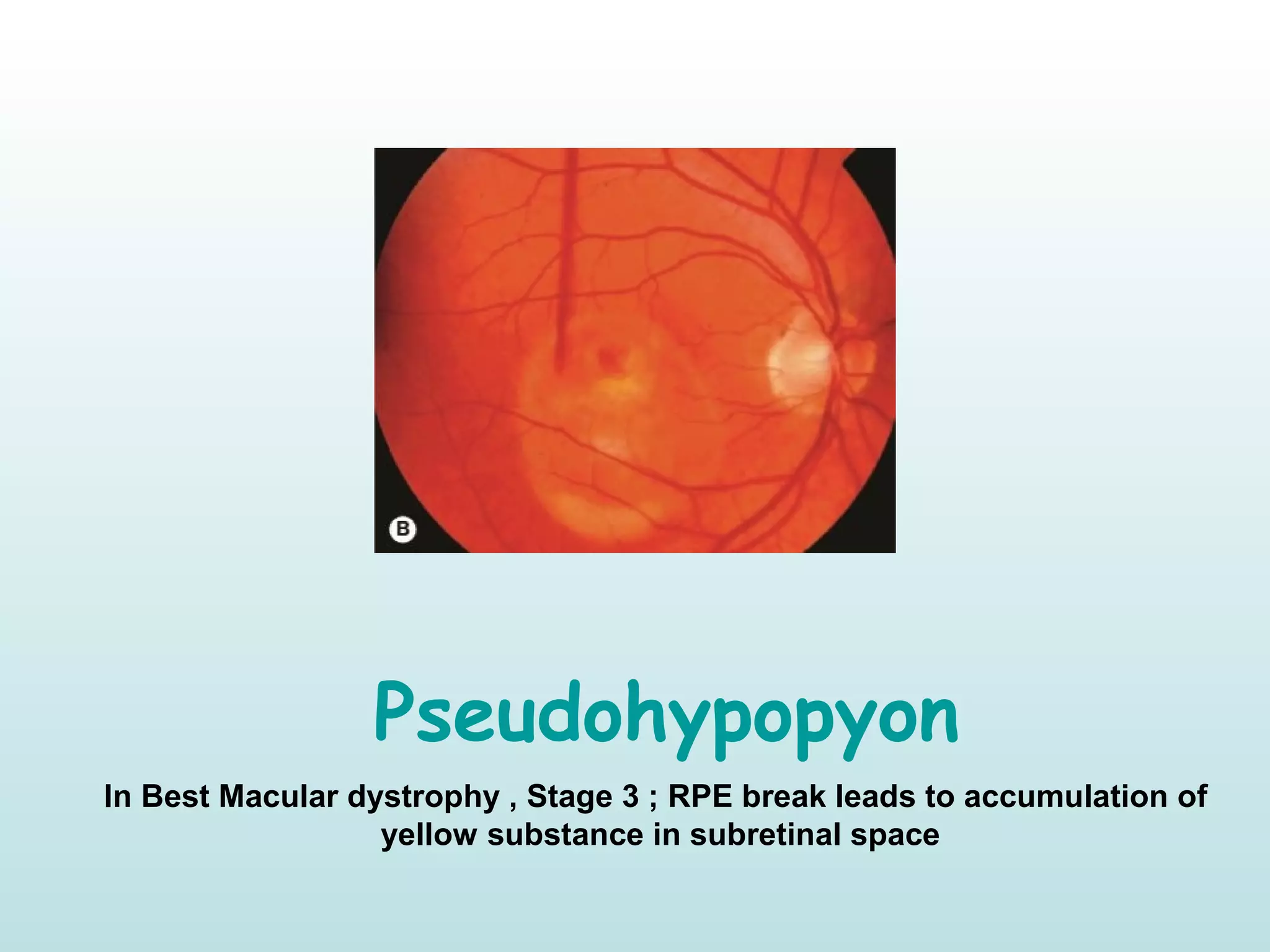
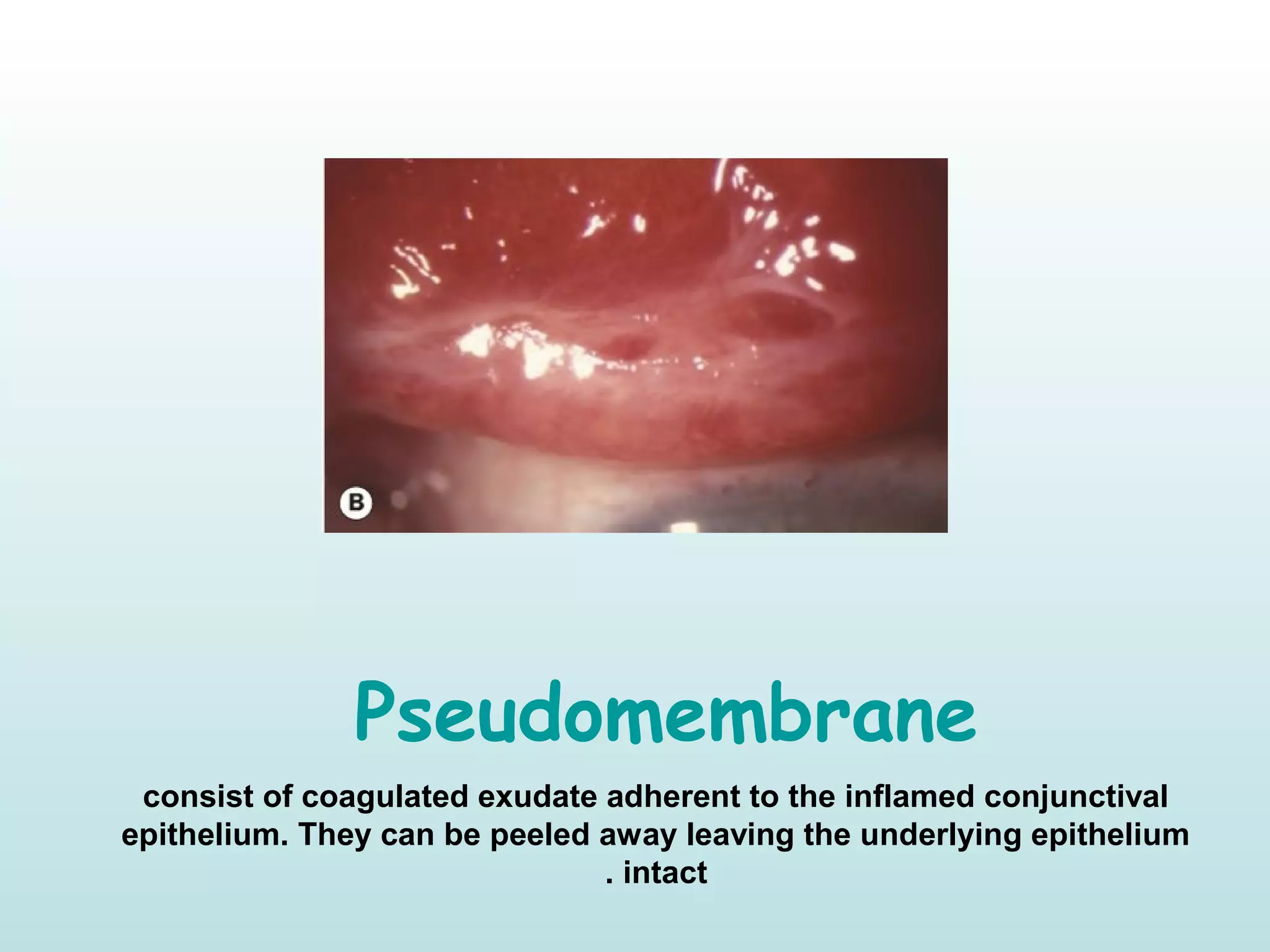
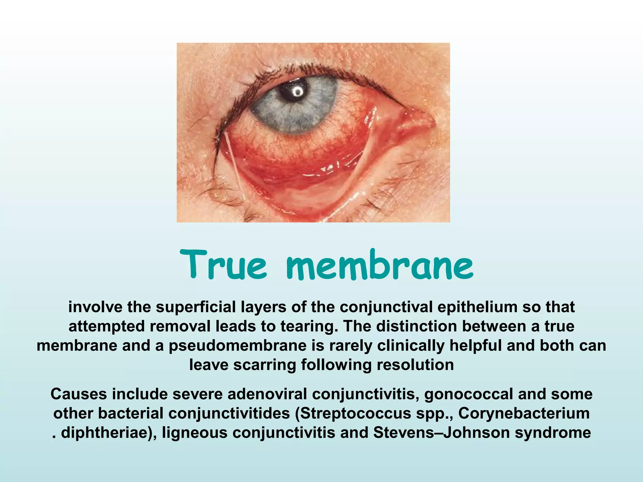
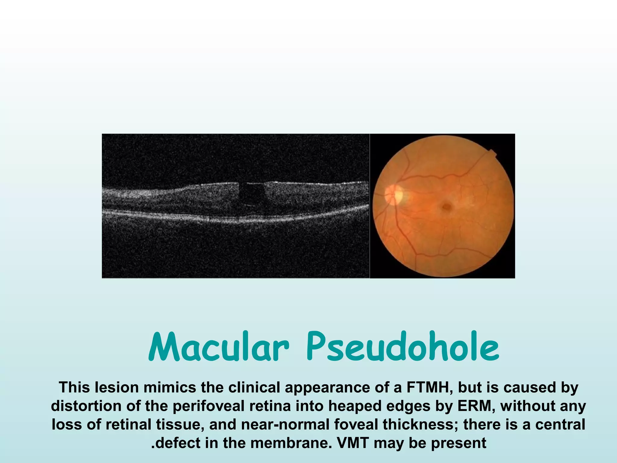
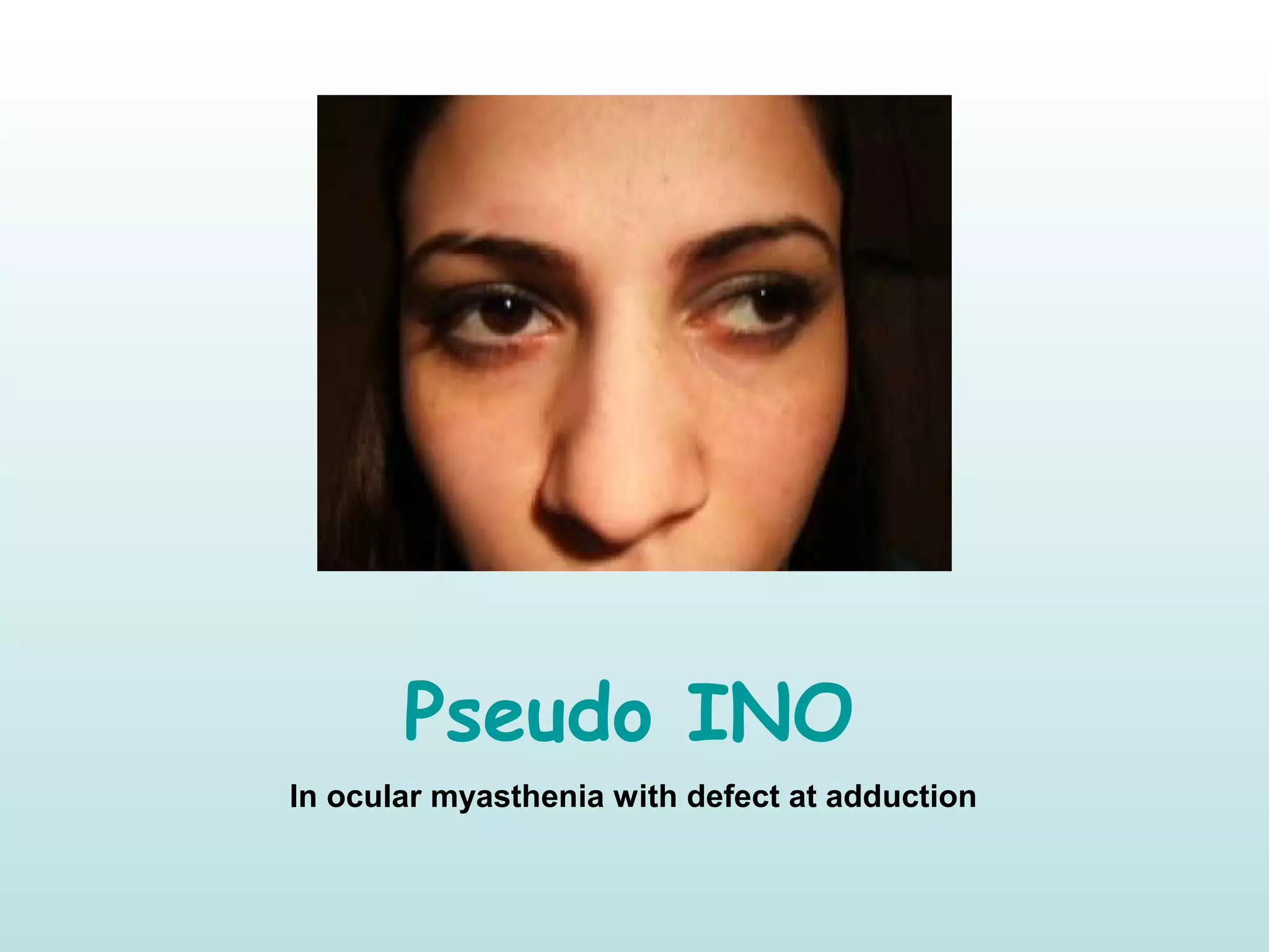
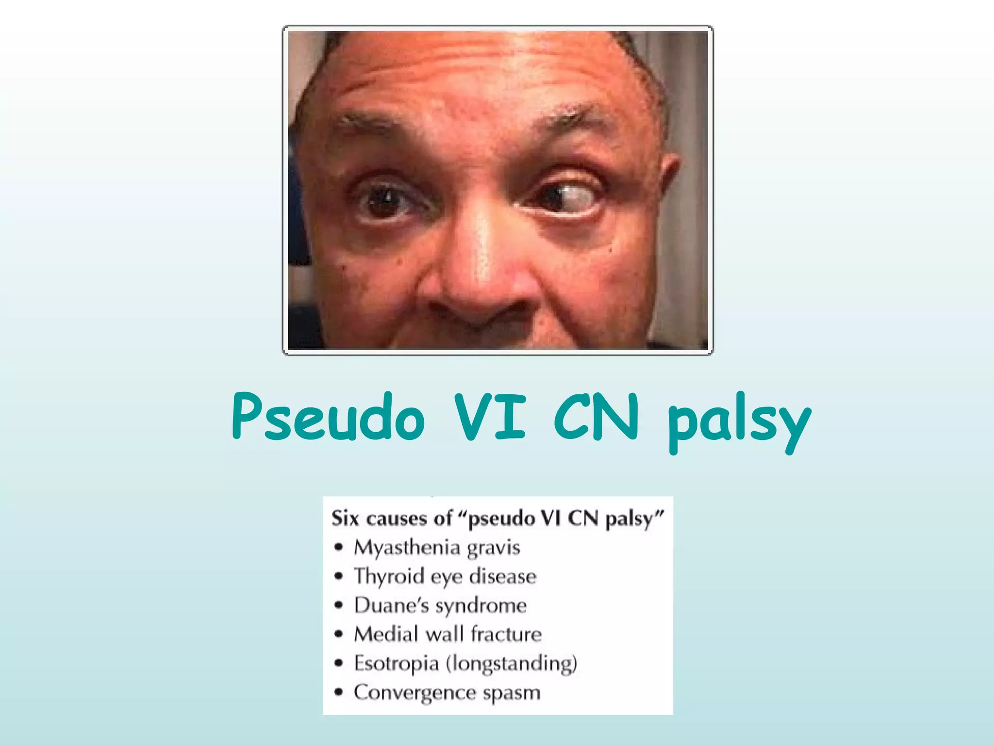
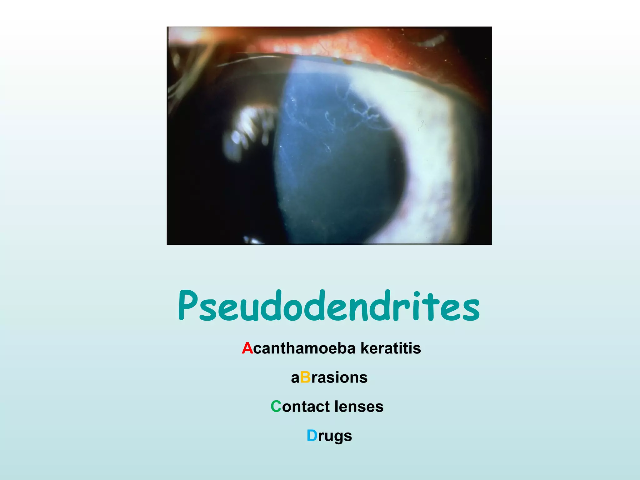
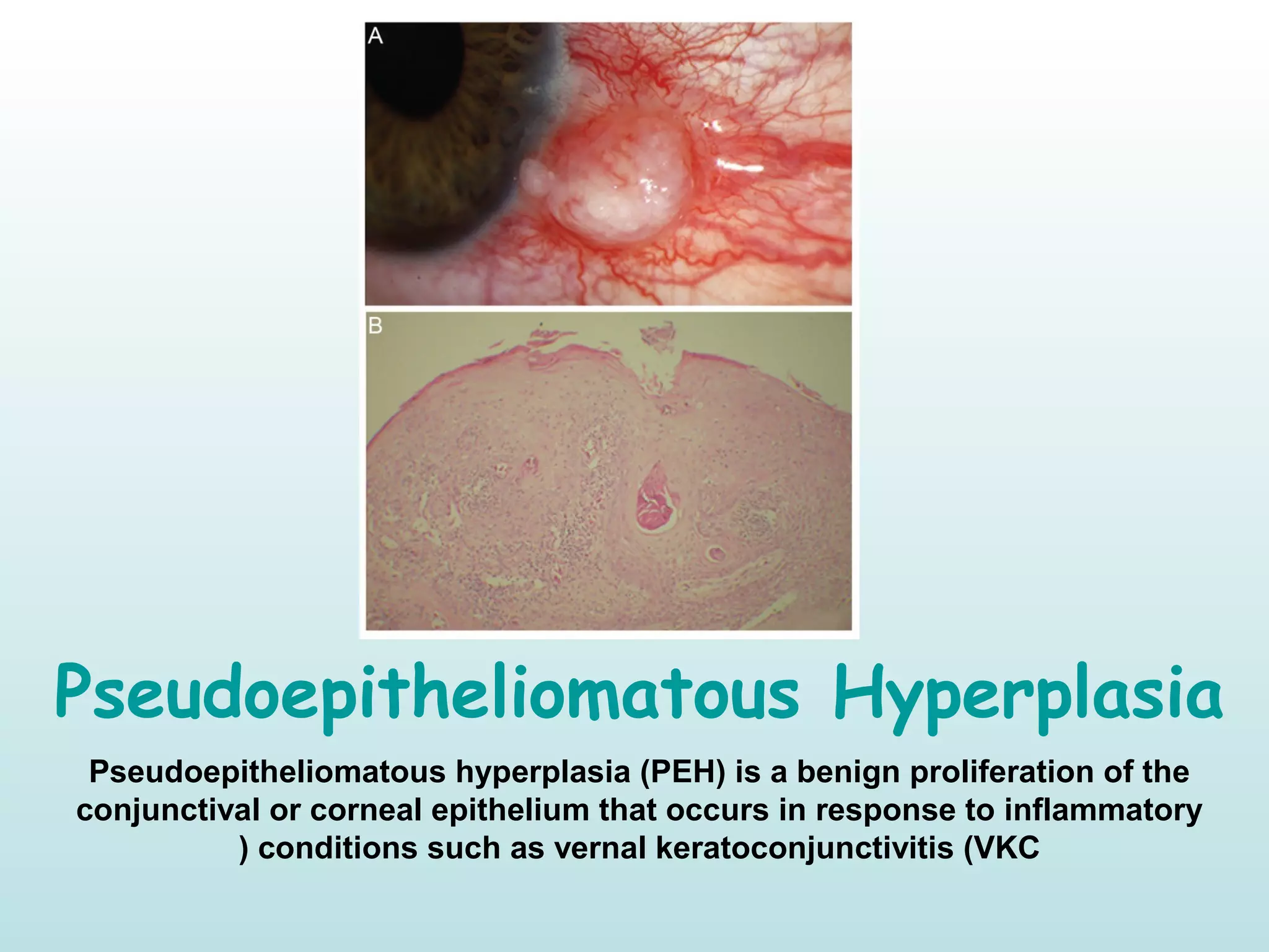
Pseudo-Ophthalmology !Pseudo-Ophthalmology !
By
Dr.Ahmed Alsherbiny
MSc. Ophth, FICO , FRCS Ophth (Glasg(
Chapter I
Pseudoptosis
A condition resembling ptosis, due to abnormalities other than those found in
the eyelid elevator muscles
Causes:
 Ipsilateral hypotropia  Enopthalmos  Dermatochalasis  Double elevator
palsy  Brow ptosis  Blepharospasm  Contralateral lid retraction 
Contralateral exopthalmos
Ptosis
is a drooping or falling of the upper eyelid
Causes:
 Myogenic Neurogenic  Mechanical  Apponeurotic (Involutional(
Pseudoproptosis
refers to the false impression of proptosis
Causes:
 Ipsilateral large globe (Buphthalmos/Myopia(  Ipsilateral lid retraction 
Shallow orbit  Facial asymmetry  Contralateral ptosis  Contralateral
enophthalmos
Proptosis
abnormal protrusion or displacement of an eye
Causes: (VEIN(
 Vascular  Endocrinal  Inflammatory  Neoplastic
Pseudopapilledema
(Optic Disc Drusen(
represent apparent optic disc swelling that simulates some features of
papilledema but is secondary to an underlying, usually benign process.
Other causes:
• Tilted optic disc • Peripapillary myelinated nerve fibres • Crowded disc                 
in hypermetropia 
Pseudoexfoliation
a systemic disease with primarily ocular manifestations characterized by
deposition of whitish-gray protein on the lens, iris, ciliary epithelium, corneal
endothelium, zonule and trabecular meshwork.
True exfoliation
Superficial zonular lamella of the capsule splits off from the deeper layer. Is
caused by heat from glassblowing or infrared radiation exposure in the anterior
lens capsule.
Pseudophakia
The term pseudophakia refers to having an artificial lens implanted after the
natural eye lens has been removed
Phakia
The presence of the natural crystalline lens of the eye.
LOOK AGAIN!!
Pseudo-Strabismus
refers to a false appearance of strabismus caused by an optical illusion
Remember : Pseudo-esotropia in Myopia due to negative angle kappa
)angle formed by pupillary axis and visual axis at the pupil(
Pseudo-exotropia in Hypermetropia due to positive angle kappa
Pseudo Foster Kennedy
The presence of pallor in one eye and disc edema in the contralateral eye in the
absence of an intracranial mass is called pseudo-Foster Kennedy syndrome. Our
patient's presentation is consistent with bilateral sequential NAION, which is the
most common cause of pseudo-Foster Kennedy syndrome
Foster Kennedy Syndrome
True Foster Kennedy syndrome is the combination of papilledema in one eye and
pallor in the other eye due to a large mass lesion causing compressive optic
neuropathy and increasing intracranial pressure. True Foster Kennedy syndrome is
very rare, and is typically caused by an olfactory groove meningioma or frontal lobe
tumors
Orbital
pseudotumor
also known as Idiopathic Orbital
Inflammatory Syndrome )IOIS( is is an
uncommon disorder characterized by non-
neoplastic, non-infective, space occupying
orbital infiltration with inflammatory
features. The process may preferentially
involve any or all of the orbital soft tissues.
Pseudotumor cerebri
is a condition in which the pressure around your brain increases, causing
headaches and vision problems. The name means “false brain tumor”
because its symptoms are similar to those caused by brain tumors. It's also
known as idiopathic intracranial hypertension
Pseudoxanthoma elasticum (PXE(
also known as Grönblad–Strandberg syndrome, is a genetic disease that causes
fragmentation and mineralization of elastic fibers in some tissues. The most
common problems arise in the skin and eyes, and later in blood vessels in the
form of premature atherosclerosis.
Pseudogerontoxon
In vernal keartoconjunctivitis , develop in recurrent limbal disease. It is
characterized by a paralimbal band of superficial scarring resembling arcus
senilis
Gerontoxon (arcus senilis(
Pseudoptrygiym
appears similar clinically but is caused by a band of conjunctiva adhering to
an area of compromised cornea at its apex. It forms as a response to an
acute inflammatory episode such as a chemical burn, corneal ulcer
)especially if marginal(, trauma and cicatrizing conjunctivitis.
A pseudopterygium is classically distinguished by both location away from
the horizontal )though this may also be seen with true pterygia( and firm
attachment to the cornea only at its apex )head(.
Pseudopolycoria
In Iridocorneal endothelial )ICE( , Progressive )essential( iris atrophy is
characterized by severe iris changes including corectopia )pupil
malposition( , pseudopolycoria )supernumerary false pupil(, ectropion
uveae, iris atrophy of varying severity
Pseudotrichiasis
The constant rubbing of the lashes on the cornea in long-standing entropion
Pseudohypopyon
B- Emusified Silicon in AC C- Phacolytic Glaucoma D- psuedohypopyon and
iris nodules in retinoblastoma
Pseudohypopyon
Pseudohypopyon formed by crystalline steroid following intravitreal             
triamcinolone injection 
Pseudohypopyon
Pseudohypopyon formed by in leukemia     
Pseudohypopyon
In Best Macular dystrophy , Stage 3 ; RPE break leads to accumulation of
yellow substance in subretinal space
Pseudoenophthalmos
may be caused by a small or shrunken eye (microphthalmos or phthisis
bulbi), by ptosis, or by contralateral proptosis or pseudoproptosis
Pseudomembrane
consist of coagulated exudate adherent to the inflamed conjunctival
epithelium. They can be peeled away leaving the underlying epithelium
intact.
True membrane
involve the superficial layers of the conjunctival epithelium so that
attempted removal leads to tearing. The distinction between a true
membrane and a pseudomembrane is rarely clinically helpful and both can
leave scarring following resolution
Causes include severe adenoviral conjunctivitis, gonococcal and some
other bacterial conjunctivitides (Streptococcus spp., Corynebacterium
diphtheriae), ligneous conjunctivitis and Stevens–Johnson syndrome.
Macular Pseudohole
This lesion mimics the clinical appearance of a FTMH, but is caused by
distortion of the perifoveal retina into heaped edges by ERM, without any
loss of retinal tissue, and near-normal foveal thickness; there is a central
defect in the membrane. VMT may be present.
Pseudo Graefe’s Sign
In III CN palsy, Aberrant regeneration may follow acute traumatic and
compressive, but not vascular, third nerve palsies ; elevation of the upper
eyelid on attempted adduction or depression (the pseudo-Graefe or pseudo-
von Graefe phenomenon), are caused by misdirected regenerating axons
that re-innervate the incorrect extraocular muscle.
Von Graefe’s Sign
Failure of the upper lid to follow a downward movement of the eyeball when
the patient changes his or her vision from looking up to looking down.
Pseudo Argyll Robertson pupil
In III CN palsy, Aberrant regeneration may follow acute traumatic and
compressive, but not vascular, third nerve palsies ; pupillary constriction
(Pseudo-Argyll Robertson Pupil) when the patient moved the eye by muscles
normally innervated by cranial nerve III
Argyll
Robertson
pupil
are bilateral small pupils that reduce
in size on a near object (i.e., they
accommodate), but do not constrict
when exposed to bright light (i.e.,
they do not react to light). They are a
highly specific sign of neurosyphilis
Pseudoepithelium
Posterior polymorphous corneal dystrophy
The pathological basis involves metaplasia of endothelial cells.
Multilayering of endothelium (pseudoepithelium)
Pseudo INO
In ocular myasthenia with defect at adduction
Pseudo Retinitis Pigmentosa
Pseudo VI CN palsy
Pseudodendrites
Acanthamoeba keratitis
aBrasions
Contact lenses
Drugs
Pseudoepitheliomatous Hyperplasia
Pseudoepitheliomatous hyperplasia (PEH) is a benign proliferation of the
conjunctival or corneal epithelium that occurs in response to inflammatory
conditions such as vernal keratoconjunctivitis (VKC)
Thank You

Pseudo-Ophthalmology !

  • 1.
    Pseudo-Ophthalmology !Pseudo-Ophthalmology ! By Dr.AhmedAlsherbiny MSc. Ophth, FICO , FRCS Ophth (Glasg(
  • 2.
  • 3.
    Pseudoptosis A condition resemblingptosis, due to abnormalities other than those found in the eyelid elevator muscles Causes:  Ipsilateral hypotropia  Enopthalmos  Dermatochalasis  Double elevator palsy  Brow ptosis  Blepharospasm  Contralateral lid retraction  Contralateral exopthalmos
  • 4.
    Ptosis is a droopingor falling of the upper eyelid Causes:  Myogenic Neurogenic  Mechanical  Apponeurotic (Involutional(
  • 5.
    Pseudoproptosis refers to thefalse impression of proptosis Causes:  Ipsilateral large globe (Buphthalmos/Myopia(  Ipsilateral lid retraction  Shallow orbit  Facial asymmetry  Contralateral ptosis  Contralateral enophthalmos
  • 6.
    Proptosis abnormal protrusion ordisplacement of an eye Causes: (VEIN(  Vascular  Endocrinal  Inflammatory  Neoplastic
  • 7.
    Pseudopapilledema (Optic Disc Drusen( representapparent optic disc swelling that simulates some features of papilledema but is secondary to an underlying, usually benign process. Other causes: • Tilted optic disc • Peripapillary myelinated nerve fibres • Crowded disc                  in hypermetropia 
  • 8.
    Pseudoexfoliation a systemic diseasewith primarily ocular manifestations characterized by deposition of whitish-gray protein on the lens, iris, ciliary epithelium, corneal endothelium, zonule and trabecular meshwork.
  • 9.
    True exfoliation Superficial zonularlamella of the capsule splits off from the deeper layer. Is caused by heat from glassblowing or infrared radiation exposure in the anterior lens capsule.
  • 10.
    Pseudophakia The term pseudophakiarefers to having an artificial lens implanted after the natural eye lens has been removed
  • 11.
    Phakia The presence ofthe natural crystalline lens of the eye. LOOK AGAIN!!
  • 12.
    Pseudo-Strabismus refers to afalse appearance of strabismus caused by an optical illusion Remember : Pseudo-esotropia in Myopia due to negative angle kappa )angle formed by pupillary axis and visual axis at the pupil( Pseudo-exotropia in Hypermetropia due to positive angle kappa
  • 13.
    Pseudo Foster Kennedy Thepresence of pallor in one eye and disc edema in the contralateral eye in the absence of an intracranial mass is called pseudo-Foster Kennedy syndrome. Our patient's presentation is consistent with bilateral sequential NAION, which is the most common cause of pseudo-Foster Kennedy syndrome
  • 14.
    Foster Kennedy Syndrome TrueFoster Kennedy syndrome is the combination of papilledema in one eye and pallor in the other eye due to a large mass lesion causing compressive optic neuropathy and increasing intracranial pressure. True Foster Kennedy syndrome is very rare, and is typically caused by an olfactory groove meningioma or frontal lobe tumors
  • 15.
    Orbital pseudotumor also known asIdiopathic Orbital Inflammatory Syndrome )IOIS( is is an uncommon disorder characterized by non- neoplastic, non-infective, space occupying orbital infiltration with inflammatory features. The process may preferentially involve any or all of the orbital soft tissues.
  • 16.
    Pseudotumor cerebri is acondition in which the pressure around your brain increases, causing headaches and vision problems. The name means “false brain tumor” because its symptoms are similar to those caused by brain tumors. It's also known as idiopathic intracranial hypertension
  • 17.
    Pseudoxanthoma elasticum (PXE( alsoknown as Grönblad–Strandberg syndrome, is a genetic disease that causes fragmentation and mineralization of elastic fibers in some tissues. The most common problems arise in the skin and eyes, and later in blood vessels in the form of premature atherosclerosis.
  • 18.
    Pseudogerontoxon In vernal keartoconjunctivitis, develop in recurrent limbal disease. It is characterized by a paralimbal band of superficial scarring resembling arcus senilis
  • 19.
  • 20.
    Pseudoptrygiym appears similar clinicallybut is caused by a band of conjunctiva adhering to an area of compromised cornea at its apex. It forms as a response to an acute inflammatory episode such as a chemical burn, corneal ulcer )especially if marginal(, trauma and cicatrizing conjunctivitis. A pseudopterygium is classically distinguished by both location away from the horizontal )though this may also be seen with true pterygia( and firm attachment to the cornea only at its apex )head(.
  • 21.
    Pseudopolycoria In Iridocorneal endothelial)ICE( , Progressive )essential( iris atrophy is characterized by severe iris changes including corectopia )pupil malposition( , pseudopolycoria )supernumerary false pupil(, ectropion uveae, iris atrophy of varying severity
  • 22.
    Pseudotrichiasis The constant rubbingof the lashes on the cornea in long-standing entropion
  • 23.
    Pseudohypopyon B- Emusified Siliconin AC C- Phacolytic Glaucoma D- psuedohypopyon and iris nodules in retinoblastoma
  • 24.
    Pseudohypopyon Pseudohypopyon formed bycrystalline steroid following intravitreal              triamcinolone injection 
  • 25.
  • 26.
    Pseudohypopyon In Best Maculardystrophy , Stage 3 ; RPE break leads to accumulation of yellow substance in subretinal space
  • 27.
    Pseudoenophthalmos may be causedby a small or shrunken eye (microphthalmos or phthisis bulbi), by ptosis, or by contralateral proptosis or pseudoproptosis
  • 28.
    Pseudomembrane consist of coagulatedexudate adherent to the inflamed conjunctival epithelium. They can be peeled away leaving the underlying epithelium intact.
  • 29.
    True membrane involve thesuperficial layers of the conjunctival epithelium so that attempted removal leads to tearing. The distinction between a true membrane and a pseudomembrane is rarely clinically helpful and both can leave scarring following resolution Causes include severe adenoviral conjunctivitis, gonococcal and some other bacterial conjunctivitides (Streptococcus spp., Corynebacterium diphtheriae), ligneous conjunctivitis and Stevens–Johnson syndrome.
  • 30.
    Macular Pseudohole This lesionmimics the clinical appearance of a FTMH, but is caused by distortion of the perifoveal retina into heaped edges by ERM, without any loss of retinal tissue, and near-normal foveal thickness; there is a central defect in the membrane. VMT may be present.
  • 31.
    Pseudo Graefe’s Sign InIII CN palsy, Aberrant regeneration may follow acute traumatic and compressive, but not vascular, third nerve palsies ; elevation of the upper eyelid on attempted adduction or depression (the pseudo-Graefe or pseudo- von Graefe phenomenon), are caused by misdirected regenerating axons that re-innervate the incorrect extraocular muscle.
  • 32.
    Von Graefe’s Sign Failureof the upper lid to follow a downward movement of the eyeball when the patient changes his or her vision from looking up to looking down.
  • 33.
    Pseudo Argyll Robertsonpupil In III CN palsy, Aberrant regeneration may follow acute traumatic and compressive, but not vascular, third nerve palsies ; pupillary constriction (Pseudo-Argyll Robertson Pupil) when the patient moved the eye by muscles normally innervated by cranial nerve III
  • 34.
    Argyll Robertson pupil are bilateral smallpupils that reduce in size on a near object (i.e., they accommodate), but do not constrict when exposed to bright light (i.e., they do not react to light). They are a highly specific sign of neurosyphilis
  • 35.
    Pseudoepithelium Posterior polymorphous cornealdystrophy The pathological basis involves metaplasia of endothelial cells. Multilayering of endothelium (pseudoepithelium)
  • 36.
    Pseudo INO In ocularmyasthenia with defect at adduction
  • 37.
  • 38.
  • 39.
  • 40.
    Pseudoepitheliomatous Hyperplasia Pseudoepitheliomatous hyperplasia(PEH) is a benign proliferation of the conjunctival or corneal epithelium that occurs in response to inflammatory conditions such as vernal keratoconjunctivitis (VKC)
  • 41.