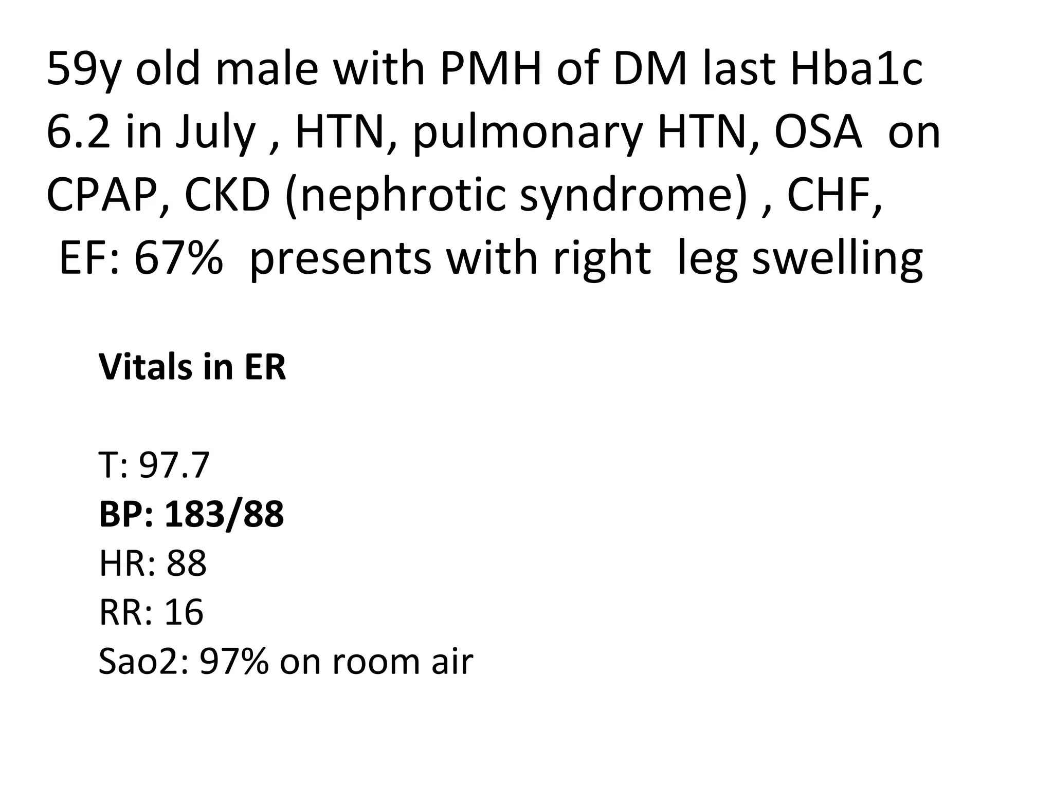
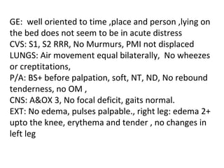
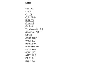
59y old male with PMH of DM last Hba1c 6.2 in July , HTN, pulmonary HTN, OSA  on CPAP, CKD (nephrotic syndrome) , CHF, EF: 67%  presents with right  leg swelling Vitals in ER  T: 97.7 BP: 183/88 HR: 88 RR: 16 Sao2: 97% on room air
GE:  well oriented to time ,place and person ,lying on the bed does not seem to be in acute distress CVS: S1, S2 RRR, No Murmurs, PMI not displaced LUNGS: Air movement equal bilaterally,  No wheezes or creptitations,  P/A: BS+ before palpation, soft, NT, ND, No rebound tenderness, no OM ,  CNS: A&OX 3, No focal deficit, gaits normal. EXT: No edema, pulses palpable., right leg: edema 2+ upto the knee, erythema and tender , no changes in left leg
Labs:   Na: 140 K: 4.6 Cl: 106 Co2:  29.9 BUN: 35 Crea: 2.2 Ca: 8 .4 Total protein:  6.2 Albumin:  2.8 Gfr:30 Anion gap:4 WBC:  8.0 HGB: 15.0 Platelets: 192 MCV:  89.4 RDW: 147 APTT: 24.3 PT: 11.0 INR: 1.06
Venous Thromboembolism Management Namrata Dass
Diagnostic Testing D-dimer: positive test require further evaluation. (highly sensitive) negative D-dimer in combination with low pretest probability can exclude almost all PE Imaging Compression ultrasound/duplex examination May fail to visualize parts of the deep femoral vein, parts of the upper extremity venous system, and the pelvic veins.
PE-specific testing Nondefinitive tests :ekg, troponin and brain natriuretic peptide (BNP) levels, blood gases Contrast-enhanced spiral (helical) chest CT: Contraindications: renal dysfunction and dye allergy. Advantages of CT scan over V/Q scan include more diagnostic results , with fewer indeterminate or inadequate studies, and the detection of alternative diagnoses, such as dissecting aortic aneurysm, pneumonia, and malignancy.
V/Q scanning Classified as normal, non-diagnostic (i.e., very low probability, low probability, intermediate probability), or high probability for PE. Pulmonary angiography (PA) : gold standard - Less invasive tests have mostly replaced PA over the past decade.
TREATMENT To prevent recurrent VTE, To prevent  consequences of VTE (i.e., post-phlebitic syndrome [i.e., pain, edema, and ulceration], pulmonary arterial hypertension, and death), and  To prevent complications of therapy (e.g., bleeding and HIT).
Medications WARFARIN: leads to depletion of the vitamin K-dependent clotting factors II, VII, IX, and X and proteins C, S, and Z. It requires 4 to 5 days to achieve the full anticoagulant effect. Because of the rapid depletion of the anticoagulant protein C and slower onset of anticoagulant effect, patients might develop increased hyper-coagulability if warfarin is not combined with a parenteral anticoagulant
For most indications, warfarin has a target INR of 2.5 and a therapeutic range of 2 to 3. INR monitoring: twice weekly for 1 to 2 weeks, then weekly for 2 weeks, then less frequently. Unfractionated heparin (UFH): inactivation of thrombin and factor Xa by antithrombin. Prolongs both  aPTT ,minimal effect on PT For DVT prophylaxis, the typical dosage is 5,000 units SC q8-12h
For therapeutic anticoagulation, UFH is usually administered IV with a bolus followed by continuous infusion Choice of anticoagulant in patient with increased risk of bleeding
LMWH Produced by chemical or enzymatic cleavage of UFH. Minimally prolongs the aPTT. In pt with renal dysfunction, obesity, or pregnancy, factor Xa level monitoring may be prudent. Because of the SC dosing route, LMWH facilitates outpatient VTE therapy. First choice in pregnant women and cancer pts
Fondaparinux Selective inhibitor of factor Xa. Does not significantly prolong the aPTT. Monitoring with factor X level not indicated except in renal insufficiency May be used for outpatient VTE therapy.
Lepirudin (Refludan, recombinant hirudin) Direct thrombin inhibitor that is used for the treatment of HIT Requires cautious use and dose adjustments in patients with renal insufficiency aPTT monitoring should occur 4 hours after a dose change Argatroban: used for HIT therapy.
Thrombolytic therapy Indications :refractory systemic hypotension and PE associated with objectively demonstrated (e.g. echocardiogram, spiral CT) acute, severe right ventricular strain alteplase or recombinant tissue plasminogen activator
IVC filters Indicated for acute DVT situations in which there are absolute contraindications to anticoagulation (e.g., active bleeding, severe thrombocytopenia, urgent surgery) or recurrent thromboemboli despite therapeutic anticoagulation.
Surgical embolectomy should be considered in patients with life-threatening massive PE that have contraindications to thrombolytic therapy

Presentationaug2011

  • 1.
    59y old malewith PMH of DM last Hba1c 6.2 in July , HTN, pulmonary HTN, OSA  on CPAP, CKD (nephrotic syndrome) , CHF, EF: 67%  presents with right leg swelling Vitals in ER T: 97.7 BP: 183/88 HR: 88 RR: 16 Sao2: 97% on room air
  • 2.
    GE:  well orientedto time ,place and person ,lying on the bed does not seem to be in acute distress CVS: S1, S2 RRR, No Murmurs, PMI not displaced LUNGS: Air movement equal bilaterally, No wheezes or creptitations, P/A: BS+ before palpation, soft, NT, ND, No rebound tenderness, no OM , CNS: A&OX 3, No focal deficit, gaits normal. EXT: No edema, pulses palpable., right leg: edema 2+ upto the knee, erythema and tender , no changes in left leg
  • 3.
    Labs:   Na:140 K: 4.6 Cl: 106 Co2:  29.9 BUN: 35 Crea: 2.2 Ca: 8 .4 Total protein:  6.2 Albumin:  2.8 Gfr:30 Anion gap:4 WBC:  8.0 HGB: 15.0 Platelets: 192 MCV:  89.4 RDW: 147 APTT: 24.3 PT: 11.0 INR: 1.06
  • 4.
  • 5.
    Diagnostic Testing D-dimer:positive test require further evaluation. (highly sensitive) negative D-dimer in combination with low pretest probability can exclude almost all PE Imaging Compression ultrasound/duplex examination May fail to visualize parts of the deep femoral vein, parts of the upper extremity venous system, and the pelvic veins.
  • 6.
    PE-specific testing Nondefinitivetests :ekg, troponin and brain natriuretic peptide (BNP) levels, blood gases Contrast-enhanced spiral (helical) chest CT: Contraindications: renal dysfunction and dye allergy. Advantages of CT scan over V/Q scan include more diagnostic results , with fewer indeterminate or inadequate studies, and the detection of alternative diagnoses, such as dissecting aortic aneurysm, pneumonia, and malignancy.
  • 7.
    V/Q scanning Classifiedas normal, non-diagnostic (i.e., very low probability, low probability, intermediate probability), or high probability for PE. Pulmonary angiography (PA) : gold standard - Less invasive tests have mostly replaced PA over the past decade.
  • 8.
    TREATMENT To preventrecurrent VTE, To prevent consequences of VTE (i.e., post-phlebitic syndrome [i.e., pain, edema, and ulceration], pulmonary arterial hypertension, and death), and To prevent complications of therapy (e.g., bleeding and HIT).
  • 9.
    Medications WARFARIN: leadsto depletion of the vitamin K-dependent clotting factors II, VII, IX, and X and proteins C, S, and Z. It requires 4 to 5 days to achieve the full anticoagulant effect. Because of the rapid depletion of the anticoagulant protein C and slower onset of anticoagulant effect, patients might develop increased hyper-coagulability if warfarin is not combined with a parenteral anticoagulant
  • 10.
    For most indications,warfarin has a target INR of 2.5 and a therapeutic range of 2 to 3. INR monitoring: twice weekly for 1 to 2 weeks, then weekly for 2 weeks, then less frequently. Unfractionated heparin (UFH): inactivation of thrombin and factor Xa by antithrombin. Prolongs both aPTT ,minimal effect on PT For DVT prophylaxis, the typical dosage is 5,000 units SC q8-12h
  • 11.
    For therapeutic anticoagulation,UFH is usually administered IV with a bolus followed by continuous infusion Choice of anticoagulant in patient with increased risk of bleeding
  • 12.
    LMWH Produced bychemical or enzymatic cleavage of UFH. Minimally prolongs the aPTT. In pt with renal dysfunction, obesity, or pregnancy, factor Xa level monitoring may be prudent. Because of the SC dosing route, LMWH facilitates outpatient VTE therapy. First choice in pregnant women and cancer pts
  • 13.
    Fondaparinux Selective inhibitorof factor Xa. Does not significantly prolong the aPTT. Monitoring with factor X level not indicated except in renal insufficiency May be used for outpatient VTE therapy.
  • 14.
    Lepirudin (Refludan, recombinanthirudin) Direct thrombin inhibitor that is used for the treatment of HIT Requires cautious use and dose adjustments in patients with renal insufficiency aPTT monitoring should occur 4 hours after a dose change Argatroban: used for HIT therapy.
  • 15.
    Thrombolytic therapy Indications:refractory systemic hypotension and PE associated with objectively demonstrated (e.g. echocardiogram, spiral CT) acute, severe right ventricular strain alteplase or recombinant tissue plasminogen activator
  • 16.
    IVC filters Indicatedfor acute DVT situations in which there are absolute contraindications to anticoagulation (e.g., active bleeding, severe thrombocytopenia, urgent surgery) or recurrent thromboemboli despite therapeutic anticoagulation.
  • 17.
    Surgical embolectomy shouldbe considered in patients with life-threatening massive PE that have contraindications to thrombolytic therapy