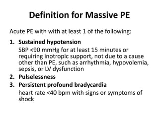
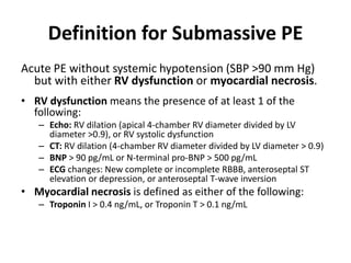
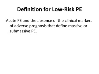
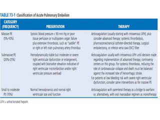
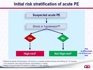
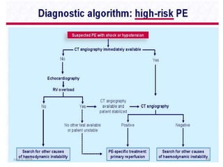
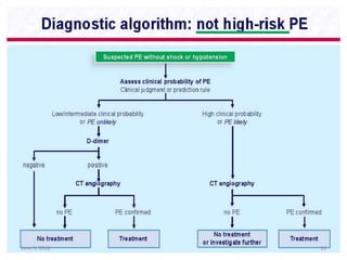
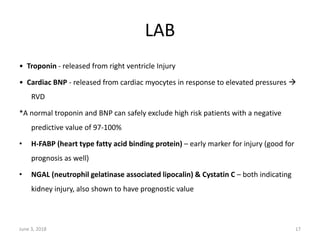
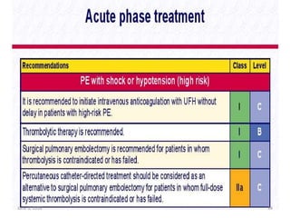
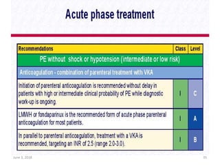
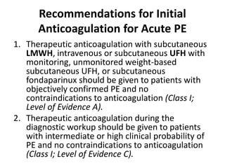
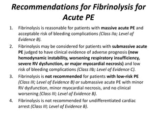
PULMONARY EMBOLISM
Definition for Massive PE
Acute PE with with at least 1 of the following:
1. Sustained hypotension
SBP <90 mmHg for at least 15 minutes or
requiring inotropic support, not due to a cause
other than PE, such as arrhythmia, hypovolemia,
sepsis, or LV dysfunction
2. Pulselessness
3. Persistent profound bradycardia
heart rate <40 bpm with signs or symptoms of
shock
Definition for Submassive PE
Acute PE without systemic hypotension (SBP >90 mm Hg)
but with either RV dysfunction or myocardial necrosis.
• RV dysfunction means the presence of at least 1 of the
following:
– Echo: RV dilation (apical 4-chamber RV diameter divided by LV
diameter >0.9), or RV systolic dysfunction
– CT: RV dilation (4-chamber RV diameter divided by LV diameter > 0.9)
– BNP > 90 pg/mL or N-terminal pro-BNP > 500 pg/mL
– ECG changes: New complete or incomplete RBBB, anteroseptal ST
elevation or depression, or anteroseptal T-wave inversion
• Myocardial necrosis is defined as either of the following:
– Troponin I > 0.4 ng/mL, or Troponin T > 0.1 ng/mL
Definition for Low-Risk PE
Acute PE and the absence of the clinical markers
of adverse prognosis that define massive or
submassive PE.
PERC
June 3, 2018 8
Kline JA, Mitchell AM, Kabrhel C, et al. Clinical criteria to prevent unnecessary diagnostic testing in emergency department patients
with suspected pulmonary embolism. J Thromb Haemost 2004;2:1247–55.
June 3, 2018 9
June 3, 2018 10
PESI
to differentiate
Int from Low
Risk
June 3, 2018 11
Pulmonary Embolism Severity Index (PESI)
June 3, 2018 12
June 3, 2018 13
14
June 3, 2018 15
June 3, 2018 16
LAB
• Troponin ‐ released from right ventricle Injury
• Cardiac BNP ‐ released from cardiac myocytes in response to elevated pressures 
RVD
*A normal troponin and BNP can safely exclude high risk patients with a negative
predictive value of 97-100%
• H-FABP (heart type fatty acid binding protein) – early marker for injury (good for
prognosis as well)
• NGAL (neutrophil gelatinase associated lipocalin) & Cystatin C – both indicating
kidney injury, also shown to have prognostic value
June 3, 2018 17
D-dimer
• patients with PE have ongoing endogenous fibrinolysis that is not
effective enough to prevent PE but that does break down fibrin clot
to d-dimers .
• they are not specific.
• negative d-dimer results, additional diagnostic testing not
necessary
• PE in low-risk patients with a negative D-dimer…
– Thrombus formation >72 hrs before blood draw (circulating dimer t1/2
= 8 hrs)
– Subsegmental PE
• False-positives = age >70, pregnancy, active malignancy, recent
surgery, liver disease, RA, infections, trauma
• False-negatives = Coumadin use, symptoms >5days, small clots or
infarction, isolated calf vein thrombosis.
• Westermark sign - focal oligemia
Hampton hump –Wedge shaped density above
diaphragm- indicates pul. infarction
Pallas sign – enlargement of descending
R pulmonary artery
Ventilation Perfusion Scan
Indications:
- Renal failure
- Pregnancy
Procedure:
- Ventilation scan with Xenon inhalation
- Perfusion scan with Tc99m labelled radioactive dye infusion
- Scan V/Q
- Result: unmatched V/Q
June 3, 2018 26
June 3, 2018 27
June 3, 2018 28
Indications:
- Suspected PE
Contra-indications:
- Renal failure
- Pregnancy
- Allergy to radio-contrast
1.7-5% risk of developing breast cancers (Hurwitz et al. 2007)
June 3, 2018 29
CT-PA
Multidetector helical CT-PA
• First line modality
• Cover all chest with high spatial resolution in one breath
• Detect peripheral smaller emboli
• Detect other pathologies
• Detect RV strain (straightening or leftward bowing of IV septum)
• Radiation Exposure
• # in renal failure and contrast allergy
June 3, 2018 32
June 3, 2018 34
June 3, 2018 35
• Heparin passive reduction of thrombus size.
• Thrombolytic agents actively promote the
hydrolysis of fibrin molecules.
• At 24 hours, patients treated with heparin have
no substantial improvement in pulmonary blood
flow, whereas patients treated with adjunctive
fibrinolysis manifest a 30% to 35% reduction in
total perfusion defect. However, by 7 days, blood
flow improves similarly (65% to 70% reduction in
total defect).
Recommendations for Initial
Anticoagulation for Acute PE
1. Therapeutic anticoagulation with subcutaneous
LMWH, intravenous or subcutaneous UFH with
monitoring, unmonitored weight-based
subcutaneous UFH, or subcutaneous
fondaparinux should be given to patients with
objectively confirmed PE and no
contraindications to anticoagulation (Class I;
Level of Evidence A).
2. Therapeutic anticoagulation during the
diagnostic workup should be given to patients
with intermediate or high clinical probability of
PE and no contraindications to anticoagulation
(Class I; Level of Evidence C).
Recommendations for Fibrinolysis for
Acute PE
1. Fibrinolysis is reasonable for patients with massive acute PE and
acceptable risk of bleeding complications (Class IIa; Level of
Evidence B).
2. Fibrinolysis may be considered for patients with submassive acute
PE judged to have clinical evidence of adverse prognosis (new
hemodynamic instability, worsening respiratory insufficiency,
severe RV dysfunction, or major myocardial necrosis) and low
risk of bleeding complications (Class IIb; Level of Evidence C).
3. Fibrinolysis is not recommended for patients with low-risk PE
(Class III; Level of Evidence B) or submassive acute PE with minor
RV dysfunction, minor myocardial necrosis, and no clinical
worsening (Class III; Level of Evidence B).
4. Fibrinolysis is not recommended for undifferentiated cardiac
arrest (Class III; Level of Evidence B).
Pharmacological Profile of Plasminogen-
Activating Fibrinolytic Agents
Fibrinolytic
FDA Indication
for PE?
Fibrinolytic Dose
Streptokinase Yes
250 000-IU IV bolus followed by
100 000-IU/h infusion for 12–24 h
Urokinase Yes
4400-IU/kg bolus, followed by 4400
IU.kg-1.h-1 for 12–24 h
Alteplase Yes 100-mg IV infusion over 2 h
Reteplase No Double 10-U IV bolus 30 min apart
Tenecteplase No
Weight-adjusted IV bolus over 5 s
(30–50 mg with a 5-mg step every
10 kg from <60 to >90 kg)
Recommendations on IVC Filters in
the Setting of Acute PE
1. Adult patients with any confirmed acute PE (or proximal
DVT) with contraindications to anticoagulation or with
active bleeding complication should receive an IVC filter
(Class I; Level of Evidence C).
2. Anticoagulation should be resumed in patients with an
IVC filter once contraindications to anticoagulation or
active bleeding complications have resolved (Class I; Level
of Evidence B).
3. Patients who receive retrievable IVC filters should be
evaluated periodically for filter retrieval within the specific
filter’s retrieval window (Class I; Level of Evidence C).
4. For patients with recurrent acute PE despite therapeutic
anticoagulation, it is reasonable to place an IVC filter
(Class IIa; Level of Evidence C).
Recommendations on IVC Filters in
the Setting of Acute PE
5. For DVT or PE patients who will require permanent IVC filtration (eg,
those with a long-term contraindication to anticoagulation), it is
reasonable to select a permanent IVC filter device (Class IIa; Level of
Evidence C).
6. For DVT or PE patients with a time-limited indication for an IVC filter
(eg, those with a short-term contraindication to anticoagulation
therapy), it is reasonable to select a retrievable IVC filter device
(Class IIa; Level of Evidence C).
7. Placement of an IVC filter may be considered for patients with acute
PE and very poor cardiopulmonary reserve, including those with
massive PE (Class IIb; Level of Evidence C).
8. An IVC filter should not be used routinely as an adjuvant to
anticoagulation and systemic fibrinolysis in the treatment of acute
PE (Class III; Level of Evidence C).
Pulmonary embolism
Pulmonary embolism
Pulmonary embolism
Pulmonary embolism
Pulmonary embolism

Pulmonary embolism

  • 1.
  • 2.
    Definition for MassivePE Acute PE with with at least 1 of the following: 1. Sustained hypotension SBP <90 mmHg for at least 15 minutes or requiring inotropic support, not due to a cause other than PE, such as arrhythmia, hypovolemia, sepsis, or LV dysfunction 2. Pulselessness 3. Persistent profound bradycardia heart rate <40 bpm with signs or symptoms of shock
  • 3.
    Definition for SubmassivePE Acute PE without systemic hypotension (SBP >90 mm Hg) but with either RV dysfunction or myocardial necrosis. • RV dysfunction means the presence of at least 1 of the following: – Echo: RV dilation (apical 4-chamber RV diameter divided by LV diameter >0.9), or RV systolic dysfunction – CT: RV dilation (4-chamber RV diameter divided by LV diameter > 0.9) – BNP > 90 pg/mL or N-terminal pro-BNP > 500 pg/mL – ECG changes: New complete or incomplete RBBB, anteroseptal ST elevation or depression, or anteroseptal T-wave inversion • Myocardial necrosis is defined as either of the following: – Troponin I > 0.4 ng/mL, or Troponin T > 0.1 ng/mL
  • 4.
    Definition for Low-RiskPE Acute PE and the absence of the clinical markers of adverse prognosis that define massive or submassive PE.
  • 8.
    PERC June 3, 20188 Kline JA, Mitchell AM, Kabrhel C, et al. Clinical criteria to prevent unnecessary diagnostic testing in emergency department patients with suspected pulmonary embolism. J Thromb Haemost 2004;2:1247–55.
  • 9.
  • 10.
  • 11.
    PESI to differentiate Int fromLow Risk June 3, 2018 11
  • 12.
    Pulmonary Embolism SeverityIndex (PESI) June 3, 2018 12
  • 13.
  • 14.
  • 15.
  • 16.
  • 17.
    LAB • Troponin ‐released from right ventricle Injury • Cardiac BNP ‐ released from cardiac myocytes in response to elevated pressures  RVD *A normal troponin and BNP can safely exclude high risk patients with a negative predictive value of 97-100% • H-FABP (heart type fatty acid binding protein) – early marker for injury (good for prognosis as well) • NGAL (neutrophil gelatinase associated lipocalin) & Cystatin C – both indicating kidney injury, also shown to have prognostic value June 3, 2018 17
  • 18.
    D-dimer • patients withPE have ongoing endogenous fibrinolysis that is not effective enough to prevent PE but that does break down fibrin clot to d-dimers . • they are not specific. • negative d-dimer results, additional diagnostic testing not necessary • PE in low-risk patients with a negative D-dimer… – Thrombus formation >72 hrs before blood draw (circulating dimer t1/2 = 8 hrs) – Subsegmental PE • False-positives = age >70, pregnancy, active malignancy, recent surgery, liver disease, RA, infections, trauma • False-negatives = Coumadin use, symptoms >5days, small clots or infarction, isolated calf vein thrombosis.
  • 21.
    • Westermark sign- focal oligemia
  • 22.
    Hampton hump –Wedgeshaped density above diaphragm- indicates pul. infarction
  • 23.
    Pallas sign –enlargement of descending R pulmonary artery
  • 26.
    Ventilation Perfusion Scan Indications: -Renal failure - Pregnancy Procedure: - Ventilation scan with Xenon inhalation - Perfusion scan with Tc99m labelled radioactive dye infusion - Scan V/Q - Result: unmatched V/Q June 3, 2018 26
  • 27.
  • 28.
  • 29.
    Indications: - Suspected PE Contra-indications: -Renal failure - Pregnancy - Allergy to radio-contrast 1.7-5% risk of developing breast cancers (Hurwitz et al. 2007) June 3, 2018 29 CT-PA
  • 32.
    Multidetector helical CT-PA •First line modality • Cover all chest with high spatial resolution in one breath • Detect peripheral smaller emboli • Detect other pathologies • Detect RV strain (straightening or leftward bowing of IV septum) • Radiation Exposure • # in renal failure and contrast allergy June 3, 2018 32
  • 34.
  • 35.
  • 36.
    • Heparin passivereduction of thrombus size. • Thrombolytic agents actively promote the hydrolysis of fibrin molecules. • At 24 hours, patients treated with heparin have no substantial improvement in pulmonary blood flow, whereas patients treated with adjunctive fibrinolysis manifest a 30% to 35% reduction in total perfusion defect. However, by 7 days, blood flow improves similarly (65% to 70% reduction in total defect).
  • 37.
    Recommendations for Initial Anticoagulationfor Acute PE 1. Therapeutic anticoagulation with subcutaneous LMWH, intravenous or subcutaneous UFH with monitoring, unmonitored weight-based subcutaneous UFH, or subcutaneous fondaparinux should be given to patients with objectively confirmed PE and no contraindications to anticoagulation (Class I; Level of Evidence A). 2. Therapeutic anticoagulation during the diagnostic workup should be given to patients with intermediate or high clinical probability of PE and no contraindications to anticoagulation (Class I; Level of Evidence C).
  • 39.
    Recommendations for Fibrinolysisfor Acute PE 1. Fibrinolysis is reasonable for patients with massive acute PE and acceptable risk of bleeding complications (Class IIa; Level of Evidence B). 2. Fibrinolysis may be considered for patients with submassive acute PE judged to have clinical evidence of adverse prognosis (new hemodynamic instability, worsening respiratory insufficiency, severe RV dysfunction, or major myocardial necrosis) and low risk of bleeding complications (Class IIb; Level of Evidence C). 3. Fibrinolysis is not recommended for patients with low-risk PE (Class III; Level of Evidence B) or submassive acute PE with minor RV dysfunction, minor myocardial necrosis, and no clinical worsening (Class III; Level of Evidence B). 4. Fibrinolysis is not recommended for undifferentiated cardiac arrest (Class III; Level of Evidence B).
  • 40.
    Pharmacological Profile ofPlasminogen- Activating Fibrinolytic Agents Fibrinolytic FDA Indication for PE? Fibrinolytic Dose Streptokinase Yes 250 000-IU IV bolus followed by 100 000-IU/h infusion for 12–24 h Urokinase Yes 4400-IU/kg bolus, followed by 4400 IU.kg-1.h-1 for 12–24 h Alteplase Yes 100-mg IV infusion over 2 h Reteplase No Double 10-U IV bolus 30 min apart Tenecteplase No Weight-adjusted IV bolus over 5 s (30–50 mg with a 5-mg step every 10 kg from <60 to >90 kg)
  • 41.
    Recommendations on IVCFilters in the Setting of Acute PE 1. Adult patients with any confirmed acute PE (or proximal DVT) with contraindications to anticoagulation or with active bleeding complication should receive an IVC filter (Class I; Level of Evidence C). 2. Anticoagulation should be resumed in patients with an IVC filter once contraindications to anticoagulation or active bleeding complications have resolved (Class I; Level of Evidence B). 3. Patients who receive retrievable IVC filters should be evaluated periodically for filter retrieval within the specific filter’s retrieval window (Class I; Level of Evidence C). 4. For patients with recurrent acute PE despite therapeutic anticoagulation, it is reasonable to place an IVC filter (Class IIa; Level of Evidence C).
  • 42.
    Recommendations on IVCFilters in the Setting of Acute PE 5. For DVT or PE patients who will require permanent IVC filtration (eg, those with a long-term contraindication to anticoagulation), it is reasonable to select a permanent IVC filter device (Class IIa; Level of Evidence C). 6. For DVT or PE patients with a time-limited indication for an IVC filter (eg, those with a short-term contraindication to anticoagulation therapy), it is reasonable to select a retrievable IVC filter device (Class IIa; Level of Evidence C). 7. Placement of an IVC filter may be considered for patients with acute PE and very poor cardiopulmonary reserve, including those with massive PE (Class IIb; Level of Evidence C). 8. An IVC filter should not be used routinely as an adjuvant to anticoagulation and systemic fibrinolysis in the treatment of acute PE (Class III; Level of Evidence C).