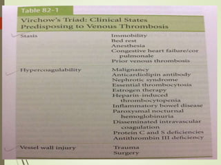
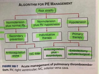
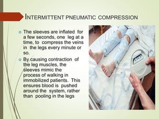
PULMONARY
EMBOLISM
PRESENTED BY
Dr. Marwan Abdel Rahman Sneymeh
DEFINITION
 It is an obstruction of the pulmonary artery or one
of its branches by a thrombus that
originates somewhere in the venous system or in
the right side of heart.
It is most common preventable cause of death
among hospitalized patient
EPIDEMOLOGY (INDIAN SCENARIO)
Overall, the annual incidence of PE
ranges between 23 and 69 cases per
100,000 population
Responsible for up to 15% of all inhospital
deaths
EPIDEMOLOGY (GLOBAL SCENARIO)
The incidence of venous thromboembolism(VTE), which
includes PE and deep venous thrombosis (DVT), has
remained relatively constant, with age- and sexadjusted
rates of 117 cases per 100 000 person-years.
VTE incidence rises sharply after age 60 in both men and
women, with PE accounting for the majority of the increase.
The mortality rate associated with PE is
underappreciated; it exceeds15% in the first 3 months
after diagnosis.
In nearly 25% of patients with PE, the initial clinical
manifestation is sudden death.
VIRCHOW’S
TRIAD
PREDISPOSING
FACTORS
PATHOPHYSIOLOGY
Virchow’s triad of
inflammation
Recruitment of
activated platelets
releasing micro
particle
Microparticle
containing
proinflammatory
mediator binds
neutrophils
Neutrophil releases
nuclear material
forming web like
extra cellular
network
Aggregation of
platelet and
platelet dependent
thrombin
generation
PATHOPHYSIOLOGY
HEREDITARY FACTORS
 Antithrombin III deficiency
 Protein C deficiency
 Protein S deficiency
 Factor V Leiden
 Plasminogen abnormality
 Fibrinogen abnormality
CLINICAL FEATURES
SYMPTOM LIST
 73% Dyspnea
 66% Pleuritc Pain
 43% Cough
 33% Leg Swelling
 30% Leg Pain
 15% Hemoptysis
 12% Palpitations
 10% Wheezing
 5% Angina-Like pain
DIAGNOSIS
 Dr. Wells demonstrated the utility of his scoring
system for determining the pre-test probability for PEs,
known now as the Wells Criteria. This study evaluated
946 patients, and based on the criteria, divided them
into low, moderate and high probability of having a
PE. These criteria included: clinical signs and
symptoms of DVT (3 points), PE as the most likely
diagnosis (3 points), tachycardia (1.5 points),
immobilization for at least 3 days or surgery within the
previous 4 weeks (1.5 points), previous objectively
diagnosed PE or DVT (1.5 points), hemoptysis (1
point), and malignancy (1point). Risk score
interpretation (probability of PE) was the following: >6
points: high risk (78.4%); 2 to 6 points: moderate risk
(27.8%);
Annals Intern Med 2001;135:98-107
ORIGINAL GENEVA CRITERIA
REVISED GENEVA CRITERIA
PERC RULE
 In 2004, Kline conducted a prospective study looking at eight
variables to rule out pulmonary embolism.
 The rule-out test (with poor specificity of 27% in low-risk
patients and 15% in very-low-risk patients)
 Pulmonary Embolism Rule out Criteria
(PERC) are as follows:
1. Age greater than or equal to 50 years
2. Heart rate greater than or equal to 100 beats per minute
3. Arterial oxygen saturation (SaO 2) on room air less than 95%
4. Venous thromboembolism
5. Recent (<28 days) trauma or surgery
6. Unilateral leg swelling
7. Hemoptysis
8. Oral hormone use
 Pulmonary embolism workup can be ruled out if
1. None of the above eight variables is positive.
2. A PERC evaluation is considered positive if any
one of the eight criteria are met.
 In 2015, pulmonary embolism guidelines were released by
the American College of Physicians and are summarizedas
follows .
1. Use either the Wells or Geneva rules to choose tests based on a patient's risk for
pulmonary embolism.
If the patient is at low risk, clinicians should use the eight PERC; if a patientdoes
not meet all eight criteria, the risks of testing are greater than the risk for
embolism, and no testing is needed.
For patients at intermediate risk, or for those at low risk who do not meet all of
the rule-out criteria, use a high-sensitivity plasma D-dimer test as the initial test.
In patients older than 50 years, use an age-adjusted threshold (age × 10 ng/mL,
rather than a blanket 500 ng/mL), because normal D-dimer levels increase with
age.
Patients with a D-dimer level below the age-adjusted cutoff should not receive
any imaging studies.
Patients with elevated D-dimer levels should then receive imaging.
Patients at high risk should skip the D-dimer test and proceed to CT pulmonary
angiography, because a negative D-dimer test will not eliminate the need for
imaging in these patients.
Clinicians should only obtain ventilation-perfusion scans in patients with a
contraindication to CT pulmonary angiography or if CT pulmonary angiographyis
unavailable.
2.
3.
4.
5.
6.
7.
8.
Skwarecki B. Pulmonary embolism guidelines
released by ACP. Medscape Medical News. WebMD
Inc. Sept 28, 2015.
CHEST X-RAY
Fleishner sign
Hampton hump
Westermark sign
FLEISHNER SIGN
ECG
D - DIMER
 D-dimer ELISA is an excellent screening test
for suspected PE
 A negative D-Dimer assay in low clinical
probability case rules out PE
 D-dimer ELISA was often elevated in the
absence of PE like sepsis ,cancer,acute medical
illness
 Low specificity and poor positive predictive
value
 Sensitivity >80% for DVT and >95% for PE
ECHO
 Right ventricular enlargement or
hypokinesis, especially free wall
hypokinesis, with sparing of the apex (the
McConnell sign)
 Interventricular septal flattening and
paradoxical motion toward the left
ventricle, resulting in a D-shaped left
ventricle in cross section
 Tricuspid regurgitation
 Direct visualization of
thrombus (more likely
with transesophageal
echocardiography
CT PULMONARY
ANGIOGRAPHY
 It is investigation of
choice
CT PULMONARY ANGIOGRAPHY
VENOUS
ULTRASONOGRAPHY
 Relies on loss of vein compressibility as the
primary criterion
 About 1/3 of pts will have no imaging
evidence of DVT
 Clot may have already embolized
 Clot present in the pelvic veins (U/S
usually inadequate)
 Workup for PE should continue even if
dopplers (-) in a pt in which you have a
high clinical suspicion
LUNG SCAN
 As many as 40% of pts with high clinical suspicion
for PE and low probability scans have a PE on
angiogram
 It has become second line diagnostic test for
patients who cannot tolerate intravenous
contrast
 Small particulate aggregates of albumin labelled
with gamma emitting radio nucleid is injected
 Ventilation can be obtained by radio labelled
inhaled gas such as xenon, krypton
 A high probability scan is defined as two or
more segmental perfusion defects in
presence of normal ventilation scan
 The diagnosis of PE is very unlikely in pt with
normal or near normal scan.
PULMONARY
ANGIOGRAM
Most specific test available for diagnosis of PE
Can detect emboli as small as 1-2 mm
Most useful when the clinical likelihood of PE
differs substantially from the lung scan result or
when the lung scan is intermediate probability
Definitive diagnosis is visualization of
an intraluminal filling defect .
Secondary sign is abrupt occlusion of
vessel, segmental oligemia,prolonged arterial
phase with slow filling .
MANAGEMENT
HEPARIN
 Choices include either intravenous
unfractionated heparin (UFH)
or subcutaneous low-molecular-weight
heparin (LMWH) preparations
 Initial intravenous bolus of 80 units of
heparin per kilogram followed by a
continuous infusion initiated at 18 units per
kilogram per hour.
 The heparin drip is adjusted based on
monitoring of the activated partial
thromboplastin time (aPPT), drawn 6hours
after initial bolus dose, then 6 hours after
each dose adjustment , with a target
aPPT ratio of 2.0 to 3.5
 More recently, an approach using a fixed
dose of subcutaneous unfractionated
heparin , administered as an initial dose of
333 U/kg followed by a dose of 250 U/kg
every 12 hours, has been demonstrated to
be as safe and effective as LMWH.
 Situations in which the use of UFH is
appropriate
1. Renal insufficiency.
2. Extremes of body weight.
3. Hypertensive crisis,
o Rapid adjustment of anticoagulation is
needed, such as…
A. Women in the late pregnancy who may need
caesarian sections,
B. Patients with recent surgery or
C. Recent history of bleeding
LMWH
 Advantages of LMWH compared with UFH include:
1. Longer half-life and ease of use
2. Ability to consistently achieve early therapeutic anticoagulation
3. No need to monitor anticoagulant effects
4. Reduced incidence of major bleeding complications
 In general, therapeutic monitoring is not needed with LMWH, but
there are situations where the therapeutic effetcts may be less
predictable and monitoring with ant-Xa levels is indicated
 Typical examples include:
1. Patients with antiphospholipid antibodies or other circulating
anticoagulants who have elevated baseline a PPT
2. Extremes of body weight(<40kgs and >50kgs)
3. Significant renal disease(creatinine clearance <30 ml/min)
4. Pregnancy
5. Unexplained bleeding or recurrent thrombosis during therapy
FONDAPARINUX
 A synthetic pentasaccharide, selective for
factor Xa
 By binding rapidly and strongly to antithrombin,
fondaparinux catalyzes specifically the inhibition
of factor Xa, which results in inhibition of
thrombin generation
 Half-life of approx 17 hours, and is exctreted
almost completely by the kidneys
 It does not cause heparin induced
thrombocytopenia
THROMBOLYTIC THERAPY
1. Streptokinase
2. Urokinase
3. rt PA
 these agents convert circulating plasminogen to plasmin.
 The preferred fibronolytic regimen is 100mg rt PAover 2
hour.
Contraindication :
 Intracranial disease
 Recent surgery
 Trauma
 The exact role of thrombolytic agents in acute
pulmonary embolim remains controversial. While
thrombolytic therapy does appear to accelerate the
rate of thrombolysis, there is no convincing evidence
to suggest that
 It decreaes mortality,
 Increases the ultimate extent of embolic resolution when
measured at 7 days,
 Reduces thromboembolic recurrence rates,
 improves symptomatic outcomes,
 Decreases the incidence of thromboembolic
pulmonary hypertension
 Catheter- directed techniques have been successfully
employed in the setting of acute ileo- femoral DVT
using doses of urokinase ranging from 1.4 to 16
million units delivered over an
average of 30 hours.
INTERVENTIONA
L
RADIOLOGIC TE
CHNIQUES
 Interventional thrombus
fragmentation.
 The devices use either
pressured saline or
a rotating impeller to
fragment central
thrombi.
 The fragments are either
aspirated through a separate
port on the catheter or
allowed to migrate distally.
 There limitations, including
a risk of
paradoxical embolism from
the clot fragments
PULMONARY EMBOLECTOMY
 It is reasonable to consider surgical embolectomy in
patients with
1. Persistent hypotension.
2. Shock.
3. Cardiac arrest.
4. Failed thrombolysis.
5. Contraindications to
thrombolytics.
ORAL
ANTICOAGULANTS
 Warfarin inhibits gamma carboxylation
activation of coagulation factors II, VII,
IX, and X as well as proteins C and S
 Use of Warfarin without heparin
is strongly discouraged
 It generally takes 3 to 5 days
of warfarin to achieve full
therapeutic eficacy.
 In patients with protein C deficiency,
skin necrosis or paradoxical
thrombosis may occur.
WARFARIN
 INR range between 2 and 3 is
recommended for most patients
 Another rare complication of warfarin
use is cholesterol microembolism
(“purple toes” syndrome), which
is thought to be due to cholesterol
crystal release from ulcerated
intravascular plaques
 an initial daily dose of 5 or 10 mg with
use of an standardized nomogram to
dose adjust based on INR values
obtained on days 3 and 5
 patients should receive atleast 5 days of
combined heparin and warfaein therapy,
including atleast 2 days in which the INR
is in a therapeutic range prior to
stopping heparin
 Because of the teratogenic potential of
warfarin, UFH or LMWH should be used
in pregnant women who developed VTE
in the first trimesters
DURATION
OF
THERAPY
 The risk of recurrent disease after 3 months
of anticoagulation is still in the region of
10%; therefore, the patients should
be treated with warfarin for 3 to 6 months
 Patients with idiopathic thromboembolism
have higher rate of recurrence therefore
treated for atleast 6 to 12 months
 Patients with antiphospholipid antibody
syndrome have considerable risk of
recurrence, therefore treated for
minimum 12 months with consideration of
life-long therapy
 In patients with >2 episodes of recurrence
life-long anticoagulation given
DEEP VEIN THROMBOSIS
 Thrombolysis
 Anticoagulation
 Vena cava filter
VENA CAVAFILTER
 Used in patients with contraindication to
anticoagulation or patients
with recurrent embolism while on adequate
anticoagulation
 Long term IVC filter increases risk of
thromboembolism
 This observation lead to recent development of
retrievable filters
 Four types of retrievable filters:
1. Gunther tulip filter
2. ALN filter
3. Recovery filter
4. OptEase filter
VENA CAVAFILTER
PROPHYLAXIS
INTERMITTENT PNEUMATIC COMPRESSION
 The sleeves are inflated for
a few seconds, one leg at a
time, to compress the veins
in the legs every minute or
so.
 By causing contraction of
the leg muscles, the
sleeves mimic the
process of walking in
immobilized patients. This
ensures blood is pushed
around the system, rather
than pooling in the legs
GRADUATED COMPRESSION STOCKING
 Compression stockings
are made of a special
elastic fabric. They are very
tight at the ankle and are
less tight as the stocking
moves up the leg. This
graduated tightness helps
the leg muscles squeeze
fluid up the leg, which
improves blood flow from the
leg back to the heart and
decreases leg swelling and
pain.
TYPE OF
EMBOLISM
Fat embolism
Venous air embolism
Amniotic fluid embolism
Septic embolism
Tumour embolism
Sickle cell disease
FAT EMBOLISM
 Due to entry of neutral at in vascular system
 Leads to dyspnea, hypoxemia, petechiae, mental confusion
 Lag time of 24 to 72 hours in the onset of syndrome following
the inciting event
 Most common inciting event, fracture of long bones
 Pathophysiology :
 Actual vascular obstruction by neutral particle of fat
 Injurious effect of free fatty acids released by the action of lipases
on the neutral fat
 Supportive treatment advised
 Other suggested treatment intravenous ethanol, albumin, dextran,
heparin
VENOUS AIR EMBOLISM
 Due to indwelling central venous catheter, positive pressure
ventilation, trauma to thorax
 Physiological consequence due to abrupt rise in pulmonary
artery pressure
 Treatment is prevention and early detection
 Patient positioning (Trendelenburg position with left side
down)
 Removal of air through central venous catheter
 Direct needle aspiration
 Closed chest cardiac massage
 Increase absorption with use of 100% oxygen
AMNIOTIC FLUID EMBOLISM
 Third leading cause of maternal mortality
 Amniotic fluid contains particulate material that can
cause pulmonary vascular obstruction
 Amniotic fluid has thromboplastic activity that leads
to extensive fibrin deposition in lung vasculature
that leads to consumptive coagulopathy leading to
hypofibrinogenemia and thrombocytopenia
 Presence of squamous cell in pulmonary arterial
blood once considered pathognomonic.
SEPTIC EMBOLISM
 Microscopically septic phlebitis consist of
purulent material mixed with fibrin thrombus
 Chest X-ray display pulmonary infiltrates
 Treatment consists of anti microbial agents
TUMOUR EMBOLISM
 Most common site of origin breast, lungs,
prostate, stomach and liver
 Clinical feature is typically subacute and involves
progressive dyspnea, tachypnea, tachycardia
 Pulmonary angiographic findings reveal
delayed vascular filling, pruning and
tortuosity
Pulmonary Embolism lecture 2022

Pulmonary Embolism lecture 2022

  • 1.
  • 2.
    DEFINITION  It isan obstruction of the pulmonary artery or one of its branches by a thrombus that originates somewhere in the venous system or in the right side of heart. It is most common preventable cause of death among hospitalized patient
  • 3.
    EPIDEMOLOGY (INDIAN SCENARIO) Overall,the annual incidence of PE ranges between 23 and 69 cases per 100,000 population Responsible for up to 15% of all inhospital deaths
  • 4.
    EPIDEMOLOGY (GLOBAL SCENARIO) Theincidence of venous thromboembolism(VTE), which includes PE and deep venous thrombosis (DVT), has remained relatively constant, with age- and sexadjusted rates of 117 cases per 100 000 person-years. VTE incidence rises sharply after age 60 in both men and women, with PE accounting for the majority of the increase. The mortality rate associated with PE is underappreciated; it exceeds15% in the first 3 months after diagnosis. In nearly 25% of patients with PE, the initial clinical manifestation is sudden death.
  • 5.
  • 6.
  • 7.
    PATHOPHYSIOLOGY Virchow’s triad of inflammation Recruitmentof activated platelets releasing micro particle Microparticle containing proinflammatory mediator binds neutrophils Neutrophil releases nuclear material forming web like extra cellular network Aggregation of platelet and platelet dependent thrombin generation
  • 8.
  • 9.
    HEREDITARY FACTORS  AntithrombinIII deficiency  Protein C deficiency  Protein S deficiency  Factor V Leiden  Plasminogen abnormality  Fibrinogen abnormality
  • 10.
  • 11.
    SYMPTOM LIST  73%Dyspnea  66% Pleuritc Pain  43% Cough  33% Leg Swelling  30% Leg Pain  15% Hemoptysis  12% Palpitations  10% Wheezing  5% Angina-Like pain
  • 12.
  • 13.
     Dr. Wellsdemonstrated the utility of his scoring system for determining the pre-test probability for PEs, known now as the Wells Criteria. This study evaluated 946 patients, and based on the criteria, divided them into low, moderate and high probability of having a PE. These criteria included: clinical signs and symptoms of DVT (3 points), PE as the most likely diagnosis (3 points), tachycardia (1.5 points), immobilization for at least 3 days or surgery within the previous 4 weeks (1.5 points), previous objectively diagnosed PE or DVT (1.5 points), hemoptysis (1 point), and malignancy (1point). Risk score interpretation (probability of PE) was the following: >6 points: high risk (78.4%); 2 to 6 points: moderate risk (27.8%); Annals Intern Med 2001;135:98-107
  • 15.
  • 16.
  • 17.
    PERC RULE  In2004, Kline conducted a prospective study looking at eight variables to rule out pulmonary embolism.  The rule-out test (with poor specificity of 27% in low-risk patients and 15% in very-low-risk patients)  Pulmonary Embolism Rule out Criteria (PERC) are as follows: 1. Age greater than or equal to 50 years 2. Heart rate greater than or equal to 100 beats per minute 3. Arterial oxygen saturation (SaO 2) on room air less than 95% 4. Venous thromboembolism 5. Recent (<28 days) trauma or surgery 6. Unilateral leg swelling 7. Hemoptysis 8. Oral hormone use
  • 18.
     Pulmonary embolismworkup can be ruled out if 1. None of the above eight variables is positive. 2. A PERC evaluation is considered positive if any one of the eight criteria are met.
  • 19.
     In 2015,pulmonary embolism guidelines were released by the American College of Physicians and are summarizedas follows . 1. Use either the Wells or Geneva rules to choose tests based on a patient's risk for pulmonary embolism. If the patient is at low risk, clinicians should use the eight PERC; if a patientdoes not meet all eight criteria, the risks of testing are greater than the risk for embolism, and no testing is needed. For patients at intermediate risk, or for those at low risk who do not meet all of the rule-out criteria, use a high-sensitivity plasma D-dimer test as the initial test. In patients older than 50 years, use an age-adjusted threshold (age × 10 ng/mL, rather than a blanket 500 ng/mL), because normal D-dimer levels increase with age. Patients with a D-dimer level below the age-adjusted cutoff should not receive any imaging studies. Patients with elevated D-dimer levels should then receive imaging. Patients at high risk should skip the D-dimer test and proceed to CT pulmonary angiography, because a negative D-dimer test will not eliminate the need for imaging in these patients. Clinicians should only obtain ventilation-perfusion scans in patients with a contraindication to CT pulmonary angiography or if CT pulmonary angiographyis unavailable. 2. 3. 4. 5. 6. 7. 8. Skwarecki B. Pulmonary embolism guidelines released by ACP. Medscape Medical News. WebMD Inc. Sept 28, 2015.
  • 22.
  • 23.
  • 29.
  • 30.
    D - DIMER D-dimer ELISA is an excellent screening test for suspected PE  A negative D-Dimer assay in low clinical probability case rules out PE  D-dimer ELISA was often elevated in the absence of PE like sepsis ,cancer,acute medical illness  Low specificity and poor positive predictive value  Sensitivity >80% for DVT and >95% for PE
  • 31.
    ECHO  Right ventricularenlargement or hypokinesis, especially free wall hypokinesis, with sparing of the apex (the McConnell sign)  Interventricular septal flattening and paradoxical motion toward the left ventricle, resulting in a D-shaped left ventricle in cross section  Tricuspid regurgitation  Direct visualization of thrombus (more likely with transesophageal echocardiography
  • 32.
    CT PULMONARY ANGIOGRAPHY  Itis investigation of choice
  • 33.
  • 34.
    VENOUS ULTRASONOGRAPHY  Relies onloss of vein compressibility as the primary criterion  About 1/3 of pts will have no imaging evidence of DVT  Clot may have already embolized  Clot present in the pelvic veins (U/S usually inadequate)  Workup for PE should continue even if dopplers (-) in a pt in which you have a high clinical suspicion
  • 38.
    LUNG SCAN  Asmany as 40% of pts with high clinical suspicion for PE and low probability scans have a PE on angiogram  It has become second line diagnostic test for patients who cannot tolerate intravenous contrast  Small particulate aggregates of albumin labelled with gamma emitting radio nucleid is injected  Ventilation can be obtained by radio labelled inhaled gas such as xenon, krypton  A high probability scan is defined as two or more segmental perfusion defects in presence of normal ventilation scan  The diagnosis of PE is very unlikely in pt with normal or near normal scan.
  • 41.
    PULMONARY ANGIOGRAM Most specific testavailable for diagnosis of PE Can detect emboli as small as 1-2 mm Most useful when the clinical likelihood of PE differs substantially from the lung scan result or when the lung scan is intermediate probability Definitive diagnosis is visualization of an intraluminal filling defect . Secondary sign is abrupt occlusion of vessel, segmental oligemia,prolonged arterial phase with slow filling .
  • 44.
  • 46.
    HEPARIN  Choices includeeither intravenous unfractionated heparin (UFH) or subcutaneous low-molecular-weight heparin (LMWH) preparations  Initial intravenous bolus of 80 units of heparin per kilogram followed by a continuous infusion initiated at 18 units per kilogram per hour.  The heparin drip is adjusted based on monitoring of the activated partial thromboplastin time (aPPT), drawn 6hours after initial bolus dose, then 6 hours after each dose adjustment , with a target aPPT ratio of 2.0 to 3.5  More recently, an approach using a fixed dose of subcutaneous unfractionated heparin , administered as an initial dose of 333 U/kg followed by a dose of 250 U/kg every 12 hours, has been demonstrated to be as safe and effective as LMWH.
  • 47.
     Situations inwhich the use of UFH is appropriate 1. Renal insufficiency. 2. Extremes of body weight. 3. Hypertensive crisis, o Rapid adjustment of anticoagulation is needed, such as… A. Women in the late pregnancy who may need caesarian sections, B. Patients with recent surgery or C. Recent history of bleeding
  • 48.
    LMWH  Advantages ofLMWH compared with UFH include: 1. Longer half-life and ease of use 2. Ability to consistently achieve early therapeutic anticoagulation 3. No need to monitor anticoagulant effects 4. Reduced incidence of major bleeding complications  In general, therapeutic monitoring is not needed with LMWH, but there are situations where the therapeutic effetcts may be less predictable and monitoring with ant-Xa levels is indicated  Typical examples include: 1. Patients with antiphospholipid antibodies or other circulating anticoagulants who have elevated baseline a PPT 2. Extremes of body weight(<40kgs and >50kgs) 3. Significant renal disease(creatinine clearance <30 ml/min) 4. Pregnancy 5. Unexplained bleeding or recurrent thrombosis during therapy
  • 49.
    FONDAPARINUX  A syntheticpentasaccharide, selective for factor Xa  By binding rapidly and strongly to antithrombin, fondaparinux catalyzes specifically the inhibition of factor Xa, which results in inhibition of thrombin generation  Half-life of approx 17 hours, and is exctreted almost completely by the kidneys  It does not cause heparin induced thrombocytopenia
  • 50.
    THROMBOLYTIC THERAPY 1. Streptokinase 2.Urokinase 3. rt PA  these agents convert circulating plasminogen to plasmin.  The preferred fibronolytic regimen is 100mg rt PAover 2 hour. Contraindication :  Intracranial disease  Recent surgery  Trauma
  • 51.
     The exactrole of thrombolytic agents in acute pulmonary embolim remains controversial. While thrombolytic therapy does appear to accelerate the rate of thrombolysis, there is no convincing evidence to suggest that  It decreaes mortality,  Increases the ultimate extent of embolic resolution when measured at 7 days,  Reduces thromboembolic recurrence rates,  improves symptomatic outcomes,  Decreases the incidence of thromboembolic pulmonary hypertension  Catheter- directed techniques have been successfully employed in the setting of acute ileo- femoral DVT using doses of urokinase ranging from 1.4 to 16 million units delivered over an average of 30 hours.
  • 52.
    INTERVENTIONA L RADIOLOGIC TE CHNIQUES  Interventionalthrombus fragmentation.  The devices use either pressured saline or a rotating impeller to fragment central thrombi.  The fragments are either aspirated through a separate port on the catheter or allowed to migrate distally.  There limitations, including a risk of paradoxical embolism from the clot fragments
  • 53.
    PULMONARY EMBOLECTOMY  Itis reasonable to consider surgical embolectomy in patients with 1. Persistent hypotension. 2. Shock. 3. Cardiac arrest. 4. Failed thrombolysis. 5. Contraindications to thrombolytics.
  • 54.
    ORAL ANTICOAGULANTS  Warfarin inhibitsgamma carboxylation activation of coagulation factors II, VII, IX, and X as well as proteins C and S  Use of Warfarin without heparin is strongly discouraged  It generally takes 3 to 5 days of warfarin to achieve full therapeutic eficacy.  In patients with protein C deficiency, skin necrosis or paradoxical thrombosis may occur.
  • 55.
    WARFARIN  INR rangebetween 2 and 3 is recommended for most patients  Another rare complication of warfarin use is cholesterol microembolism (“purple toes” syndrome), which is thought to be due to cholesterol crystal release from ulcerated intravascular plaques  an initial daily dose of 5 or 10 mg with use of an standardized nomogram to dose adjust based on INR values obtained on days 3 and 5  patients should receive atleast 5 days of combined heparin and warfaein therapy, including atleast 2 days in which the INR is in a therapeutic range prior to stopping heparin  Because of the teratogenic potential of warfarin, UFH or LMWH should be used in pregnant women who developed VTE in the first trimesters
  • 56.
    DURATION OF THERAPY  The riskof recurrent disease after 3 months of anticoagulation is still in the region of 10%; therefore, the patients should be treated with warfarin for 3 to 6 months  Patients with idiopathic thromboembolism have higher rate of recurrence therefore treated for atleast 6 to 12 months  Patients with antiphospholipid antibody syndrome have considerable risk of recurrence, therefore treated for minimum 12 months with consideration of life-long therapy  In patients with >2 episodes of recurrence life-long anticoagulation given
  • 57.
    DEEP VEIN THROMBOSIS Thrombolysis  Anticoagulation  Vena cava filter
  • 58.
    VENA CAVAFILTER  Usedin patients with contraindication to anticoagulation or patients with recurrent embolism while on adequate anticoagulation  Long term IVC filter increases risk of thromboembolism  This observation lead to recent development of retrievable filters  Four types of retrievable filters: 1. Gunther tulip filter 2. ALN filter 3. Recovery filter 4. OptEase filter
  • 59.
  • 60.
  • 61.
    INTERMITTENT PNEUMATIC COMPRESSION The sleeves are inflated for a few seconds, one leg at a time, to compress the veins in the legs every minute or so.  By causing contraction of the leg muscles, the sleeves mimic the process of walking in immobilized patients. This ensures blood is pushed around the system, rather than pooling in the legs
  • 62.
    GRADUATED COMPRESSION STOCKING Compression stockings are made of a special elastic fabric. They are very tight at the ankle and are less tight as the stocking moves up the leg. This graduated tightness helps the leg muscles squeeze fluid up the leg, which improves blood flow from the leg back to the heart and decreases leg swelling and pain.
  • 64.
    TYPE OF EMBOLISM Fat embolism Venousair embolism Amniotic fluid embolism Septic embolism Tumour embolism Sickle cell disease
  • 65.
    FAT EMBOLISM  Dueto entry of neutral at in vascular system  Leads to dyspnea, hypoxemia, petechiae, mental confusion  Lag time of 24 to 72 hours in the onset of syndrome following the inciting event  Most common inciting event, fracture of long bones  Pathophysiology :  Actual vascular obstruction by neutral particle of fat  Injurious effect of free fatty acids released by the action of lipases on the neutral fat  Supportive treatment advised  Other suggested treatment intravenous ethanol, albumin, dextran, heparin
  • 66.
    VENOUS AIR EMBOLISM Due to indwelling central venous catheter, positive pressure ventilation, trauma to thorax  Physiological consequence due to abrupt rise in pulmonary artery pressure  Treatment is prevention and early detection  Patient positioning (Trendelenburg position with left side down)  Removal of air through central venous catheter  Direct needle aspiration  Closed chest cardiac massage  Increase absorption with use of 100% oxygen
  • 67.
    AMNIOTIC FLUID EMBOLISM Third leading cause of maternal mortality  Amniotic fluid contains particulate material that can cause pulmonary vascular obstruction  Amniotic fluid has thromboplastic activity that leads to extensive fibrin deposition in lung vasculature that leads to consumptive coagulopathy leading to hypofibrinogenemia and thrombocytopenia  Presence of squamous cell in pulmonary arterial blood once considered pathognomonic.
  • 68.
    SEPTIC EMBOLISM  Microscopicallyseptic phlebitis consist of purulent material mixed with fibrin thrombus  Chest X-ray display pulmonary infiltrates  Treatment consists of anti microbial agents
  • 69.
    TUMOUR EMBOLISM  Mostcommon site of origin breast, lungs, prostate, stomach and liver  Clinical feature is typically subacute and involves progressive dyspnea, tachypnea, tachycardia  Pulmonary angiographic findings reveal delayed vascular filling, pruning and tortuosity