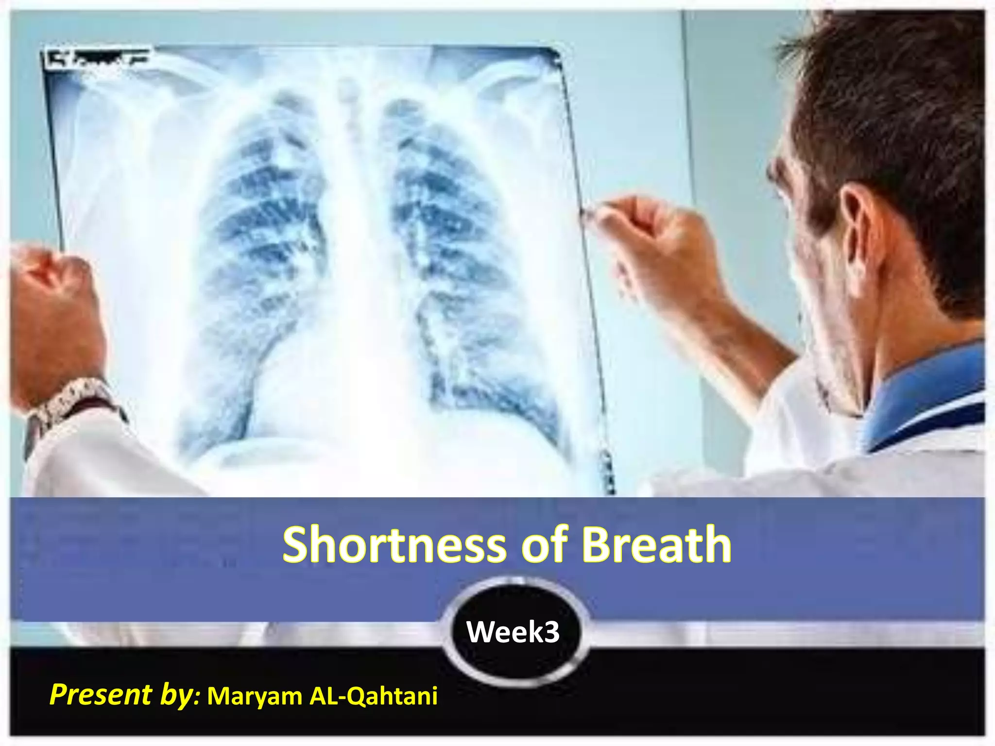
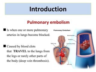
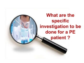
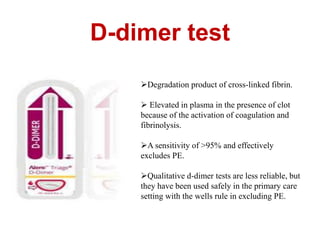
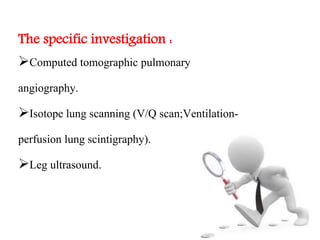
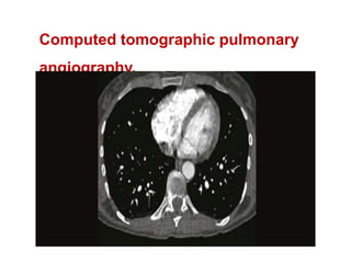
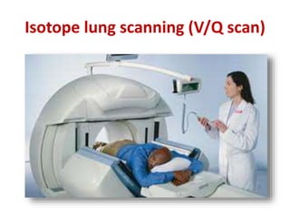
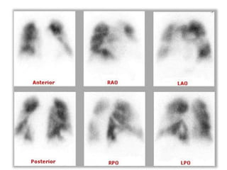
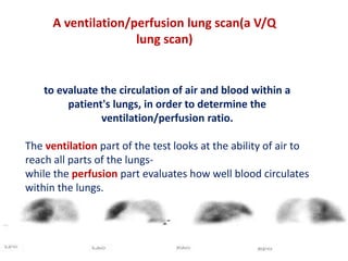
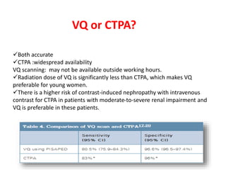
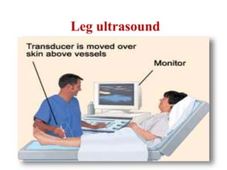
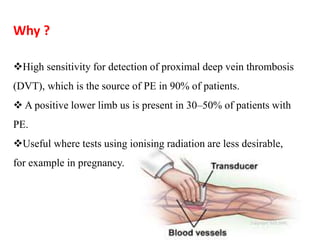
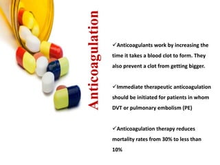
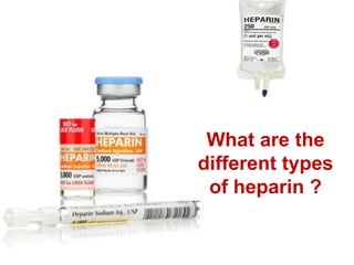
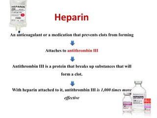
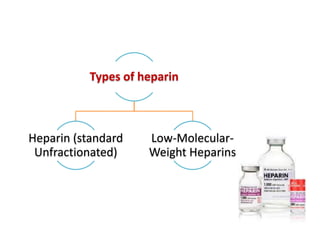
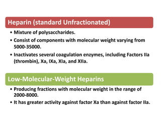
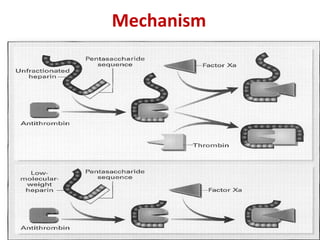
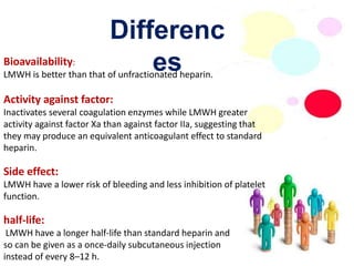
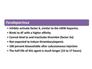
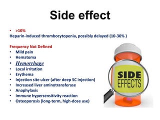
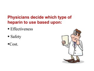
Present by: Maryam AL-Qahtani 
Week3
Introduction 
Pulmonary embolism 
Is when one or more pulmonary 
arteries in lungs become blocked. 
Caused by blood clots 
that TRAVEL to the lungs from 
the legs or rarely other parts of 
the body (deep vein thrombosis).
Objectiv 
eWsha:t are the specific investigation to be done for a PE 
patient ? 
What are the anticoagulation agent which can be given 
in pulmonary embolism as a prophylactic or therapeutic ? 
What are the different types of heparin ?
What are the 
specific 
investigation to be 
done for a PE 
patient ?
The choice and order of investigations will 
depend on 
The clinical likelihood of PE. 
How ill the patient is (stable or unstable). 
Availability of the test.
D-dimer test 
Degradation product of cross-linked fibrin. 
 Elevated in plasma in the presence of clot 
because of the activation of coagulation and 
fibrinolysis. 
A sensitivity of >95% and effectively 
excludes PE. 
Qualitative d-dimer tests are less reliable, but 
they have been used safely in the primary care 
setting with the wells rule in excluding PE.
The specific investigation : 
Computed tomographic pulmonary 
angiography. 
Isotope lung scanning (V/Q scan;Ventilation-perfusion 
lung scintigraphy). 
Leg ultrasound.
Computed tomographic pulmonary 
angiography.
CTPA has become the method of choice for imaging the 
pulmonary vasculature in patients with suspected PE 
Why ? 
Its wider availability 
Ability to demonstrate alternative causes of symptoms. 
Fast and generally well tolerated 
Has superior sensitivity for the detection of small 
subsegmental emboli (detects clots in smaller vessels.)
Isotope lung scanning (V/Q scan)
A ventilation/perfusion lung scan(a V/Q 
to evaluate the circulation of air and blood within a 
patient's lungs, in order to determine the 
ventilation/perfusion ratio. 
The ventilation part of the test looks at the ability of air to 
reach all parts of the lungs-while 
the perfusion part evaluates how well blood circulates 
within the lungs. 
lung scan)
VQ or CTPA? 
Both accurate 
CTPA :widespread availability 
VQ scanning: may not be available outside working hours. 
Radiation dose of VQ is significantly less than CTPA, which makes VQ 
preferable for young women. 
There is a higher risk of contrast-induced nephropathy with intravenous 
contrast for CTPA in patients with moderate-to-severe renal impairment and 
VQ is preferable in these patients.
Leg ultrasound
Why ? 
High sensitivity for detection of proximal deep vein thrombosis 
(DVT), which is the source of PE in 90% of patients. 
 A positive lower limb us is present in 30–50% of patients with 
PE. 
Useful where tests using ionising radiation are less desirable, 
for example in pregnancy.
What are the anticoagulation agent 
which can be given in pulmonary 
embolism as a prophylactic or 
therapeutic ?
Anticoagulation 
Anticoagulants work by increasing the 
time it takes a blood clot to form. They 
also prevent a clot from getting bigger. 
Immediate therapeutic anticoagulation 
should be initiated for patients in whom 
DVT or pulmonary embolism (PE) 
Anticoagulation therapy reduces 
mortality rates from 30% to less than 
10%
Prophylactic anticoagulation agents: 
• Most hospitalized patients developed VTE. 
• The estimated risk factor is high as 50% when 
thromboporphylaxis is not used. 
Prophylaxis measurements 
• Early mobilization. 
• Elevation of the legs. 
• Compression stocking. 
• Intermittent compression device. 
• Use of drug such as : LMWH , and Anti-thrombin.
Therapeutics anticoagulation agents: 
Aim of Treatment: 
prevent further thrombosis and PE while resolution of venous 
thrombi occur by natural fibrinolytic activity. 
1. Start w. : Heparin as its produced direct effect, For 5 Days 
2. LMWH e.g. (Tinzaparine 175 U/kg daily, Delteparin 200 U/kg daily, 
Enoxaprain 1.5 mg/kg daily) is effective and safe for immediate treatment as 
unfractionated heparin. 
3. At the follow up : For Long –term medication will use warfarin to maintain a 
target INR of 2.0-3.0
Length of Anti-Coagulation 
• 6 weeks for isolated calf vein thrombosis. 
• 3 months after participated proximal DVT or PE in 
patient with temporary risk factors. 
• 3 to 6 months in idiopathic VTE or permanent risk 
factors.
Outpatient 
• Best to be supervised in anticoagulant clinics. 
• IVC filters are an important tool to prevent PE in patients that 
have a contraindication to anticoagulation.
Summary : 
• Start the therapy by Heparin or low molecular 
weight heparin (LMWH) followed by … 
• 6 months Warfarin with an international normal 
ratio of 2 to 3.
What are the 
different types 
of heparin ?
Heparin 
An anticoagulant or a medication that prevents clots from forming 
Attaches to antithrombin III 
Antithrombin III is a protein that breaks up substances that will 
form a clot. 
With heparin attached to it, antithrombin III is 1,000 times more 
effective
Types of heparin 
Heparin (standard 
Unfractionated) 
Low-Molecular- 
Weight Heparins
Heparin (standard Unfractionated) 
• Mixture of polysaccharides. 
• Consist of components with molecular weight varying from 
5000-35000. 
• Inactivates several coagulation enzymes, including Factors IIa 
(thrombin), Xa, IXa, XIa, and XIIa. 
Low-Molecular-Weight Heparins 
• Producing fractions with molecular weight in the range of 
2000-8000. 
• It has greater activity against factor Xa than against factor IIa.
Mechanism
Differenc 
Bioavailability: es 
LMWH is better than that of unfractionated heparin. 
Activity against factor: 
Inactivates several coagulation enzymes while LMWH greater 
activity against factor Xa than against factor IIa, suggesting that 
they may produce an equivalent anticoagulant effect to standard 
heparin. 
Side effect: 
LMWH have a lower risk of bleeding and less inhibition of platelet 
function. 
half-life: 
LMWH have a longer half-life than standard heparin and 
so can be given as a once-daily subcutaneous injection 
instead of every 8–12 h.
Fondaparinux 
• Inhibits activate factor X, similar to the LMW heparins. 
• Binds to AT with a higher affinity 
• Cannot bind to and inactivate thrombin (factor IIa) 
• Not expected to induce thrombocytopenia 
• 100 percent bioavailable after subcutaneous injection 
• The half-life of this agent is much longer (15 to 17 hours)
Side effect 
• >10% 
Heparin-induced thrombocytopenia, possibly delayed (10-30% ) 
Frequency Not Defined 
• Mild pain 
• Hematoma 
• Hemorrhage 
• Local irritation 
• Erythema 
• Injection site ulcer (after deep SC injection) 
• Increased liver aminotransferase 
• Anaphylaxis 
• Immune hypersensitivity reaction 
• Osteoporosis (long-term, high-dose use)
Physicians decide which type of 
heparin to use based upon: 
 Effectiveness 
 Safety 
Cost.
References 
 Merck Manual. 
 .Up to data.website 
 Kumar and Clark book 
 http://atvb.ahajournals.org/content/21/7/1094.long 
 http://www.livestrong.com/article/252722-types-of-heparin/ 
 http://www.thrombosisadviser.com/en/vte-prevention/ 
heparins/ 
 http://emedicine.medscape.com/article/300901-treatment 
 http://reference.medscape.com/drug/calciparine-monoparin- 
heparin-342169#4
Pulmonary embolism

Pulmonary embolism

  • 1.
    Present by: MaryamAL-Qahtani Week3
  • 2.
    Introduction Pulmonary embolism Is when one or more pulmonary arteries in lungs become blocked. Caused by blood clots that TRAVEL to the lungs from the legs or rarely other parts of the body (deep vein thrombosis).
  • 3.
    Objectiv eWsha:t arethe specific investigation to be done for a PE patient ? What are the anticoagulation agent which can be given in pulmonary embolism as a prophylactic or therapeutic ? What are the different types of heparin ?
  • 4.
    What are the specific investigation to be done for a PE patient ?
  • 5.
    The choice andorder of investigations will depend on The clinical likelihood of PE. How ill the patient is (stable or unstable). Availability of the test.
  • 6.
    D-dimer test Degradationproduct of cross-linked fibrin.  Elevated in plasma in the presence of clot because of the activation of coagulation and fibrinolysis. A sensitivity of >95% and effectively excludes PE. Qualitative d-dimer tests are less reliable, but they have been used safely in the primary care setting with the wells rule in excluding PE.
  • 7.
    The specific investigation: Computed tomographic pulmonary angiography. Isotope lung scanning (V/Q scan;Ventilation-perfusion lung scintigraphy). Leg ultrasound.
  • 8.
  • 9.
    CTPA has becomethe method of choice for imaging the pulmonary vasculature in patients with suspected PE Why ? Its wider availability Ability to demonstrate alternative causes of symptoms. Fast and generally well tolerated Has superior sensitivity for the detection of small subsegmental emboli (detects clots in smaller vessels.)
  • 10.
  • 12.
    A ventilation/perfusion lungscan(a V/Q to evaluate the circulation of air and blood within a patient's lungs, in order to determine the ventilation/perfusion ratio. The ventilation part of the test looks at the ability of air to reach all parts of the lungs-while the perfusion part evaluates how well blood circulates within the lungs. lung scan)
  • 13.
    VQ or CTPA? Both accurate CTPA :widespread availability VQ scanning: may not be available outside working hours. Radiation dose of VQ is significantly less than CTPA, which makes VQ preferable for young women. There is a higher risk of contrast-induced nephropathy with intravenous contrast for CTPA in patients with moderate-to-severe renal impairment and VQ is preferable in these patients.
  • 14.
  • 15.
    Why ? Highsensitivity for detection of proximal deep vein thrombosis (DVT), which is the source of PE in 90% of patients.  A positive lower limb us is present in 30–50% of patients with PE. Useful where tests using ionising radiation are less desirable, for example in pregnancy.
  • 16.
    What are theanticoagulation agent which can be given in pulmonary embolism as a prophylactic or therapeutic ?
  • 17.
    Anticoagulation Anticoagulants workby increasing the time it takes a blood clot to form. They also prevent a clot from getting bigger. Immediate therapeutic anticoagulation should be initiated for patients in whom DVT or pulmonary embolism (PE) Anticoagulation therapy reduces mortality rates from 30% to less than 10%
  • 18.
    Prophylactic anticoagulation agents: • Most hospitalized patients developed VTE. • The estimated risk factor is high as 50% when thromboporphylaxis is not used. Prophylaxis measurements • Early mobilization. • Elevation of the legs. • Compression stocking. • Intermittent compression device. • Use of drug such as : LMWH , and Anti-thrombin.
  • 19.
    Therapeutics anticoagulation agents: Aim of Treatment: prevent further thrombosis and PE while resolution of venous thrombi occur by natural fibrinolytic activity. 1. Start w. : Heparin as its produced direct effect, For 5 Days 2. LMWH e.g. (Tinzaparine 175 U/kg daily, Delteparin 200 U/kg daily, Enoxaprain 1.5 mg/kg daily) is effective and safe for immediate treatment as unfractionated heparin. 3. At the follow up : For Long –term medication will use warfarin to maintain a target INR of 2.0-3.0
  • 20.
    Length of Anti-Coagulation • 6 weeks for isolated calf vein thrombosis. • 3 months after participated proximal DVT or PE in patient with temporary risk factors. • 3 to 6 months in idiopathic VTE or permanent risk factors.
  • 21.
    Outpatient • Bestto be supervised in anticoagulant clinics. • IVC filters are an important tool to prevent PE in patients that have a contraindication to anticoagulation.
  • 22.
    Summary : •Start the therapy by Heparin or low molecular weight heparin (LMWH) followed by … • 6 months Warfarin with an international normal ratio of 2 to 3.
  • 23.
    What are the different types of heparin ?
  • 24.
    Heparin An anticoagulantor a medication that prevents clots from forming Attaches to antithrombin III Antithrombin III is a protein that breaks up substances that will form a clot. With heparin attached to it, antithrombin III is 1,000 times more effective
  • 25.
    Types of heparin Heparin (standard Unfractionated) Low-Molecular- Weight Heparins
  • 26.
    Heparin (standard Unfractionated) • Mixture of polysaccharides. • Consist of components with molecular weight varying from 5000-35000. • Inactivates several coagulation enzymes, including Factors IIa (thrombin), Xa, IXa, XIa, and XIIa. Low-Molecular-Weight Heparins • Producing fractions with molecular weight in the range of 2000-8000. • It has greater activity against factor Xa than against factor IIa.
  • 27.
  • 28.
    Differenc Bioavailability: es LMWH is better than that of unfractionated heparin. Activity against factor: Inactivates several coagulation enzymes while LMWH greater activity against factor Xa than against factor IIa, suggesting that they may produce an equivalent anticoagulant effect to standard heparin. Side effect: LMWH have a lower risk of bleeding and less inhibition of platelet function. half-life: LMWH have a longer half-life than standard heparin and so can be given as a once-daily subcutaneous injection instead of every 8–12 h.
  • 29.
    Fondaparinux • Inhibitsactivate factor X, similar to the LMW heparins. • Binds to AT with a higher affinity • Cannot bind to and inactivate thrombin (factor IIa) • Not expected to induce thrombocytopenia • 100 percent bioavailable after subcutaneous injection • The half-life of this agent is much longer (15 to 17 hours)
  • 30.
    Side effect •>10% Heparin-induced thrombocytopenia, possibly delayed (10-30% ) Frequency Not Defined • Mild pain • Hematoma • Hemorrhage • Local irritation • Erythema • Injection site ulcer (after deep SC injection) • Increased liver aminotransferase • Anaphylaxis • Immune hypersensitivity reaction • Osteoporosis (long-term, high-dose use)
  • 31.
    Physicians decide whichtype of heparin to use based upon:  Effectiveness  Safety Cost.
  • 33.
    References  MerckManual.  .Up to data.website  Kumar and Clark book  http://atvb.ahajournals.org/content/21/7/1094.long  http://www.livestrong.com/article/252722-types-of-heparin/  http://www.thrombosisadviser.com/en/vte-prevention/ heparins/  http://emedicine.medscape.com/article/300901-treatment  http://reference.medscape.com/drug/calciparine-monoparin- heparin-342169#4

Editor's Notes

  • #6 The choice and order of investigations will depend on the clinical likelihood of PE, how ill the patient is and availability of the test. The 'gold standard' test is conventional pulmonary angiography, but this is invasive and usually unavailable urgently. Investigation strategies are detailed as part of the initial management and pregnancy sections below. An explanation of the scope of each test helps in understanding these strategies
  • #10 its wider availability ability to demonstrate alternative causes of symptoms. fast and generally well tolerated has superior sensitivity for the detection of small subsegmental emboli (detects clots in smaller vessels.)
  • #14 Both accurate CTPA may have the advantage of widespread availability where VQ scanning may not be available outside working hours. Radiation dose of VQ is significantly less than CTPA, which makes VQ preferable for young women. There is a higher risk of contrast-induced nephropathy with intravenous contrast for CTPA in patients with moderate-to-severe renal impairment and VQ is preferable in these patients
  • #17 therapeutic anticoagulation agent
  • #25 unfractionated heparin (UFH) and low-molecular-weight heparins (LMWHs) 
  • #27 LMWHs in current use globally include enoxaparin, dalteparin, nadroparin, tinzaparin, certoparin, reviparin, ardeparin, panaparin and bemiparin104
  • #30 the extent to which a drug or other substance is taken up by a specifictissue or organ after administration; the proportion of the dose of adrug that reaches the systemic circulation intact after administration bya route other than intravenous Also called systemic availability
  • #34 Kumar and Clark book