Downloaded 292 times

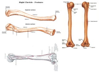
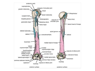
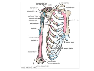
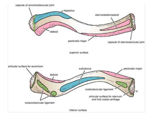
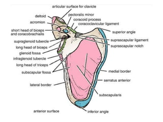
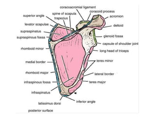
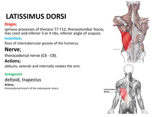
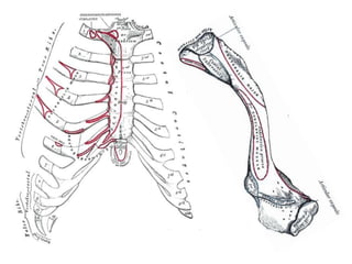
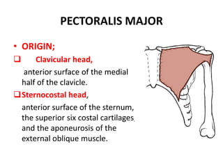
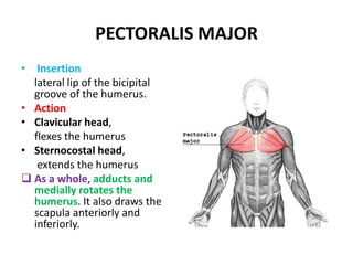
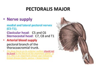
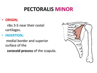
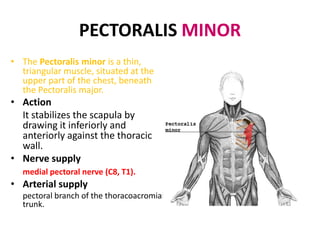
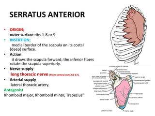
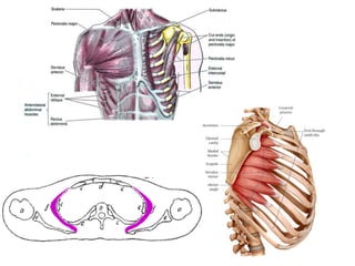
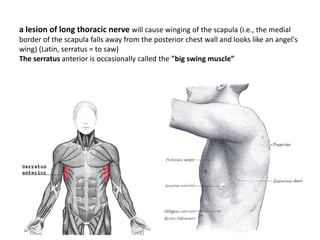
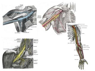
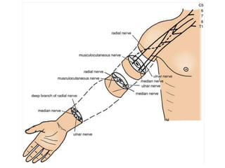
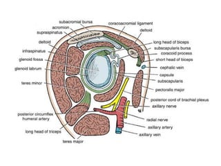
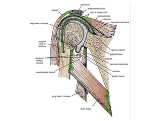
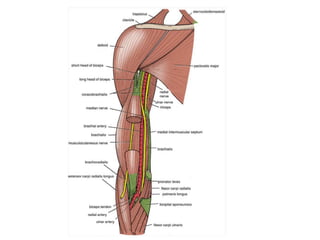
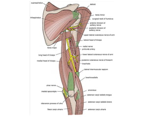
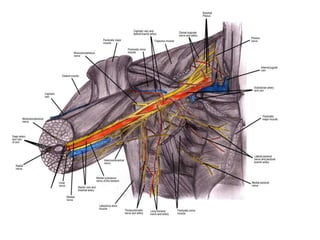
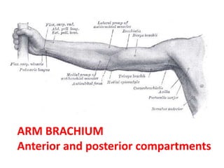
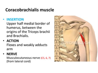
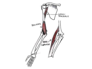
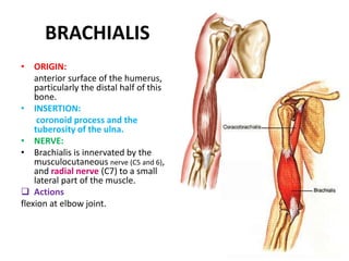
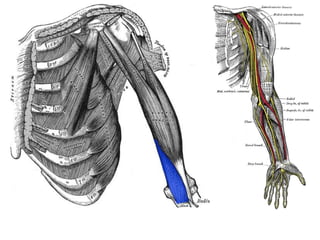
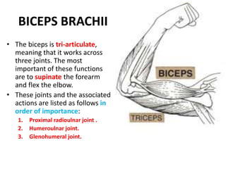
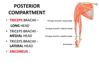
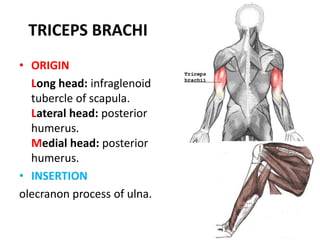
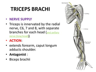
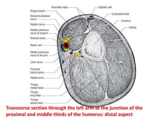
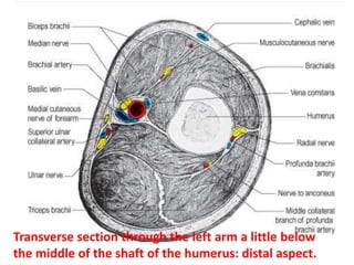
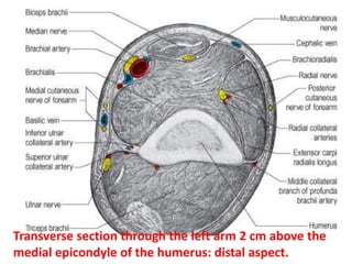
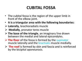
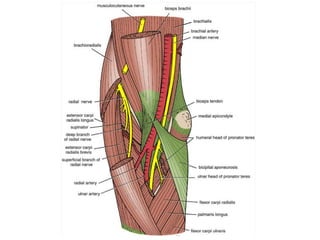
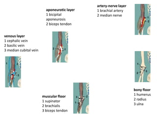
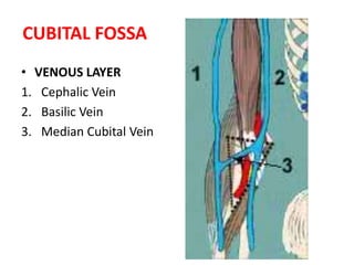
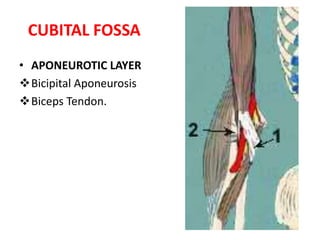
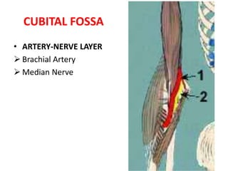
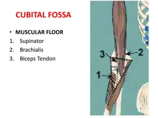
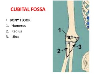
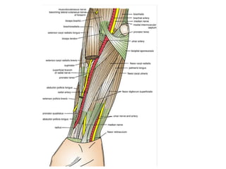


This document provides an overview of the anatomy of the arm and pectoral region. It describes the origins, insertions, innervations, and actions of muscles like the latissimus dorsi, pectoralis major and minor, serratus anterior, biceps brachii, triceps brachii, brachialis, and coracobrachialis. It also details the fascia, compartments, and structures of the arm, forearm, cubital fossa, and pectoral region including bones, muscles, vessels and nerves.


![Hypothalamus short ppt by Dr. Neha [PT].pptx](https://cdn.slidesharecdn.com/ss_thumbnails/hypothalamusbydr-260124145759-b9f94a93-thumbnail.jpg?width=640&height=640&fit=bounds)