Downloaded 163 times


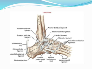


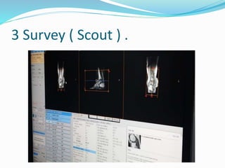
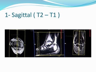
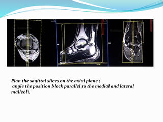


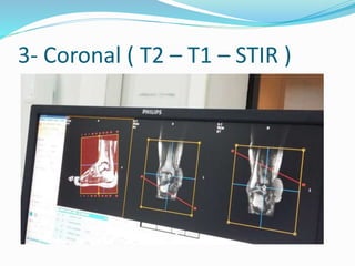

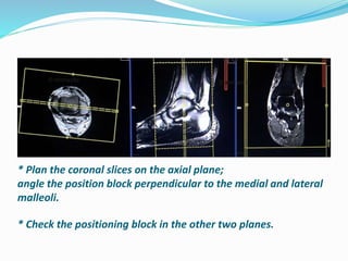
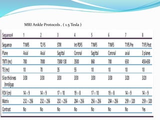
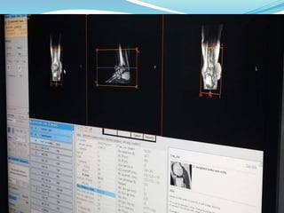
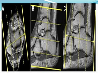


An MRI of the ankle provides images of ligaments, tendons, bones and soft tissues. The document outlines the major ligaments of the ankle joint and indications for an MRI such as fractures, tendon disorders and infections. It describes positioning the patient with their ankle in the coil at a 90 degree angle and securing it to prevent movement. Three scan planes are discussed - sagittal, axial and coronal - with specific sequences and positioning for each view.


![ankle MRI.pptx [Repaired].pptx](https://cdn.slidesharecdn.com/ss_thumbnails/anklemri-230430061225-0fe22f3a-thumbnail.jpg?width=640&height=640&fit=bounds)


