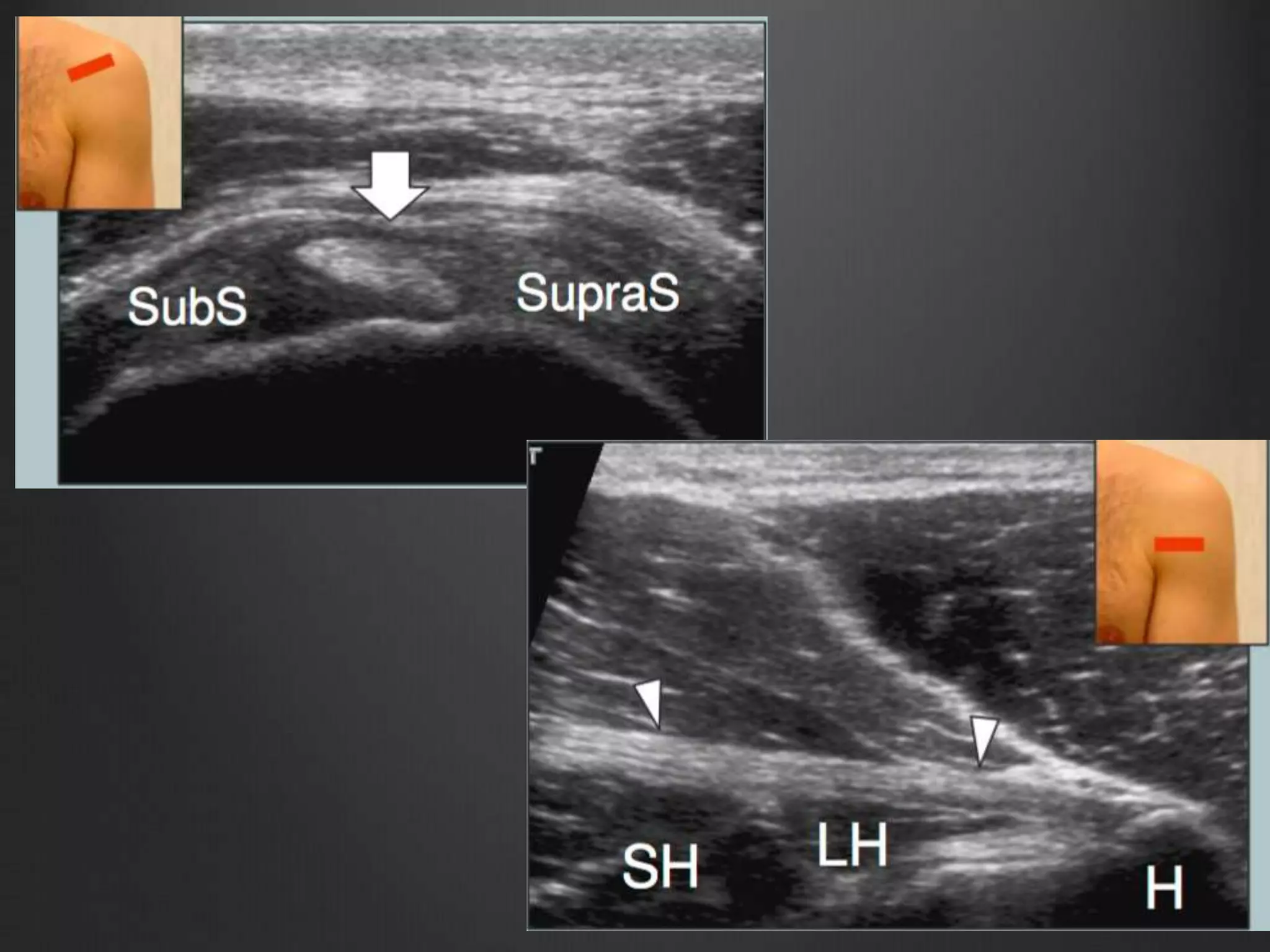
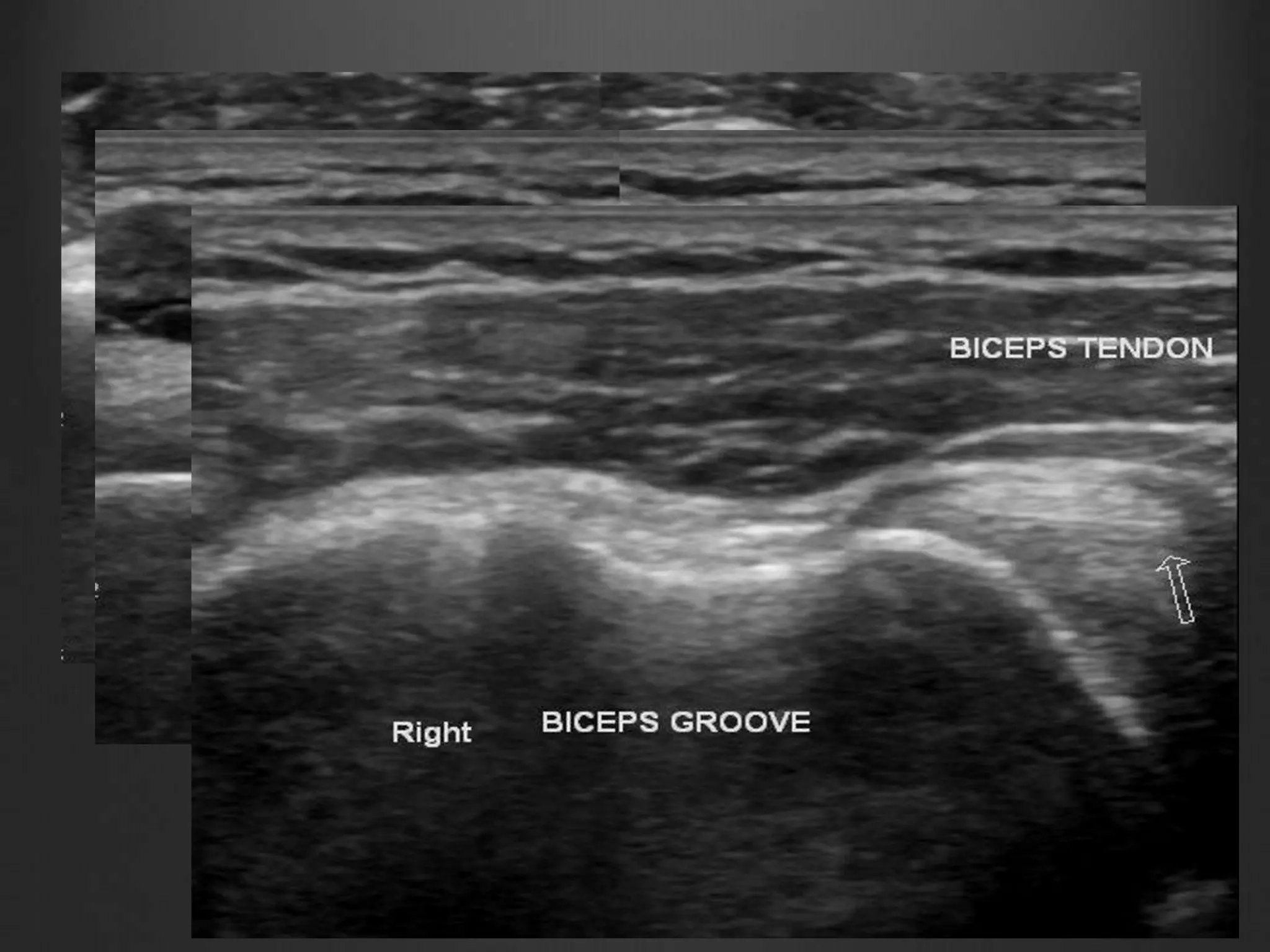
USG SHOULDER
Presented by : Dr. Kanishka Aggarwal R2/RD
Guide : Dr. Abhishek R3/RD
References : Rumack
Radiographics
ESSR Musculoskeletal Guidelines
Radiology Assistant
POINTS TO BE DISCUSSED
Anatomy
Scanning technique
Normal shoulder imaging
Pathological Conditions
Departmental cases
SHOULDER JOINT : BALL & SOCKET JOINT.
3 BONES : HUMERUS , SCAPULA, CLAVICLE
4 JOINTS: GLENOHUMERAL, ACROMIOCLAVICULAR,
STERNOCLAVICULAR, SCAPULOTHORACIC
MUSCLES OF ROTATOR CUFF : SUPRASPINATUS, INFRASPINATUS,
SUBSCAPULARIS, TERES MINOR
ROTATOR CUFF MUSCLES
SHOUDER ULTRASOUND
EVALUATION
Long head of biceps brachii tendon
Subscapularis tendon, excludes transient biceps tendon
subluxation or dislocation
Supraspinatus tendon and rotator interval
Acromioclavicular joint, subacromial-subdeltoid bursa, including
dynamic evaluation for subacromial impingement
Infraspinatus, teres minor and posterior labrum.
 All the tendons are viewed in short and long axis.
 A high frequency (7-12 MHz) probe is used.
LONG HEAD OF BICEPS
BRACHII
Place the arm in slight internal rotation (directed towards
the contralateral knee) with elbow flexed 90o, palm up.
Identify the biceps tendon in the bicipital groove.
Shift probe up to examine biceps in its intraarticular
course course and down to reach the myotendinous
junction.
Short axis view of LHBT
Long axis view of LHBT
SUBSCAPULARIS TENDON
Position:
• Rotate the arm externally fixing the elbow on iliac crest
to show subscapularis tendon and its insertion at lesser
tuberosity (slight supination of the hand may be helpful
to neutralize the tendency to lift and abduct the elbow
from the lateral chest wall) .
• This tendon should be evaluated along its long-
(transverse planes) and short- (sagittal planes) axis
during passive external and internal rotation with
hanging arm.
Insertion Of Subscapularis Tendon
Muscle tissue interposed between tendon
fascicles
SUBSCAPULARIS
SUPRASPINATUS TENDON
Place the patient’s arm posteriorly, placing the palmar side
of the hand on the superior aspect of the iliac wing with
the elbow flexed and directed posteriorly.
Long Axis
Short Axis
ROTATOR INTERVAL
It is a triangular space between the tendons of
subscapularis and supraspinatus and the base of the
coracoid process.
It is roofed by the rotator interval capsule, principally made
of coracohumeral ligament. It contains the tendon of the
long head of the biceps and the superior glenohumeral
ligament.
Clinical Importance : Coracohumeral and superior
glenohumeral ligament prevent long head of biceps
tendon from subluxation or dislocation.
ACROMIO-CLAVICULAR
JOINT
Place the transducer in the coronal plane over the
shoulder to examine the acromioclavicular joint.
Sweep the transducer anteriorly and posteriorly over this
joint to assess the presence of an os acromiale. Shifting
the probe posterior to the acromioclavicular joint, it is
possible to assess the status of the supraspinatus muscle.
IMPINGEMENT TEST
Dynamic assessment of subacromial impingment can
be attempted by placing the tranducer in coronal plane
with its medial margin at the lateral margin of the
acromion.
Patient is ask to abduct his arm while in internal
rotation.With this manoeuvre,the supraspinatus and
bursa can be seen passing deep to coracoacromial
arch.
SUBACROMIAL IMPINGEMENT
Repititive friction results bursal thickening and is often
accompanied by minimal fluid accumulation.
During dynamic testing the thickened bursal tissue may be
seen to bunch up against the outer edge of the
acromion,failing to pass beneath it.
INFRASPINATUS & TERES
MINOR TENDONS
Place the transducer over the posterior aspect of the
glenohumeral joint with the hand on the opposite shoulder
and increase the depth to include the structures of the
posterior fossa within the field-of-view of the US image.
Use the spine of the scapula as the landmark to
distinguish the supraspinous fossa (transducer shifted-up)
from the infraspinous fossa (transducer shifted-down) on
sagittal planes.
INFRASPINATUS
TERES MINOR
POSTERIOR STRUCTURES &
POSTERIOR GLENOHUMERAL
JOINT RECESS
Examine these infraspinatus and teres minor tendons separately
on their long-axis (transverse planes) during external and
internal rotation of the arm in neutral position by placing the
probe over the posterior aspect of the glenohumeral joint.
Look at the posterior labrum-capsular complex and check the
posterior recess of the joint for effusion during scanning. In thin
subjects the posterior labrum can be clearly seen.
Move the transducer medial to the labrum on transverse plane
to visualize the spinoglenoid notch. It is often necessary to
increase the depth of the field-of-view not to miss this area. A
paralabral cyst originating in this area should be sought.
HH : Humeral Head
Curved Arrow : Bony glenoid
Asterisk : Spinoglenoid notch
Arrow heads : Posterior Labrum
POSTERIOR LABRUM
ROTATOR CUFF
PATHOLOGIES
Rotator cuff tears
Biceps tendon subluxation/dislocation
Tendinosis
Calcific tendinitis
Labral cysts
ROTATOR CUFF TEARS
2 types : Partial thickness and full thickness tear.
Etiology : acute or chronic trauma, sports related injuries,
collagen vascular diseases.
Most common : Supraspinatus tendon
Most common age group : >40 years
USG CRITERIA OF ROTATOR
CUFF TEARS
Non- visualisation of the cuff
Focal non-visualisation of the cuff
Focal abnormal echogenicity
Discontinuity in the cuff
Partial thickness tears :
Articular
Interstitial
Bursal
Associated with impingement and degenerative changes.
On X-ray:
Acromial spurs
Type III acromion
Humeral head arthritic changes at greater tuberosity
AC degenerative changes
Partial tear on USG:
Decreased echogenicity and thinning of the tendon in affected
region.
Loss of convexity of tendon / bursal interface in bursal surface
tear.
Calcific foci in tendon in chronic cases.
Full Thickness Tears :
ON USG:
1.Focal tendon interruption
2.Fluid filled gap
3.Loss of convexity of tendon/bursal interface
4.Cartilage interface sign.
5.Tendon retraction.
Partial Thickness Tear of
Supraspinatus
Full Thickness Tear of
Supraspinatus
BICEPS TENDON
SUBLUXATION/DISLOCATION
Long head of biceps brachii travels
through the bicipital groove and is
stabilized by 2 main ligaments
coracohumeral and superior
glenohumeral, forming the rotator cuff
interval.
Disruption of the stabilizing ligament or
shallow bicipital groove can cause
subluxation/dislocation of biceps tendon.
TENDINOSIS
Degeneration of long head of biceps brachii.
Etiology:
1.Chronic microtrauma
2.Accompanies with rotator cuff disease
3.Associated with subacromial impingment
4.May accompany tenosynovitis
On USG:
-- Thickening of long head of biceps
-- Fluid surrounding biceps tendon
tendinosis
CALCIFIC TENDINITIS
Deposition of calcium hydroxyapatite crystals in various
shoulder tendons.
Most common site is within the supraspinatus tendon near
its insertion.
At US, calcium deposits may have a fluffy appearance,
with echogenic foci without posterior shadowing, or may
appear as typical discrete, well-circumscribed
calcifications with posterior shadowing.
US is very sensitive for detecting even tiny calcium
deposits.
PARALABRAL CYSTS
May represent a synovial cyst, ganglion cyst or
pseudocyst.
Etiology : A/W glenoid labral tears
MC Location : posterior > anterior > superior aspects of
the glenohumeral joint. The postero-superior location is
most common.
DEPARTMENTAL CASE
A 40 year old female presented with the complaint of left
shoulder pain with restricted movement since 12 days with
history of fall 12 days back.
Long head of biceps tendon
Long axis - LHBT
SUBSCAPULARIS
DEPARTMENTAL CASE
A 47 year old male presented with the complaint of right
shoulder pain with restricted shoulder movement since 25-
30 days.
No history of trauma.
Discontinuity along fibers of supraspinatus tendon
Calcific focus – calcific tendinitis
Fluid collection around the biceps tendon
Fluid collection around the tendon
Fluid collection around the tendon near articular surface
Interstitial tear of subscapularis
DEPARTMENTAL CASE
A 40 year old female came with the complaint of left
shoulder pain with restricted movement of the shoulder
and with history of RTA 15 days back.
Fluid collection around the LHBT with tear in LHBT with medial subluxation
Subdeltoid effusion
Full thickness tear in supraspinatus
THANK YOU !

usg shoulder by dr. kanishka.pptx

  • 1.
    USG SHOULDER Presented by: Dr. Kanishka Aggarwal R2/RD Guide : Dr. Abhishek R3/RD References : Rumack Radiographics ESSR Musculoskeletal Guidelines Radiology Assistant
  • 2.
    POINTS TO BEDISCUSSED Anatomy Scanning technique Normal shoulder imaging Pathological Conditions Departmental cases
  • 3.
    SHOULDER JOINT :BALL & SOCKET JOINT. 3 BONES : HUMERUS , SCAPULA, CLAVICLE 4 JOINTS: GLENOHUMERAL, ACROMIOCLAVICULAR, STERNOCLAVICULAR, SCAPULOTHORACIC MUSCLES OF ROTATOR CUFF : SUPRASPINATUS, INFRASPINATUS, SUBSCAPULARIS, TERES MINOR
  • 4.
  • 5.
    SHOUDER ULTRASOUND EVALUATION Long headof biceps brachii tendon Subscapularis tendon, excludes transient biceps tendon subluxation or dislocation Supraspinatus tendon and rotator interval Acromioclavicular joint, subacromial-subdeltoid bursa, including dynamic evaluation for subacromial impingement Infraspinatus, teres minor and posterior labrum.  All the tendons are viewed in short and long axis.  A high frequency (7-12 MHz) probe is used.
  • 6.
    LONG HEAD OFBICEPS BRACHII Place the arm in slight internal rotation (directed towards the contralateral knee) with elbow flexed 90o, palm up. Identify the biceps tendon in the bicipital groove. Shift probe up to examine biceps in its intraarticular course course and down to reach the myotendinous junction.
  • 9.
    Short axis viewof LHBT Long axis view of LHBT
  • 10.
    SUBSCAPULARIS TENDON Position: • Rotatethe arm externally fixing the elbow on iliac crest to show subscapularis tendon and its insertion at lesser tuberosity (slight supination of the hand may be helpful to neutralize the tendency to lift and abduct the elbow from the lateral chest wall) . • This tendon should be evaluated along its long- (transverse planes) and short- (sagittal planes) axis during passive external and internal rotation with hanging arm.
  • 12.
    Insertion Of SubscapularisTendon Muscle tissue interposed between tendon fascicles
  • 13.
  • 15.
    SUPRASPINATUS TENDON Place thepatient’s arm posteriorly, placing the palmar side of the hand on the superior aspect of the iliac wing with the elbow flexed and directed posteriorly.
  • 16.
  • 17.
  • 19.
    ROTATOR INTERVAL It isa triangular space between the tendons of subscapularis and supraspinatus and the base of the coracoid process. It is roofed by the rotator interval capsule, principally made of coracohumeral ligament. It contains the tendon of the long head of the biceps and the superior glenohumeral ligament. Clinical Importance : Coracohumeral and superior glenohumeral ligament prevent long head of biceps tendon from subluxation or dislocation.
  • 21.
    ACROMIO-CLAVICULAR JOINT Place the transducerin the coronal plane over the shoulder to examine the acromioclavicular joint. Sweep the transducer anteriorly and posteriorly over this joint to assess the presence of an os acromiale. Shifting the probe posterior to the acromioclavicular joint, it is possible to assess the status of the supraspinatus muscle.
  • 24.
    IMPINGEMENT TEST Dynamic assessmentof subacromial impingment can be attempted by placing the tranducer in coronal plane with its medial margin at the lateral margin of the acromion. Patient is ask to abduct his arm while in internal rotation.With this manoeuvre,the supraspinatus and bursa can be seen passing deep to coracoacromial arch.
  • 26.
    SUBACROMIAL IMPINGEMENT Repititive frictionresults bursal thickening and is often accompanied by minimal fluid accumulation. During dynamic testing the thickened bursal tissue may be seen to bunch up against the outer edge of the acromion,failing to pass beneath it.
  • 27.
    INFRASPINATUS & TERES MINORTENDONS Place the transducer over the posterior aspect of the glenohumeral joint with the hand on the opposite shoulder and increase the depth to include the structures of the posterior fossa within the field-of-view of the US image. Use the spine of the scapula as the landmark to distinguish the supraspinous fossa (transducer shifted-up) from the infraspinous fossa (transducer shifted-down) on sagittal planes.
  • 29.
  • 30.
  • 32.
    POSTERIOR STRUCTURES & POSTERIORGLENOHUMERAL JOINT RECESS Examine these infraspinatus and teres minor tendons separately on their long-axis (transverse planes) during external and internal rotation of the arm in neutral position by placing the probe over the posterior aspect of the glenohumeral joint. Look at the posterior labrum-capsular complex and check the posterior recess of the joint for effusion during scanning. In thin subjects the posterior labrum can be clearly seen. Move the transducer medial to the labrum on transverse plane to visualize the spinoglenoid notch. It is often necessary to increase the depth of the field-of-view not to miss this area. A paralabral cyst originating in this area should be sought.
  • 33.
    HH : HumeralHead Curved Arrow : Bony glenoid Asterisk : Spinoglenoid notch Arrow heads : Posterior Labrum
  • 34.
  • 36.
    ROTATOR CUFF PATHOLOGIES Rotator cufftears Biceps tendon subluxation/dislocation Tendinosis Calcific tendinitis Labral cysts
  • 37.
    ROTATOR CUFF TEARS 2types : Partial thickness and full thickness tear. Etiology : acute or chronic trauma, sports related injuries, collagen vascular diseases. Most common : Supraspinatus tendon Most common age group : >40 years
  • 38.
    USG CRITERIA OFROTATOR CUFF TEARS Non- visualisation of the cuff Focal non-visualisation of the cuff Focal abnormal echogenicity Discontinuity in the cuff
  • 39.
    Partial thickness tears: Articular Interstitial Bursal Associated with impingement and degenerative changes. On X-ray: Acromial spurs Type III acromion Humeral head arthritic changes at greater tuberosity AC degenerative changes
  • 40.
    Partial tear onUSG: Decreased echogenicity and thinning of the tendon in affected region. Loss of convexity of tendon / bursal interface in bursal surface tear. Calcific foci in tendon in chronic cases.
  • 41.
    Full Thickness Tears: ON USG: 1.Focal tendon interruption 2.Fluid filled gap 3.Loss of convexity of tendon/bursal interface 4.Cartilage interface sign. 5.Tendon retraction.
  • 42.
    Partial Thickness Tearof Supraspinatus
  • 43.
    Full Thickness Tearof Supraspinatus
  • 44.
    BICEPS TENDON SUBLUXATION/DISLOCATION Long headof biceps brachii travels through the bicipital groove and is stabilized by 2 main ligaments coracohumeral and superior glenohumeral, forming the rotator cuff interval. Disruption of the stabilizing ligament or shallow bicipital groove can cause subluxation/dislocation of biceps tendon.
  • 46.
    TENDINOSIS Degeneration of longhead of biceps brachii. Etiology: 1.Chronic microtrauma 2.Accompanies with rotator cuff disease 3.Associated with subacromial impingment 4.May accompany tenosynovitis On USG: -- Thickening of long head of biceps -- Fluid surrounding biceps tendon
  • 47.
  • 48.
    CALCIFIC TENDINITIS Deposition ofcalcium hydroxyapatite crystals in various shoulder tendons. Most common site is within the supraspinatus tendon near its insertion. At US, calcium deposits may have a fluffy appearance, with echogenic foci without posterior shadowing, or may appear as typical discrete, well-circumscribed calcifications with posterior shadowing. US is very sensitive for detecting even tiny calcium deposits.
  • 50.
    PARALABRAL CYSTS May representa synovial cyst, ganglion cyst or pseudocyst. Etiology : A/W glenoid labral tears MC Location : posterior > anterior > superior aspects of the glenohumeral joint. The postero-superior location is most common.
  • 52.
    DEPARTMENTAL CASE A 40year old female presented with the complaint of left shoulder pain with restricted movement since 12 days with history of fall 12 days back.
  • 53.
    Long head ofbiceps tendon
  • 54.
  • 60.
  • 63.
    DEPARTMENTAL CASE A 47year old male presented with the complaint of right shoulder pain with restricted shoulder movement since 25- 30 days. No history of trauma.
  • 64.
    Discontinuity along fibersof supraspinatus tendon
  • 65.
    Calcific focus –calcific tendinitis
  • 68.
    Fluid collection aroundthe biceps tendon
  • 69.
  • 70.
    Fluid collection aroundthe tendon near articular surface
  • 71.
    Interstitial tear ofsubscapularis
  • 73.
    DEPARTMENTAL CASE A 40year old female came with the complaint of left shoulder pain with restricted movement of the shoulder and with history of RTA 15 days back.
  • 74.
    Fluid collection aroundthe LHBT with tear in LHBT with medial subluxation
  • 76.
  • 77.
    Full thickness tearin supraspinatus
  • 79.

Editor's Notes

  • #49 Location : Supraspinatus>>infraspinatus>>tere minor>>subscapularis
  • #54 ANECHOIC FLUID COLLECTION AROUND THE TENDON
  • #55 DISCONTINUITY OF FIBERS ALONG THE BURSAL SURFACE
  • #56 Fiber discontinuity along bursal surface with fluid filled gap with irregularity of bursal surface, dipping into the space.
  • #57 Mild hyperemia