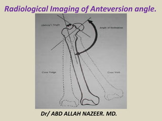
Presentation1.pptx, radiological imaging of anteversion angle.
•Download as PPTX, PDF•
21 likes•4,233 views
Femoral anteversion is defined as the angle between the femoral neck and knee joint. In adults, the normal range is 15-20 degrees, but some individuals have angles outside this range which can contribute to orthopedic problems. CT scans are used to precisely measure the femoral anteversion angle by assessing the alignment of the femoral neck and condyles. The summary describes how femoral anteversion is measured and implications of abnormal angles.
Report
Share
Report
Share

Recommended
Radiological evaluation of TKR by Dr. D. P. Swami

preoperative planning and postoperative analysis of TKR
Acetabular Fracture Radiology: Xrays, CT scan & 3D printing

The talk details how to assess various types of acetabular fracture. Combination of X-rays, CT Scan and 3D reconstruction and 3D printing also known as 3DGraphy. Basic 8 patterns and importance of various radiological parameter are explained.
Recommended
Radiological evaluation of TKR by Dr. D. P. Swami

preoperative planning and postoperative analysis of TKR
Acetabular Fracture Radiology: Xrays, CT scan & 3D printing

The talk details how to assess various types of acetabular fracture. Combination of X-rays, CT Scan and 3D reconstruction and 3D printing also known as 3DGraphy. Basic 8 patterns and importance of various radiological parameter are explained.
Rotator Cuff Injuries - Dr.CHINTAN N. PATEL

ROTATOR CUFF INJURIES - WITH FULL DESCRIPTION OF TESTS FOR SHOULDER EXAMINATION
Normal limb alignment

The exact anatomy of the bones and joints is of great importance to the clinician when examining the limbs and to the surgeon when operating on the bones and joints.
To understand deformities of the extremities, it is important to first understand and establish the parameters and limits of normal alignment.
Each long bone has a mechanical and an anatomic axis
both frontal and sagittal planes axis lines are applicable to any longitudinal projection of a bone.
The corresponding radiographic projections are the anteroposterior (AP) and lateral (LAT) views, respectively.
Shoulder joint instability

Shoulder joint instabilities ,Brief anatomy , Types of lesions in Gleno humeral ligaments, Classifications , Dislocations , closed reduction methods, Complications , operative and non operative management .
MCL,LCL & ALL injuries of the knee 

MCL. LCL.ALL injuries
To understand the relevant anatomy of the side ligaments of the knee
To study the mechanism of injury of each ligament and how to diagnose such injury
To highlight the different treatment options in acute or chronic situations
Acquired Bone Deformities

Acquired Bone DeformitiesDepartment of Orthopaedics & Trauma, Faculty of Medicine / King AbdulAziz University
امراض القدم عند الاطفال Pediatric foot 2 , البروفيسور فريح ابوحسان - استشاري...

امراض القدم عند الاطفال Pediatric foot 2 , البروفيسور فريح ابوحسان - استشاري...Prof Freih Abu Hassan البروفيسور فريح ابوحسان
جراحة العظام / علاج العظام في الاردن / افضل دكتور عظام في الاردن / افضل اخصائي عظام في الاردن / استشاري عظام/افضل استشاري عظام في الاردن /جراحة عظام / /عمليات تطويل العظام في الاردن / اطباء العظام في الاردن / دكتور طب عظام في الاردن / الاطباء في الاردن / خلع ورك / عمليات اليزاروف في الاردن /علاج الكسور /خلع الولادة / تركيب المفصل / اوجاع العظام /افضل طبيب عظام اطفال في الاردن / استشاري اطفال عظام في الاردن / /علاج خلع الكتف / علاج التواء الكاحل / التواء الكاحل / علاج الام العظام / علاج هشاشة العظام / ارقام اطباء عظام في الاردن / مشاكل العظام والمفاصل /مستشار جراحة العظام والمفاصل والكسور/مستشار جراحة عظام الأطفال.More Related Content
What's hot
Rotator Cuff Injuries - Dr.CHINTAN N. PATEL

ROTATOR CUFF INJURIES - WITH FULL DESCRIPTION OF TESTS FOR SHOULDER EXAMINATION
Normal limb alignment

The exact anatomy of the bones and joints is of great importance to the clinician when examining the limbs and to the surgeon when operating on the bones and joints.
To understand deformities of the extremities, it is important to first understand and establish the parameters and limits of normal alignment.
Each long bone has a mechanical and an anatomic axis
both frontal and sagittal planes axis lines are applicable to any longitudinal projection of a bone.
The corresponding radiographic projections are the anteroposterior (AP) and lateral (LAT) views, respectively.
Shoulder joint instability

Shoulder joint instabilities ,Brief anatomy , Types of lesions in Gleno humeral ligaments, Classifications , Dislocations , closed reduction methods, Complications , operative and non operative management .
MCL,LCL & ALL injuries of the knee 

MCL. LCL.ALL injuries
To understand the relevant anatomy of the side ligaments of the knee
To study the mechanism of injury of each ligament and how to diagnose such injury
To highlight the different treatment options in acute or chronic situations
What's hot (20)
Viewers also liked
امراض القدم عند الاطفال Pediatric foot 2 , البروفيسور فريح ابوحسان - استشاري...

امراض القدم عند الاطفال Pediatric foot 2 , البروفيسور فريح ابوحسان - استشاري...Prof Freih Abu Hassan البروفيسور فريح ابوحسان
جراحة العظام / علاج العظام في الاردن / افضل دكتور عظام في الاردن / افضل اخصائي عظام في الاردن / استشاري عظام/افضل استشاري عظام في الاردن /جراحة عظام / /عمليات تطويل العظام في الاردن / اطباء العظام في الاردن / دكتور طب عظام في الاردن / الاطباء في الاردن / خلع ورك / عمليات اليزاروف في الاردن /علاج الكسور /خلع الولادة / تركيب المفصل / اوجاع العظام /افضل طبيب عظام اطفال في الاردن / استشاري اطفال عظام في الاردن / /علاج خلع الكتف / علاج التواء الكاحل / التواء الكاحل / علاج الام العظام / علاج هشاشة العظام / ارقام اطباء عظام في الاردن / مشاكل العظام والمفاصل /مستشار جراحة العظام والمفاصل والكسور/مستشار جراحة عظام الأطفال.Rotational deformities of lower extremity in children

Rotational deformities of lower limb in children
SPORTS INJURY I Dr.RAJAT JANGIR JAIPUR

SPORTS INJURY
#aclsurgeryjaipur #aclsurgeryhindia #aclsurgerytaekwondo
Acl reconstruction in jaipur | Acl reconstruction in taekwondo | Acl injury in football player surgery | Acl reconstruction surgery in football | acl surgery | Acl surgery ke baad physiotherapy | Acl surgery in jaipur | acl surgery recovery | Best acl surgeon in jaipur | Best ligament doctor in hindi | Best acl surgeon in india | Meniscus repair surgery in jaipur | Sports injury doctor | Acl injury in football players | Acl injury in taekwondo | acl tear | Best knee surgeon in jaipur
#allinsideacl #internalbrace #drrajatjangir #bestaclsurgeon #aclexpert #bestkneesurgeon
To Know more about ACL Injury, Click the links below:
1. ACL surgery 7 different Techniques we do at our center - "Not single technique best for all"
https://youtu.be/oWkIr8IXvr8
2. Everything about ACL Injury tear surgery in Hindi I
https://youtu.be/bqpjkAkwZ14
3. Best Screw for ACL tear surgery in Hindi
https://youtu.be/1LGpU1NHiIs
4. ACL Injury Tear Surgery Recovery : All your questions & queries solved by Dr.Rajat Jangir
https://youtu.be/SIAPWiMbOqs
5. Partial ACL Tear Surgery or not ! ACL आधा टूटा हो तो क्या करें ?
https://youtu.be/NEJRPKskJTI
6. 5 Symptoms of ACL Injury tear इंजरी के पांच लक्षण ?
https://youtu.be/EXpgy19Jxzw
7. PRP injection therapy in Partial ACL TEARs
https://youtu.be/qyG1EYgS87E
Dr.RAJAT JANGIR(Asso Prof.)
Senior Consultant Arthroscopy and Joint Replacement
(Specialist in Shoulder Knee Hip Surgery)
Ligament and Joints Clinic
67/34 Mansarovar Jaipur
Whatsapp: shorturl.at/gnAEP
Appointment: +91 8104855900
Email: ligamentsurgeon@gmail.com
Google Page: https://g.page/KNEE-Shoulder-SURGERY?...
Facebook: https://www.facebook.com/Ligamentandj...
* Vast experience and specialisation in the field of Arthroscopy and sports surgery.
* M.S. orthopaedics from BJ Medical College, Civil hospital, Ahmedabad
* Fellowship in Arthroscopy and Sports injury with Prof Joon Ho Wang at Samsung Medical Center, South Korea
* Diploma in Sports Medicine from InternationaI Olympic Committee
* Invited as Athlete Medical Doctor at Rio Olympic 2016
* Done Rajasthan's first "All Inside Physeal Preserving ACL reconstruction" in 13 year old Athlete
Dr.Rajat is rated as one of the best orthopedic surgeon with with excellence in Knee Shoulder Arthroscopy surgeries as replacements'
Mystery of Knee pain Black Hole of Orthopedics- Patellofemoral joint dr.san...

Mystery of Knee pain Black Hole of Orthopedics- Patellofemoral joint dr.san...AGRASEN Fracture Arthritis Hospital, Ganesh Nagar,Gondia,Maharashtra,INDIA
Knee Pain. Patellofemoral pain syndrome,Chondromalacia patellae,Iliotibial Band fRICTION Syndrome,Patella Alta,Patellofemoral Arthritis, Patellar Tilt,Patellar Displacement,Anterior Knee Pain,Lateral Retinaculum,VMO Dysplasia.Excessive Lateral Patellar Pressure Syndrome,Patella Malalignment,Patellar Instabilty,Patella Maltracking,Patella Subluxation,Patella Dislocation,Synovial Plica,Tendinitis,Jumpers Knee,Congruence Angle,Malalignment of Extensor Mechanism,Patella Arthritis,Patella Fracture. Mystery of knee pain black hole of orthopedics- patellofemoral joint dr.sandeep c agrawal agrasen hospital gondia indiaOrthopedic

Orthopedics is a branch of surgery that deals with the conditions of the musculoskeletal system.
Avascular necrosis and Osteochondritis

Avascular necrosis and osteochondritis explained with radiological features in easy language and lots and lots of images.
Sports Injuries

Musculoskeletal Disorders Part 7
Sports Injuries : Sprain, Strain, Tennis elbow, Knee injuries
Spinal cord injuries | spine fracture | thoracolumbar fracture | colorado spi...

Spinal cord injuries | spine fracture | thoracolumbar fracture | colorado spi...Dr. Donald Corenman, M.D., D.C.
Colorado spine surgeon, Dr. Donald Corenman, M.D., D.C. (http://neckandback.com), is an expert in treating spinal cord injuries associated with a traumatic fall, sports related injury or accident. Many spine fractures include a thoracolumbar fracture, which is a break in one or more of the thoracic and lumbar vertebrae. Spine fractures can be very serious but are also treatable in many cases. This presentation on spinal cord injuries, spine fractures and thoracolumbar fractures details events that can lead to this injury, symptoms and treatment options.
Dr. Corenman is a renowned Colorado spine surgeon and also is an expert at all spine conditions and disorders including scoliosis, degenerative disc disease, spinal stenosis, sciatica, herniated disc, slipped disc and spondylolythesis. He is also a sports medicine specialist and treats athletes with traumatic sports related injuries. He recently launched his own website (http://neckandback.com) to educate patients on spine disorders and to offer second opinions to physicians and colleagues who are seeking additional information on specific spine injuries and treatment options.
Screws and plates fixation

Screw and plates are most common used devices in orthopedics. However, sometimes we forget their principles, so this presentation hopes to review most their problems. Thank you for your attention!
Viewers also liked (20)
امراض القدم عند الاطفال Pediatric foot 2 , البروفيسور فريح ابوحسان - استشاري...

امراض القدم عند الاطفال Pediatric foot 2 , البروفيسور فريح ابوحسان - استشاري...
Rotational deformities of lower extremity in children

Rotational deformities of lower extremity in children
Mystery of Knee pain Black Hole of Orthopedics- Patellofemoral joint dr.san...

Mystery of Knee pain Black Hole of Orthopedics- Patellofemoral joint dr.san...
Spinal cord injuries | spine fracture | thoracolumbar fracture | colorado spi...

Spinal cord injuries | spine fracture | thoracolumbar fracture | colorado spi...
Similar to Presentation1.pptx, radiological imaging of anteversion angle.
Developmental Dysplasia of Hip Radiological findings

Developmental Dysplasia of Hip
Radiological findings
Graf classifications
The andren-von rosen line
Severin classification
Arthrography
PRE OPERATIVE TEMPLATING IN TOTAL HIP ARTHROPLASTY

PRE OPERATIVE TEMPLATING IN TOTAL HIP ARTHROPLASTY
Subperiosteal resection of mid-clavicle in sprengel's.pdf

Subperiosteal resection of mid-clavicle in sprengel's.pdfProf Freih Abu Hassan البروفيسور فريح ابوحسان
#مركز_عظام_في_الاردن
#علاج_أمراض_العظام_والمفاصل_في_الاردن
#علاج_الديسك_الاردن_بالمنظار
#الديسك
#الم_الظهر
#العمود_الفقري
#منظار_الديسك
#علاج_تضيق_قناة_الاعصاب_بالمنظار_في_الاردنDevelopmental dysplasia of the hip 

Orthopedic course
overview
Introduction
Epidemiology
Etiology and pathogenesis
Pathology
Clinical Features and tests
Imaging
Treatment
Complications
Questions
References
Hip involvement negatively impact the postoperative radiographic outcomes aft...

Hip involvement negatively impact the postoperative radiographic outcomes aft...Clinical Surgery Research Communications
Clinical Surgery Research CommunicationsAdult Hip Dysplasia Presentation

Adult hip dysplasia describes a condition where the hip’s ball (femoral head) and socket (acetabulum) are misaligned. The condition is common in children but is also found in adolescents and adults who have had no history of problems in childhood. Treatment options include temporizing with medication and/or physical therapy but surgery is often required to fix the problem.
http://www.davidsfeldmanmd.com/specialties/adult-hip-dysplasia
Recent advances in imaging of scoliosis final

Presentation discussing everything the radiologist and clinician want to know about different etiological scoliosis and follow up after surgery.
Slipped Capital Femoral Epiphysis

slipped femoral epiphysis, SCFE, screw fixation, CT scan in SCFE, Fixation in situ
Similar to Presentation1.pptx, radiological imaging of anteversion angle. (20)
Developmental Dysplasia of Hip Radiological findings

Developmental Dysplasia of Hip Radiological findings
PRE OPERATIVE TEMPLATING IN TOTAL HIP ARTHROPLASTY

PRE OPERATIVE TEMPLATING IN TOTAL HIP ARTHROPLASTY
Subperiosteal resection of mid-clavicle in sprengel's.pdf

Subperiosteal resection of mid-clavicle in sprengel's.pdf
Presentation1, radiological imaging of developmental dysplasia of the hip joint.

Presentation1, radiological imaging of developmental dysplasia of the hip joint.
Hip involvement negatively impact the postoperative radiographic outcomes aft...

Hip involvement negatively impact the postoperative radiographic outcomes aft...
More from Abdellah Nazeer
Presentation1, Ultrasound of the bowel loops and the lymph nodes..pptx

Ultrasound of the bowel loops and the lymph nodes..pptx
More from Abdellah Nazeer (20)
Presentation1, Ultrasound of the bowel loops and the lymph nodes..pptx

Presentation1, Ultrasound of the bowel loops and the lymph nodes..pptx
Presentation1, radiological imaging of lateral hindfoot impingement.

Presentation1, radiological imaging of lateral hindfoot impingement.
Presentation2, radiological anatomy of the liver and spleen.

Presentation2, radiological anatomy of the liver and spleen.
Presentation1, artifacts and pitfalls of the wrist and elbow joints.

Presentation1, artifacts and pitfalls of the wrist and elbow joints.
Presentation1, artifact and pitfalls of the knee, hip and ankle joints.

Presentation1, artifact and pitfalls of the knee, hip and ankle joints.
Presentation1, radiological imaging of artifact and pitfalls in shoulder join...

Presentation1, radiological imaging of artifact and pitfalls in shoulder join...
Presentation1, radiological imaging of internal abdominal hernia.

Presentation1, radiological imaging of internal abdominal hernia.
Presentation11, radiological imaging of ovarian torsion.

Presentation11, radiological imaging of ovarian torsion.
Presentation1, new mri techniques in the diagnosis and monitoring of multiple...

Presentation1, new mri techniques in the diagnosis and monitoring of multiple...
Presentation1, radiological application of diffusion weighted mri in neck mas...

Presentation1, radiological application of diffusion weighted mri in neck mas...
Presentation1, radiological application of diffusion weighted images in breas...

Presentation1, radiological application of diffusion weighted images in breas...
Presentation1, radiological application of diffusion weighted images in abdom...

Presentation1, radiological application of diffusion weighted images in abdom...
Presentation1, radiological application of diffusion weighted imges in neuror...

Presentation1, radiological application of diffusion weighted imges in neuror...
Presentation1.pptx, radiological imaging of anteversion angle.
- 1. Dr/ ABD ALLAH NAZEER. MD. Radiological Imaging of Anteversion angle.
- 2. Femoral neck anteversion is defined as the angle between an imaginary transverse line that runs medially to laterally through the knee joint and an imaginary transverse line passing through the center of the femoral head and neck . In adults without pathology, the femur is twisted so the head and neck of the femur are angled forward between 15 and 20 degrees from the frontal plane of the body. In some instances, the FNA angle is directed forward or backward well beyond this angle. Some researchers suggest that FNA angles outside this 15- to 20-degree average are a contributing factor in many different orthopedic problems in the lower extremity that are commonly seen by physical therapists. The purpose of this Update is to describe how FNA is related to hip rotation, how hip rotation range of motion can be used to predict abnormal FNA, and how asymmetries in hip rotation may be used to identify patients who may be at risk for developing various orthopedic problems in the hip and lower extremity.
- 4. What causes femoral anteversion? Femoral anteversion can be the result of stiff hip muscles due to the position of the baby in the uterus. It also has a tendency to run in families. Typically, a child's walking style looks like that of his or her parents. When the child is first learning how to walk, femoral anteversion can create an intoeing appearance. As the knees and feet turn in, the legs look like they are bowed. The bowed leg stance actually helps the child achieve greater balance as they stand. Balance is not as steady when they try to stand and walk with their feet close together or with their feet turned out. This may cause them to trip and fall. How is femoral anteversion diagnosed? The diagnosis of femoral anteversion is made by a history and physical examination by your child's doctor. During the examination, the doctor obtains a complete prenatal and birth history of the child and asks if other family members are known to have femoral anteversion. Generally, no X-rays are necessary.
- 5. CT protocols: Femoral anteversion: This limited study allow the radiologist to measure the angle of rotation of femoral neck relative to the femoral condyles bilaterally. A secondary measurement is femoral lengths, made by calculating the difference in table position at the ends of the bones. Radiographic Scanogram: This is a radiographic study in which coned-down images of both hips, knees &ankles are shot on a single conventional-sized film(or CR plate) with a radiopaque ruler in place. The sole purpose of this study is to measure leg- length. This is a radiographic study in which both leg are images in their entities from hip to ankle on a single long film using a scoliosis cassette. Typically, these are used by orthopedic surgeons for planning purposes.
- 8. CT protocol for femoral anteversion measurement. Measure right and left side individually, Find the slice that reveals the alignment of the femoral neck, Measure the neck-Horizontal angle, Find the slice that best reveals the alignment of the femoral condyles, measure the condyle- Horizontal angle and calculate the angle of the neck relative to the condyles.
- 10. Thank You.
