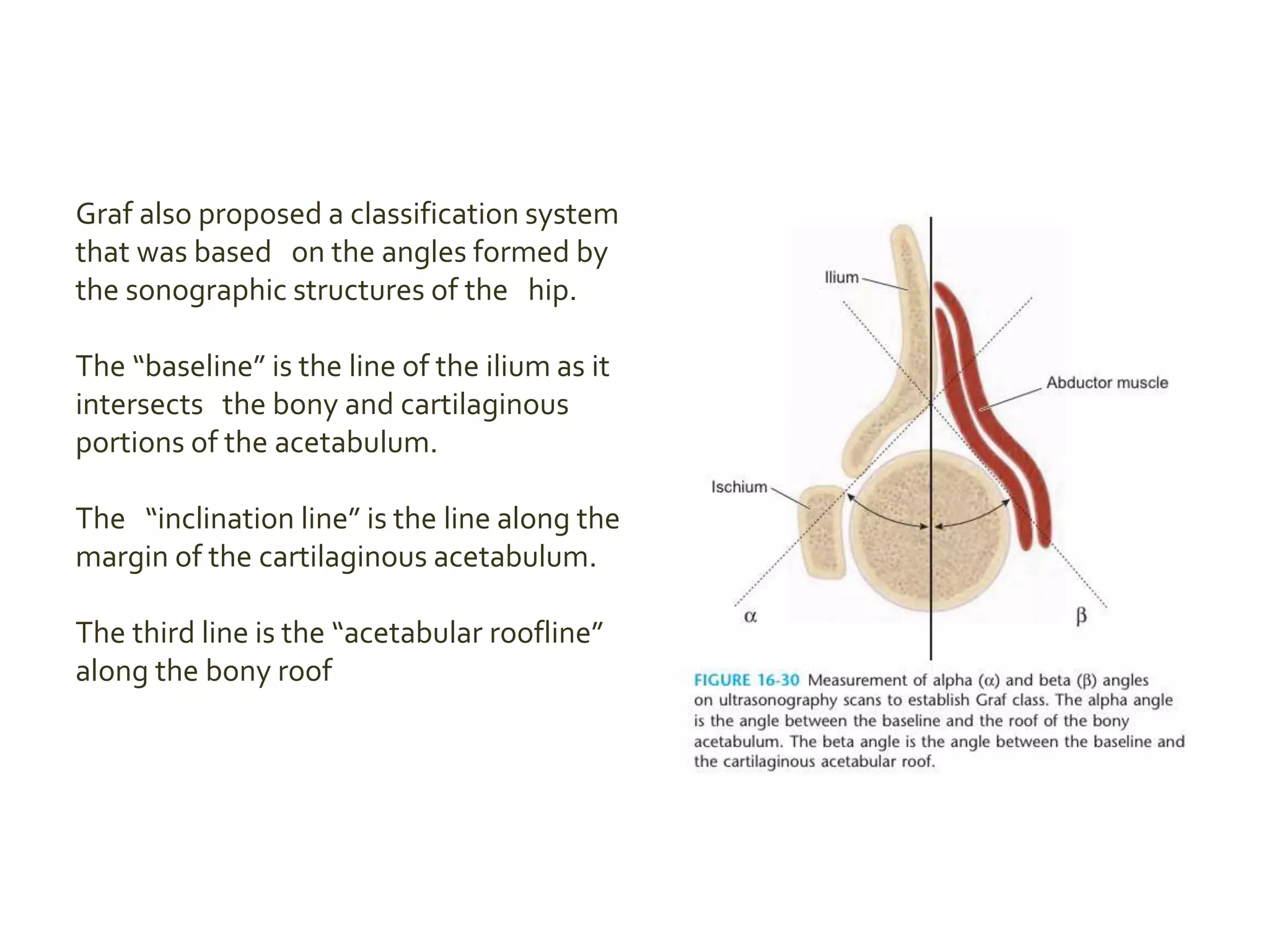
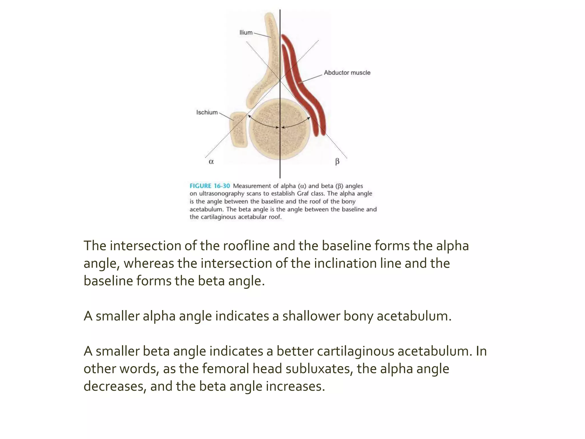
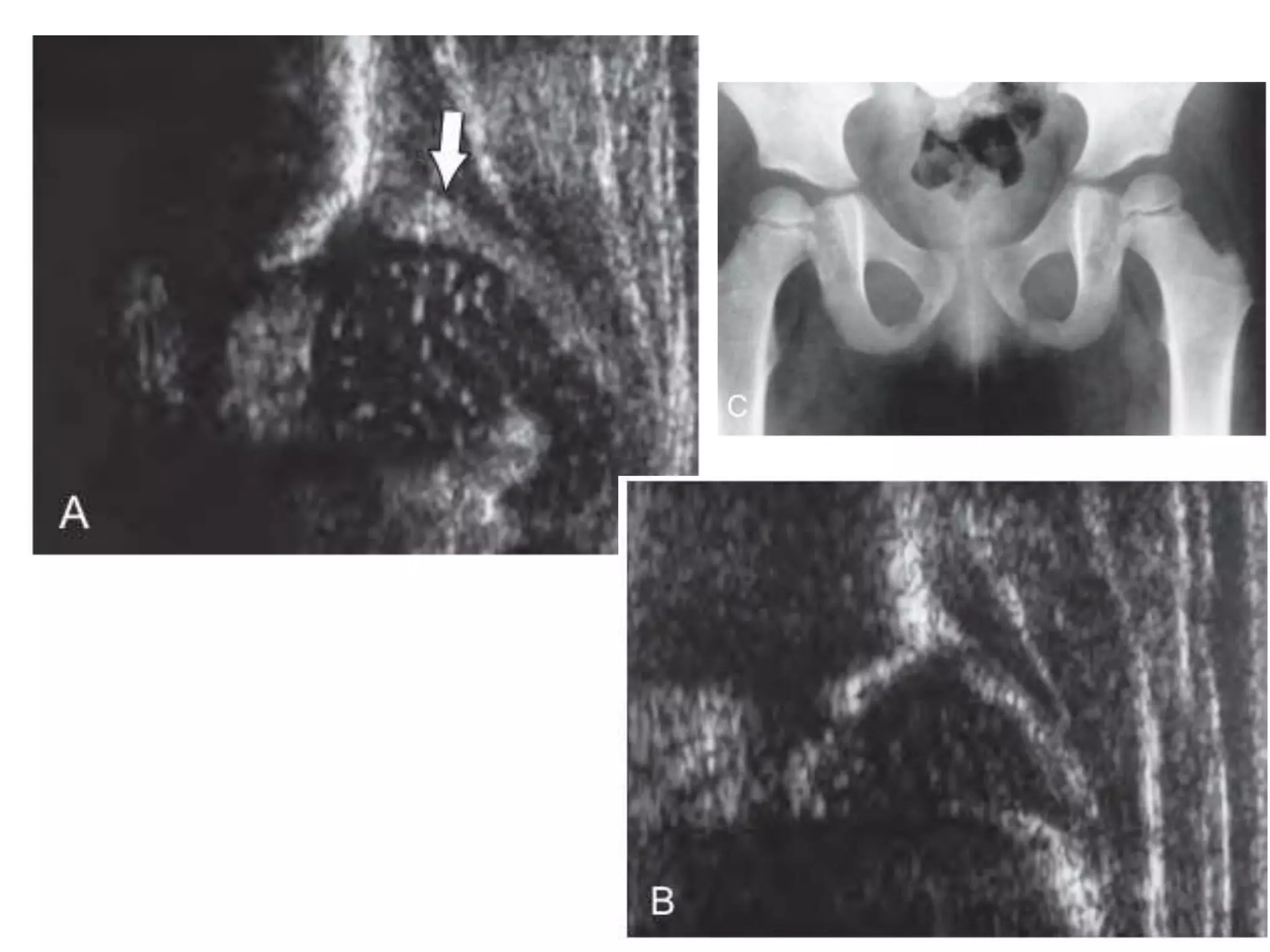
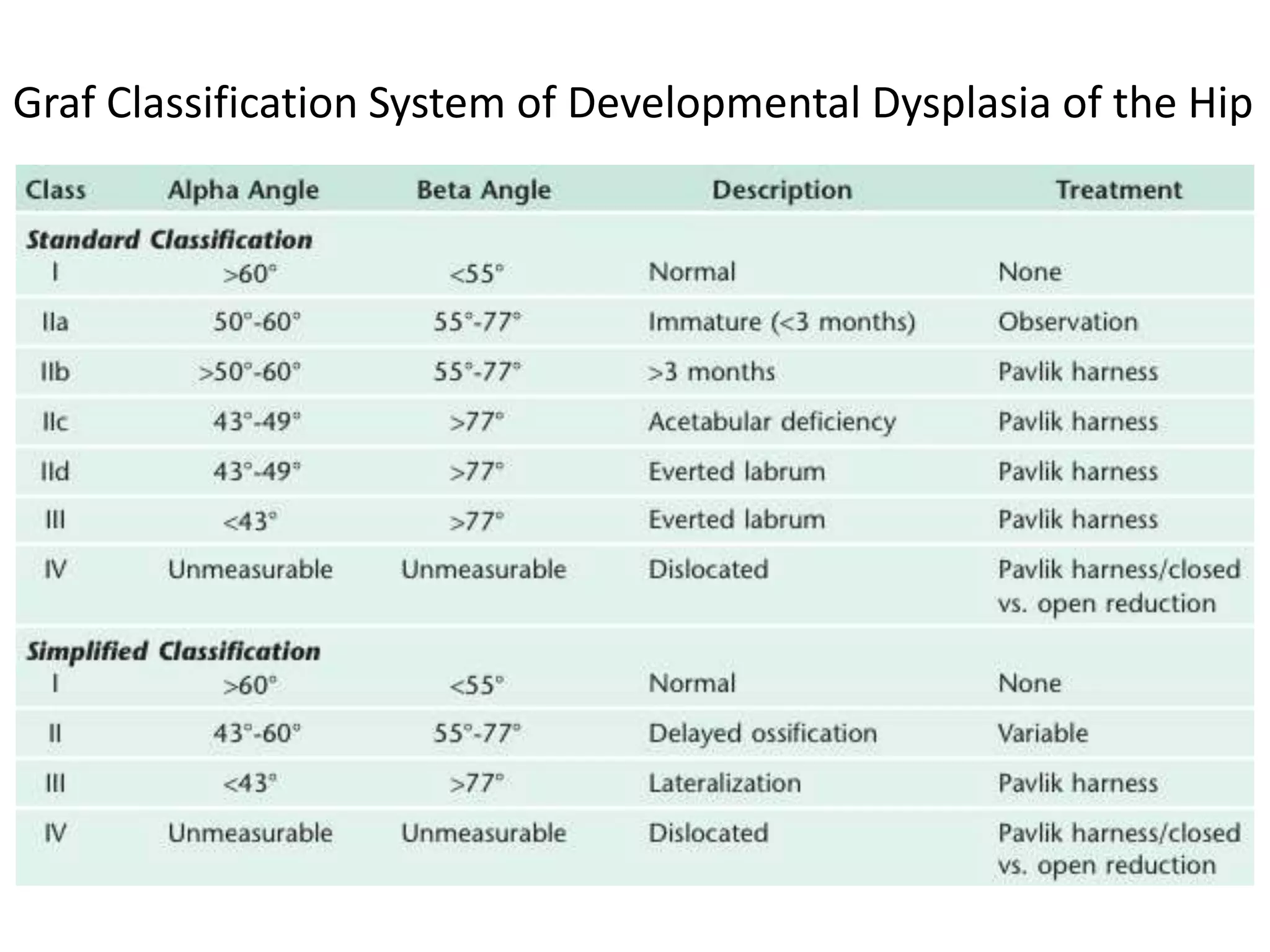
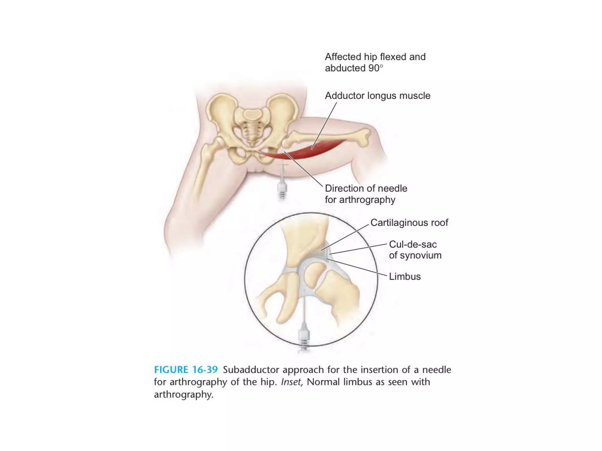
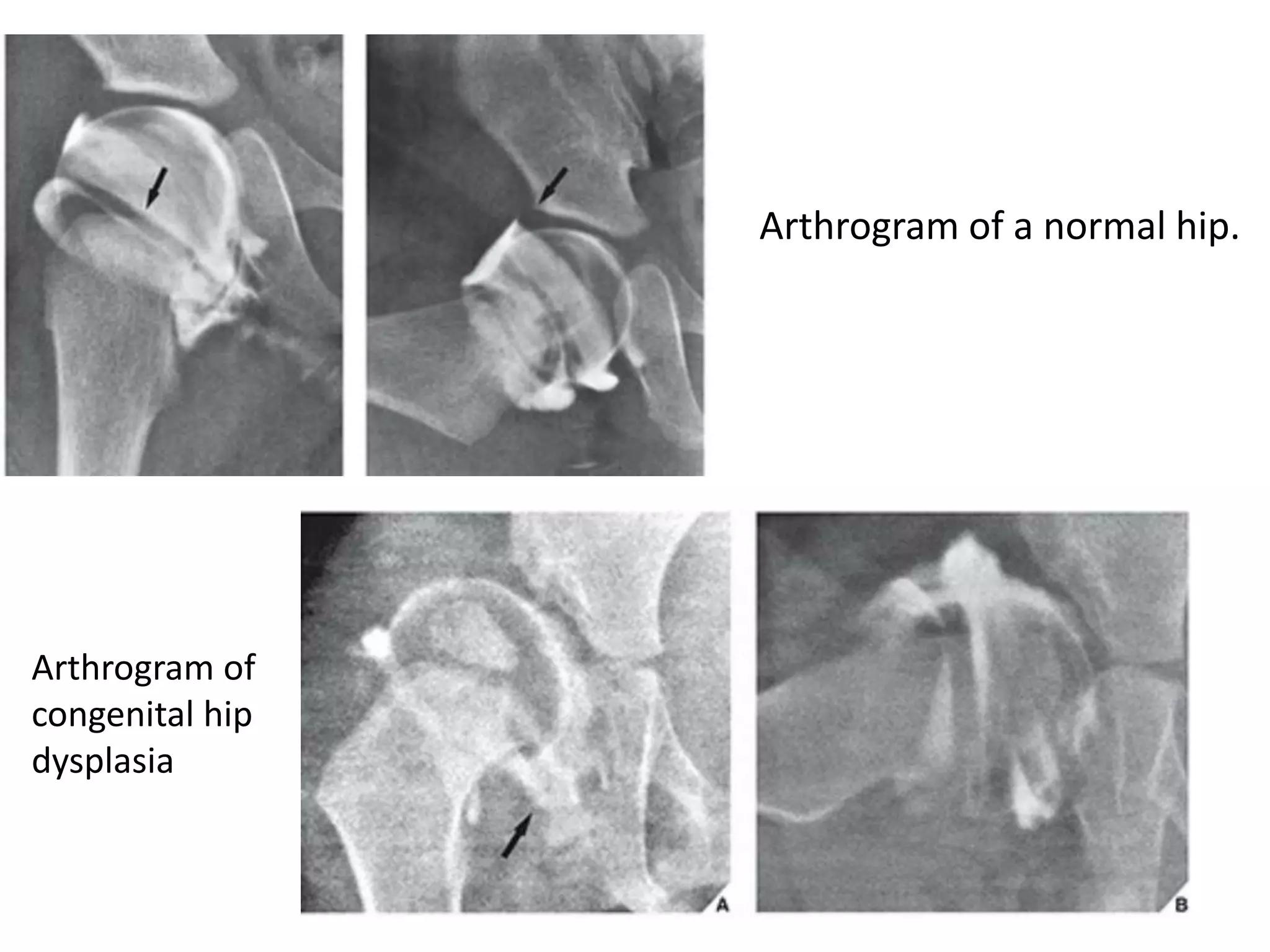
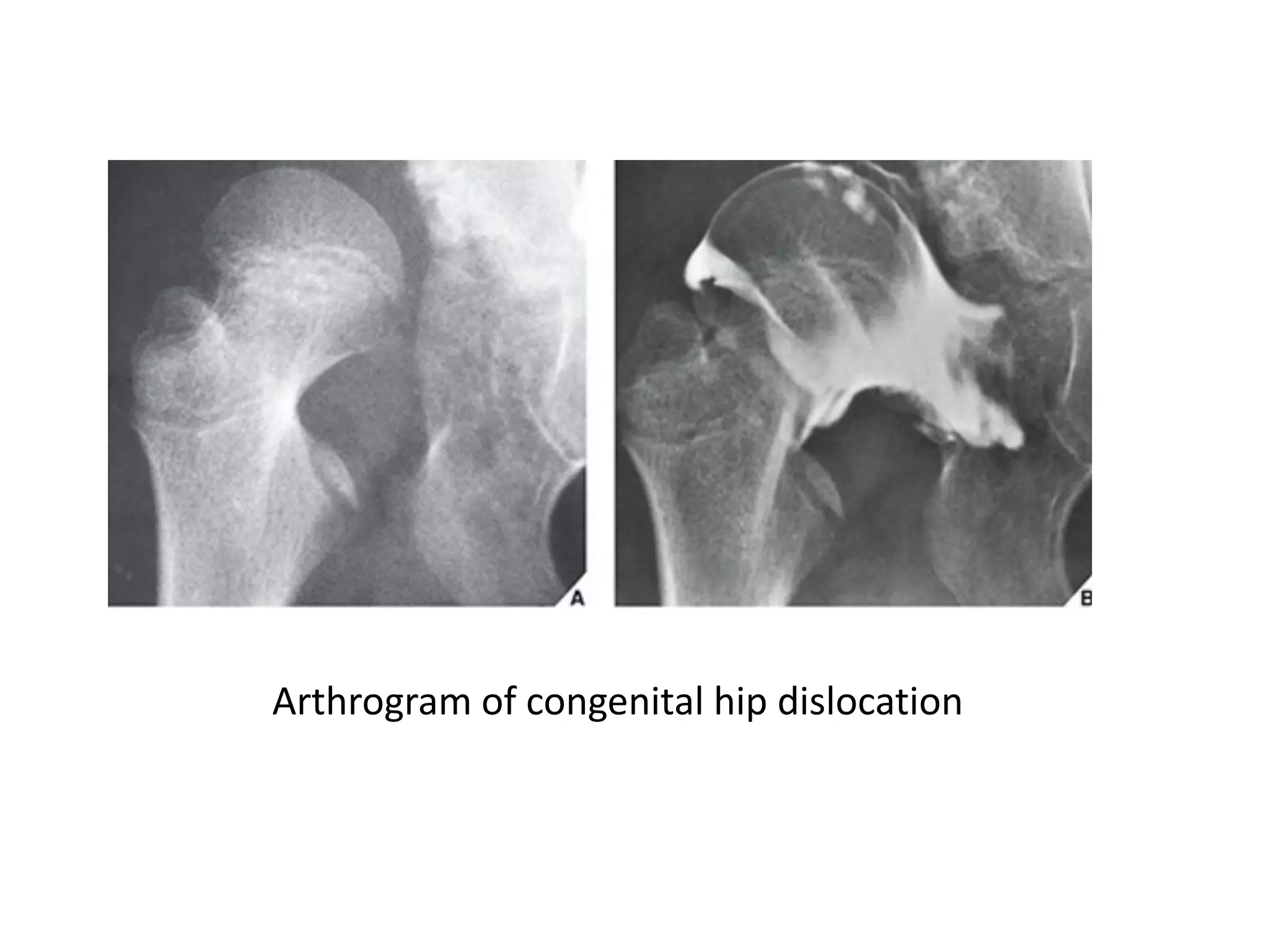
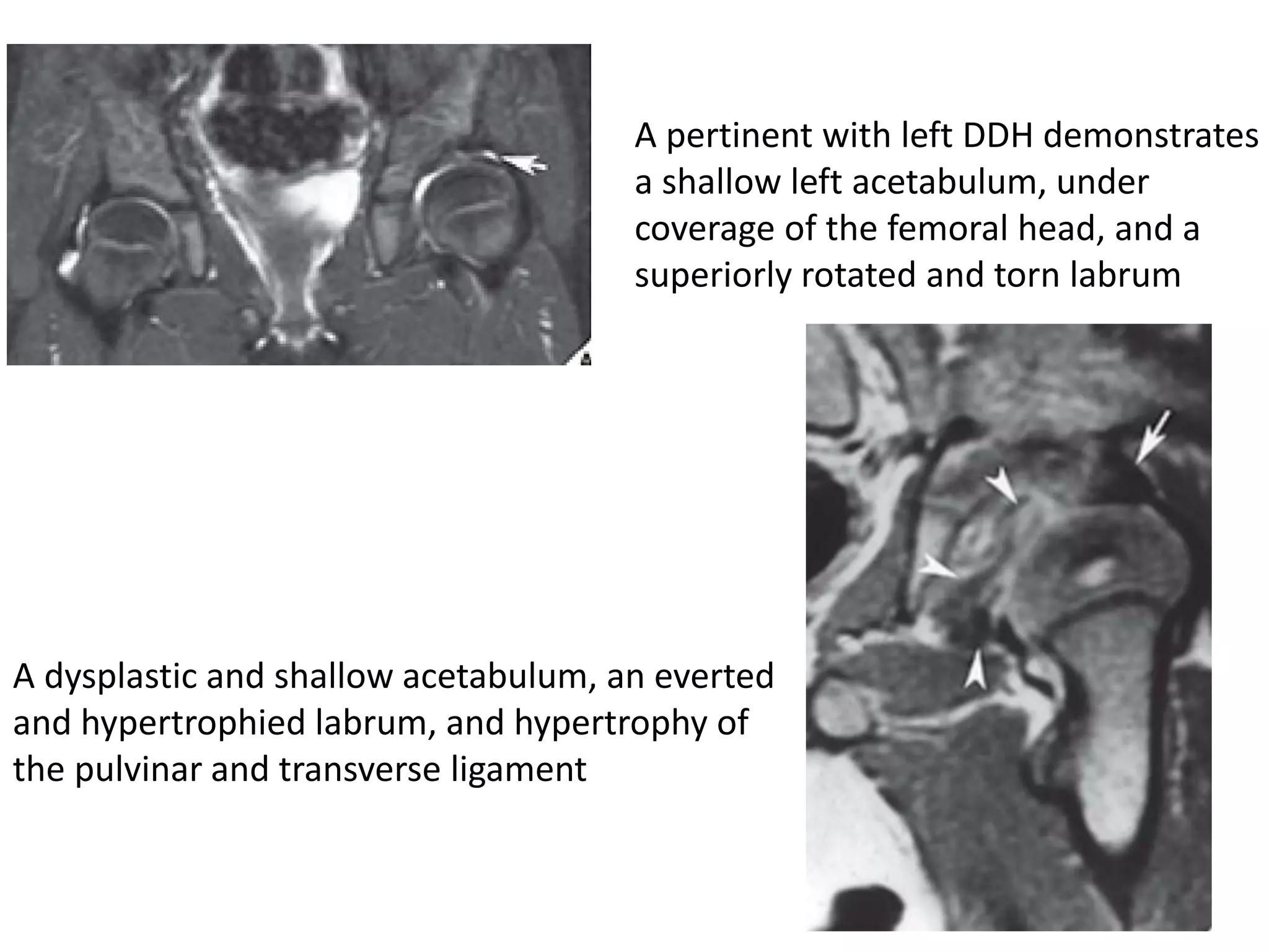
Developmental Dysplasia of Hip
Radiological findings
By Dr Kota Gandhi
II yr PG Orthopaedics
Kamineni Institute of Medical Sciences
Ultrasonography
The neonate’s hip is a difficult structure to image with standard
radiographic techniques because the hip is composed primarily of
cartilage.
Ultrasonography shows the soft anatomy of the hip and the
relationship of the femoral head and the acetabulum very well.
Four important questions about the use of ultrasonography
need to be resolved
First, how often does sonography identify a “silent” hip (i.e., one with a
negative clinical examination) that would become abnormal if it was not
treated?
Second, which sonographic findings indicate that the hip must be
treated?
Third, does the use of ultrasonography increase the rate of the treatment of hips
that would stabilize without such treatment?
Fourth, are there hips with normal ultrasound findings that later
become abnormal?
The hyaline articular cartilage of the hip had little echo, the capsule and
muscles had moderate echo, and the fibrocartilaginous labrum (as well as
the juncture of the femoral neck and the cartilaginous upper femur) had
strong echo.
Graf, who pioneered the use of ultrasonography for the evaluation of the infant
hip
Graf recommended a lateral imaging technique with the transducer
placed over the greater trochanter, and he noted that the examination
should take no more than 2 to 3 minutes.
Graf also proposed a classification system
that was based on the angles formed by
the sonographic structures of the hip.
The “baseline” is the line of the ilium as it
intersects the bony and cartilaginous
portions of the acetabulum.
The “inclination line” is the line along the
margin of the cartilaginous acetabulum.
The third line is the “acetabular roofline”
along the bony roof
The intersection of the roofline and the baseline forms the alpha
angle, whereas the intersection of the inclination line and the
baseline forms the beta angle.
A smaller alpha angle indicates a shallower bony acetabulum.
A smaller beta angle indicates a better cartilaginous acetabulum. In
other words, as the femoral head subluxates, the alpha angle
decreases, and the beta angle increases.
Graf Classification System of Developmental Dysplasia of the Hip
A number of authors believe that ultrasonography is too
sensitive and results in the over treatment of hips that would
otherwise develop normally.
Several authors have noted that universal screening
with ultrasonography results in an increase in the
number of infants who require splinting but does not
reduce the number of late dislocations.
Perhaps we should conclude that ultrasonography is a
valuable adjunct to the detection of neonatal hip
abnormalities but that it should be used judiciously to
avoid the over treatment of minor abnormalities.
A “normal” ultrasound at 6 weeks of age does not
guarantee a normal hip later in life.
Radiography
Plain radiography of the pelvis usually demonstrates a frankly
dislocated hip in individuals of any age. In newborns with
typical DDH, however, the unstable hip may appear
radiographically normal.
In the infant, the upper femur is not ossified, and most of the
acetabulum is cartilaginous.The triradiate cartilage lies between
the ilium, the ischium, and the pubis.
Several classic lines are helpful when evaluating the immature hip
The Hilgenreiner line is a line through the triradiate cartilages.
The Perkin line, which is drawn at the lateral margin of the acetabulum
perpendicular to the Hilgenreiner line.
The Shenton line is a curved line that begins at the lesser trochanter,
goes up the femoral neck, and connects with a line along the inner
margin of the pubis.
In a normal hip, the medial beak of the femoral metaphysis lies
in the lower, inner quadrant produced by the juncture of the
Perkin and Hilgenreiner lines.
The Shenton line is smooth in the normal hip.
In the dislocated hip, the metaphysis lies lateral to the Perkin
line; the Shenton line is broken because the femoral neck lies
cephalic to the line from the pubis.
Another useful measurement is the acetabular index, which is an angle formed by the
juncture of the Hilgenreiner line and a line drawn along the acetabular surface
In normal newborns, the acetabular index averages 27.5 degrees. At 6 months of age,
the mean is 23.5 degrees. By 2 years of age, the index usually decreases to 20 degrees.
Thirty degrees is considered the upper limit of normal.
In the older child, the center–edge angle is a useful measure of hip position.This
angle is formed at the juncture of the Perkin line with a line that connects the
lateral margin of the acetabulum to the center of the femoral head.
In children who are 6 to 13 years old,
an angle of more than 19 degrees has been
reported as normal;
In children who are 14 years old and older,
an angle of more than 25 degrees is
considered normal.
The acetabular teardrop figure, as seen on an anteroposterior (AP)
radiograph of the pelvis, is formed by severallines. It is derived from the
wall of the acetabulum laterally, the wall of the lesser pelvis medially,
and a curved line inferiorly, and it is formed by the acetabular notch.
The teardrop appears between 6 and 24 months of age in a
normal hip and later in a dislocated hip.
When the hip is dislocated or subluxated, the acetabular portion of the
teardrop loses its convexity, and the teardrop is wider from the superior to
the inferior directions.The reduced hip remodels the acetabulum, and the
teardrop gradually narrows.
The pelvis of a 1-year-old boy shows a slightly
flattened acetabulum
1-year-old girl shows congenital
superolateral subluxation of the left hip
2-year-old boy demonstrates complete
superolateral dislocation of the right hip
The Andrén-von Rosen line
The false-profile radiographic view represents a lateral view of
the acetabulum, and it is especially useful for evaluating anterior
acetabular dysplasia.
The patient is positioned 65 degrees obliquely to the x-ray
beam, with the foot parallel to the cassette.
The Severin classification has been used for many years to specify outcome in hips that
have been treated for DDH However, in 1997, Ward and associates reported poor levels of
intraobserver and interobserver reliability when the system was used
Arthrography
The arthrographic anatomy of the hip was well described by Severin in
1941. In the normal hip, the free border of the labrum is easily seen as a
sharp “thorn” overlying the femoral head.A recess of joint capsule
overlies this thorn.
• The needle should be directed toward the joint space.
• A small amount of contrast material is injected to be certain that the
joint has been entered;
• The contrast agent should flow freely around the femoral head.
• Another 1 mL of contrast agent is injected, and the needle is removed.
• Permanent films should be obtained for each significant position of the
hip. It is important to note the positions of maximum stability and
instability.
Arthrogram of a normal hip.
Arthrogram of
congenital hip
dysplasia
Arthrogram of congenital hip dislocation
Magnetic Resonance Imaging
MRI affords excellent anatomic visualization of the infant hip, but it is
not commonly used because of the expense involved and the need for
sedation.
Kashiwagi and associates proposed an MRI-based classification of hips
with DDH
• Group 1 hips had a sharp acetabular rim, and all were reducible with a
Pavlik harness.
• Group 2 hips had a rounded acetabular rim, and almost all could be
reduced with a Pavlik harness.
• Group 3 hips had an inverted acetabular rim, and none was reducible
with the harness.
A pertinent with left DDH demonstrates
a shallow left acetabulum, under
coverage of the femoral head, and a
superiorly rotated and torn labrum
A dysplastic and shallow acetabulum, an everted
and hypertrophied labrum, and hypertrophy of
the pulvinar and transverse ligament
MRI with gadolinium-contrast arthrography is an important tool
for the evaluation of the adolescent patient with hip dysplasia and
pain.
This technique allows for the evaluation of the condition of the
labrum and the articular cartilage of the hip joint.
Disruption and tears of the labrum, cartilage delamination, and
articular cartilage loss can be identified with this technique.
Screening Criteria
• All neonates should undergo a clinical examination for hip instability.
• Beyond that recommendation, there is a lack of consensus with regard
regard to further screening criteria.
• Most authors agree that infants with risk factors associated with DDH
should receive more careful screening that includes at least an
examination by an experienced examiner and possibly
ultrasonography.
• These risk factors include a family history of DDH, breech birth
position, torticollis, metatarsus adductus, and oligohydramnios.
• Because the incidence is higher among girls, these factors assume
greater importance in female infants.
• First-born whites also have an increased risk for DDH.
Thank You!

Developmental Dysplasia of Hip Radiological findings

  • 1.
    Developmental Dysplasia ofHip Radiological findings By Dr Kota Gandhi II yr PG Orthopaedics Kamineni Institute of Medical Sciences
  • 2.
    Ultrasonography The neonate’s hipis a difficult structure to image with standard radiographic techniques because the hip is composed primarily of cartilage. Ultrasonography shows the soft anatomy of the hip and the relationship of the femoral head and the acetabulum very well.
  • 3.
    Four important questionsabout the use of ultrasonography need to be resolved First, how often does sonography identify a “silent” hip (i.e., one with a negative clinical examination) that would become abnormal if it was not treated? Second, which sonographic findings indicate that the hip must be treated? Third, does the use of ultrasonography increase the rate of the treatment of hips that would stabilize without such treatment? Fourth, are there hips with normal ultrasound findings that later become abnormal?
  • 4.
    The hyaline articularcartilage of the hip had little echo, the capsule and muscles had moderate echo, and the fibrocartilaginous labrum (as well as the juncture of the femoral neck and the cartilaginous upper femur) had strong echo. Graf, who pioneered the use of ultrasonography for the evaluation of the infant hip Graf recommended a lateral imaging technique with the transducer placed over the greater trochanter, and he noted that the examination should take no more than 2 to 3 minutes.
  • 5.
    Graf also proposeda classification system that was based on the angles formed by the sonographic structures of the hip. The “baseline” is the line of the ilium as it intersects the bony and cartilaginous portions of the acetabulum. The “inclination line” is the line along the margin of the cartilaginous acetabulum. The third line is the “acetabular roofline” along the bony roof
  • 6.
    The intersection ofthe roofline and the baseline forms the alpha angle, whereas the intersection of the inclination line and the baseline forms the beta angle. A smaller alpha angle indicates a shallower bony acetabulum. A smaller beta angle indicates a better cartilaginous acetabulum. In other words, as the femoral head subluxates, the alpha angle decreases, and the beta angle increases.
  • 8.
    Graf Classification Systemof Developmental Dysplasia of the Hip
  • 9.
    A number ofauthors believe that ultrasonography is too sensitive and results in the over treatment of hips that would otherwise develop normally. Several authors have noted that universal screening with ultrasonography results in an increase in the number of infants who require splinting but does not reduce the number of late dislocations. Perhaps we should conclude that ultrasonography is a valuable adjunct to the detection of neonatal hip abnormalities but that it should be used judiciously to avoid the over treatment of minor abnormalities. A “normal” ultrasound at 6 weeks of age does not guarantee a normal hip later in life.
  • 10.
    Radiography Plain radiography ofthe pelvis usually demonstrates a frankly dislocated hip in individuals of any age. In newborns with typical DDH, however, the unstable hip may appear radiographically normal. In the infant, the upper femur is not ossified, and most of the acetabulum is cartilaginous.The triradiate cartilage lies between the ilium, the ischium, and the pubis.
  • 11.
    Several classic linesare helpful when evaluating the immature hip The Hilgenreiner line is a line through the triradiate cartilages. The Perkin line, which is drawn at the lateral margin of the acetabulum perpendicular to the Hilgenreiner line. The Shenton line is a curved line that begins at the lesser trochanter, goes up the femoral neck, and connects with a line along the inner margin of the pubis.
  • 13.
    In a normalhip, the medial beak of the femoral metaphysis lies in the lower, inner quadrant produced by the juncture of the Perkin and Hilgenreiner lines. The Shenton line is smooth in the normal hip. In the dislocated hip, the metaphysis lies lateral to the Perkin line; the Shenton line is broken because the femoral neck lies cephalic to the line from the pubis.
  • 14.
    Another useful measurementis the acetabular index, which is an angle formed by the juncture of the Hilgenreiner line and a line drawn along the acetabular surface In normal newborns, the acetabular index averages 27.5 degrees. At 6 months of age, the mean is 23.5 degrees. By 2 years of age, the index usually decreases to 20 degrees. Thirty degrees is considered the upper limit of normal.
  • 15.
    In the olderchild, the center–edge angle is a useful measure of hip position.This angle is formed at the juncture of the Perkin line with a line that connects the lateral margin of the acetabulum to the center of the femoral head. In children who are 6 to 13 years old, an angle of more than 19 degrees has been reported as normal; In children who are 14 years old and older, an angle of more than 25 degrees is considered normal.
  • 16.
    The acetabular teardropfigure, as seen on an anteroposterior (AP) radiograph of the pelvis, is formed by severallines. It is derived from the wall of the acetabulum laterally, the wall of the lesser pelvis medially, and a curved line inferiorly, and it is formed by the acetabular notch. The teardrop appears between 6 and 24 months of age in a normal hip and later in a dislocated hip. When the hip is dislocated or subluxated, the acetabular portion of the teardrop loses its convexity, and the teardrop is wider from the superior to the inferior directions.The reduced hip remodels the acetabulum, and the teardrop gradually narrows.
  • 17.
    The pelvis ofa 1-year-old boy shows a slightly flattened acetabulum 1-year-old girl shows congenital superolateral subluxation of the left hip 2-year-old boy demonstrates complete superolateral dislocation of the right hip
  • 18.
  • 19.
    The false-profile radiographicview represents a lateral view of the acetabulum, and it is especially useful for evaluating anterior acetabular dysplasia. The patient is positioned 65 degrees obliquely to the x-ray beam, with the foot parallel to the cassette.
  • 20.
    The Severin classificationhas been used for many years to specify outcome in hips that have been treated for DDH However, in 1997, Ward and associates reported poor levels of intraobserver and interobserver reliability when the system was used
  • 21.
    Arthrography The arthrographic anatomyof the hip was well described by Severin in 1941. In the normal hip, the free border of the labrum is easily seen as a sharp “thorn” overlying the femoral head.A recess of joint capsule overlies this thorn. • The needle should be directed toward the joint space. • A small amount of contrast material is injected to be certain that the joint has been entered; • The contrast agent should flow freely around the femoral head. • Another 1 mL of contrast agent is injected, and the needle is removed. • Permanent films should be obtained for each significant position of the hip. It is important to note the positions of maximum stability and instability.
  • 23.
    Arthrogram of anormal hip. Arthrogram of congenital hip dysplasia
  • 24.
    Arthrogram of congenitalhip dislocation
  • 25.
    Magnetic Resonance Imaging MRIaffords excellent anatomic visualization of the infant hip, but it is not commonly used because of the expense involved and the need for sedation. Kashiwagi and associates proposed an MRI-based classification of hips with DDH • Group 1 hips had a sharp acetabular rim, and all were reducible with a Pavlik harness. • Group 2 hips had a rounded acetabular rim, and almost all could be reduced with a Pavlik harness. • Group 3 hips had an inverted acetabular rim, and none was reducible with the harness.
  • 26.
    A pertinent withleft DDH demonstrates a shallow left acetabulum, under coverage of the femoral head, and a superiorly rotated and torn labrum A dysplastic and shallow acetabulum, an everted and hypertrophied labrum, and hypertrophy of the pulvinar and transverse ligament
  • 27.
    MRI with gadolinium-contrastarthrography is an important tool for the evaluation of the adolescent patient with hip dysplasia and pain. This technique allows for the evaluation of the condition of the labrum and the articular cartilage of the hip joint. Disruption and tears of the labrum, cartilage delamination, and articular cartilage loss can be identified with this technique.
  • 28.
    Screening Criteria • Allneonates should undergo a clinical examination for hip instability. • Beyond that recommendation, there is a lack of consensus with regard regard to further screening criteria. • Most authors agree that infants with risk factors associated with DDH should receive more careful screening that includes at least an examination by an experienced examiner and possibly ultrasonography. • These risk factors include a family history of DDH, breech birth position, torticollis, metatarsus adductus, and oligohydramnios. • Because the incidence is higher among girls, these factors assume greater importance in female infants. • First-born whites also have an increased risk for DDH.
  • 29.