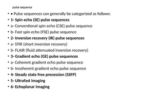
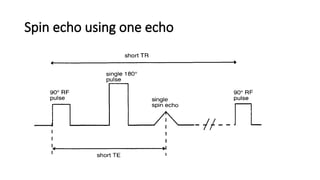
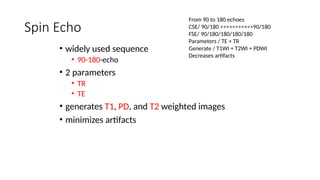
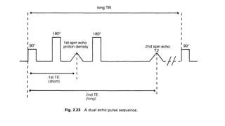
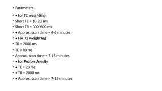
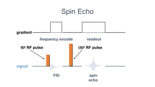
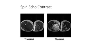
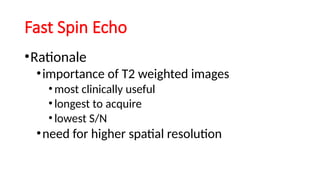
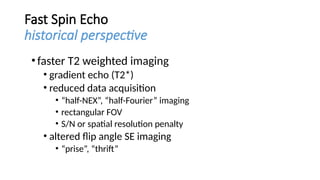
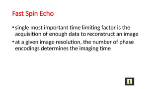
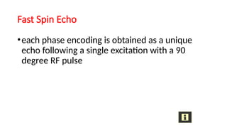
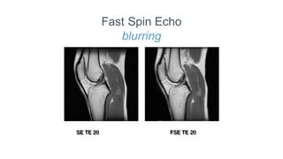
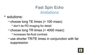
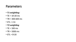
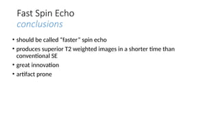
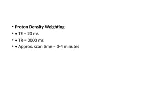
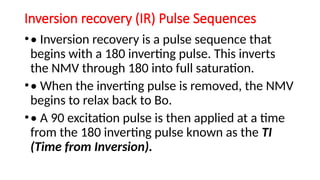
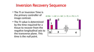
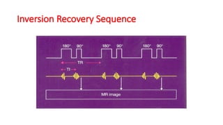
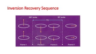
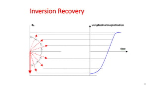
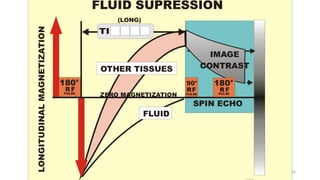
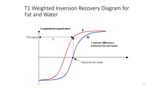
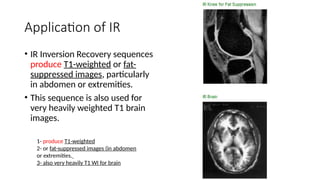
MRI
Pulse Sequence
pulse sequence
• • Pulse sequences can generally be categorized as follows:
• 1- Spin echo (SE) pulse sequences
• a- Conventional spin echo (CSE) pulse sequence
• b- Fast spin echo (FSE) pulse sequence
• 2- Inversion recovery (IR) pulse sequences
• a- STIR (short inversion recovery)
• b- FLAIR (fluid attenuated inversion recovery)
• 3- Gradient echo (GE) pulse sequences
• a- Coherent gradient echo pulse sequence
• b- Incoherent gradient echo pulse sequence
• 4- Steady state free precession (SSFP)
• 5- Ultrafast imaging
• 6- Echoplanar imaging
Spin echo using one echo
Spin Echo
• widely used sequence
• 90-180-echo
• 2 parameters
• TR
• TE
• generates T1, PD, and T2 weighted images
• minimizes artifacts
From 90 to 180 echoes
CSE/ 90/180 >>>>>>>>>>>90/180
FSE/ 90/180/180/180/180
Parameters / TE + TR
Generate / T1WI + T2WI + PDWI
Decreases artifacts
Conventional spin echo pulse sequence
Uses, Advantages & Disadvantages
• Uses:
• 1- These are the most commonly used pulse sequences
• 2- optimum signal-to-noise ratio (SNR) and contrast-to-noise ratio (CNR)
• 3- T1 weighted images are useful for demonstrating anatomy.
• 4- T2 weighted images also demonstrate pathology.
• Advantages:
• 1- Good Image quality
• 2- True T2 Weighting sensitive to pathology
• Disadvantages:
• 1- Scan times relatively long.
• 2- More RF power deposition in the body.
• Parameters
• • for T1 weighting
• Short TE = 10-20 ms
• Short TR = 300-600 ms
• • Approx. scan time = 4-6 minutes
• • For T2 weighting
• TR = 2000 ms
• TE = 80 ms
• Approx. scan time = 7-15 minutes
• • for Proton density
• • TE = 20 ms
• • TR = 2000 ms
• • Approx. scan time = 7-15 minutes
Spin Echo
FID spin
echo

RF pulse
readout
frequency encode
signal
gradient

RF pulse
Spin Echo Contrast
T1 weighted T2 weighted
Spin Echo Contrast
PD weighted T2 weighted
Spin echo using two echoes
•• This can be used to produce both a proton density
and a T2 weighted image in the TR time .
•• The first spin echo therefore has a short TE and a
long TR and is proton density weighted.
•• The second spin echo has a long TE and a long TR
and is T2 weighted
•Summary
•simultaneously generates PD and T2 weighted
images no time penalty for acquisition of PD
weighted image no mis-registration between echos
Multi Echo Spin Echo
Fast spin echo (FSE)
• • Fast spin echo (FSE) uses a 90° flip angle followed by
several 180° rephasing pulses to produce several spin
echoes in a given TR.
•• In conventional spin echo (CSE), only one line of K-space
is filled per TR. So the CSE takes longer scan time.
•• But in fast spin echo several lines of K-space will be filled
per TR.
•• Because of this reason, the scan times are reduced in fast
spin echo.
•The echo train length (ETL) refers to the
number of 180 rephasing pulses that
correspond to the number of lines of K
space filled per TR.
•• The longer the ETL, the shorter the scan
time as more lines of K space are filled per
TR.
Fast Spin Echo
•Rationale
•importance of T2 weighted images
•most clinically useful
•longest to acquire
•lowest S/N
•need for higher spatial resolution
Fast Spin Echo
historical perspective
•faster T2 weighted imaging
• gradient echo (T2*)
• reduced data acquisition
• “half-NEX”, “half-Fourier” imaging
• rectangular FOV
• S/N or spatial resolution penalty
• altered flip angle SE imaging
• “prise”, “thrift”
Fast Spin Echo
•single most important time limiting factor is the
acquisition of enough data to reconstruct an image
•at a given image resolution, the number of phase
encodings determines the imaging time
Fast Spin Echo
•each phase encoding is obtained as a unique
echo following a single excitation with a 90
degree RF pulse
Fast Spin Echo
pulse timing
RF
signal
readout
phase
slice
echo train





Fast spin echo pulse sequences
Uses- Advantages & Disadvantages
• • Uses:
• • In the central nervous system, pelvis and musculoskeletal regions.
• • Advantages:
• 1- Scan times greatly reduced
• 2- High resolution matrices
• 3- Image quality improved
• • Disadvantages:
• 1- motion affects increased (image blurring)
• 2- Fat bright on T2 weighted images
Fast Spin Echo
disadvantages
• each echo “belongs” to a different TE image
• combining the echos to form a single image
creates artifacts
• worse with shorter effective TE times
Fast Spin Echo
disadvantages
• reduced number of slices for equivalent TR SE scan
• MT effects alter image contrast
• TE time imprecise
• image blurring may occur
• fat remains relatively bright on long TR/long TE scans
• each echo “belongs” to a different TE image combining the
echos to form a single image creates artifacts
• worse with shorter effective TE times
Fast Spin Echo
blurring
SE TE 20 FSE TE 20
Fast Spin Echo
limitations
• solutions:
–choose long TE times (> 100 msec)
• don’t do PD imaging for detail
–choose long TR times (> 4000 msec)
• increases fat-fluid contrast
–use shorter TR/TE times in conjunction with fat
suppression
Parameters
• T1 weighting:
• TE = 10-20 ms
• TR = 300-600 ms
• ETL = 2-6
• T2 weighting
• TE = 100 ms
• TR = 5000 ms
• ETL = 8-20
Fast Spin Echo
conclusions
• should be called “faster” spin echo
• produces superior T2 weighted images in a shorter time than
conventional SE
• great innovation
• artifact prone
• Proton Density Weighting
• • TE = 20 ms
• • TR = 3000 ms
• • Approx. scan time = 3-4 minutes
Inversion recovery (IR) Pulse Sequences
•• Inversion recovery is a pulse sequence that
begins with a 180 inverting pulse. This inverts
the NMV through 180 into full saturation.
•• When the inverting pulse is removed, the NMV
begins to relax back to Bo.
•• A 90 excitation pulse is then applied at a time
from the 180 inverting pulse known as the TI
(Time from Inversion).
Inversion Recovery Sequence
• The TI or Inversion Time is
the primary controller of
image contrast.
• The TI value is determined
by the time required for a
tissue to recover from the
negative longitudinal axis to
the transverse plane. This
time is the null point.
Inversion Recovery Sequence
Inversion Recovery
32
33
T1 Weighted Inversion Recovery Diagram for
Fat and Water
34
Application of IR
• IR Inversion Recovery sequences
produce T1-weighted or fat-
suppressed images, particularly
in abdomen or extremities.
• This sequence is also used for
very heavily weighted T1 brain
images.
1- produce T1-weighted
2- or fat-suppressed images (in abdomen
or extremities.
3- also very heavily T1 WI for brain
Inversion recovery
Uses- Advantages & Disadvantages
• • Uses:
• • Inversion recovery was conventionally used to
• produce heavily T1 weighted images to
• demonstrate anatomy
• • Advantages
• 1- Very good SNR as the TR is long
• 2- Excellent T1 contrast
• • Disadvantages
• Long scan times unless used in conjunction with fast spin echo
1-STIR (short TI inversion recovery)
•• STIR
•• is an (IR)pulse sequence that uses a TI that
corresponds to the time it takes fat to recover from
full inversion to the transverse plane so that there is
no longitudinal magnetization corresponding to fat.
STIR
•• When the 90° excitation pulse is applied, the fat
vector is flipped through 90° to 180° and into full
saturation.
•• so that the signal from fat is nulled.
•• STIR is used to achieve suppression of the fat signal
in a T1 weighted image.
•• Generally, a TI value of around 100-200 ms is used.
STIR
STIR
• • Uses
• • Used to suppress the fat signal in T1 weighted
• image.
• • Disadvantage
• • Should not be used with contrast enhancement.
• • Parameters
• • TE = 10-30 ms
• • TR = 2000 ms
• • TI = 150-200 ms
• • Average scan time = 5-15 minutes.
STIR
41
2-FLAIR (fluid attenuated inversion recovery)
• • In FLAIR, the signal from CSF is nulled by selecting a TI corresponding to the
time of recovery of CSF from 180° to the transverse plane and there is no
longitudinal magnetization present in CSF.
• • When the 90° excitation pulse is applied, the CSF vector is flipped through
90° into full saturation again.
• • FLAIR is used to suppress the high CSF signal in T2 and proton density
weighted images so that pathology adjacent to the CSF is seen more clearly.
FLAIR
• Parameters
• • TE = Short or long depending on weighting
• • TR = 6000-8000 ms
• • TI = 2000 ms
• • Average scan time = 13-20 min
Coronal angulated slice of a T2
weighted, This MR image of the brain
shows white white matter and grey
matter of the cortex, brainstem, pons,
lateral ventricles and mastoid.
Brain MRI Coronal FLAIR
Coronal angulated slice of a T1
weigthed turbo inversion
recovery sequence of the brain,
showing cortex, pons, and
temporal lobes with
hippocampal regions in line with
the inner ears.
Brain MRI Inversion Recovery

4- MRI pulse sequences 1.pptx MRI pulse sequences MRI pulse sequences

  • 1.
  • 2.
    pulse sequence • •Pulse sequences can generally be categorized as follows: • 1- Spin echo (SE) pulse sequences • a- Conventional spin echo (CSE) pulse sequence • b- Fast spin echo (FSE) pulse sequence • 2- Inversion recovery (IR) pulse sequences • a- STIR (short inversion recovery) • b- FLAIR (fluid attenuated inversion recovery) • 3- Gradient echo (GE) pulse sequences • a- Coherent gradient echo pulse sequence • b- Incoherent gradient echo pulse sequence • 4- Steady state free precession (SSFP) • 5- Ultrafast imaging • 6- Echoplanar imaging
  • 3.
  • 4.
    Spin Echo • widelyused sequence • 90-180-echo • 2 parameters • TR • TE • generates T1, PD, and T2 weighted images • minimizes artifacts From 90 to 180 echoes CSE/ 90/180 >>>>>>>>>>>90/180 FSE/ 90/180/180/180/180 Parameters / TE + TR Generate / T1WI + T2WI + PDWI Decreases artifacts
  • 6.
    Conventional spin echopulse sequence Uses, Advantages & Disadvantages • Uses: • 1- These are the most commonly used pulse sequences • 2- optimum signal-to-noise ratio (SNR) and contrast-to-noise ratio (CNR) • 3- T1 weighted images are useful for demonstrating anatomy. • 4- T2 weighted images also demonstrate pathology. • Advantages: • 1- Good Image quality • 2- True T2 Weighting sensitive to pathology • Disadvantages: • 1- Scan times relatively long. • 2- More RF power deposition in the body.
  • 7.
    • Parameters • •for T1 weighting • Short TE = 10-20 ms • Short TR = 300-600 ms • • Approx. scan time = 4-6 minutes • • For T2 weighting • TR = 2000 ms • TE = 80 ms • Approx. scan time = 7-15 minutes • • for Proton density • • TE = 20 ms • • TR = 2000 ms • • Approx. scan time = 7-15 minutes
  • 8.
    Spin Echo FID spin echo  RFpulse readout frequency encode signal gradient  RF pulse
  • 9.
    Spin Echo Contrast T1weighted T2 weighted
  • 10.
    Spin Echo Contrast PDweighted T2 weighted
  • 11.
    Spin echo usingtwo echoes •• This can be used to produce both a proton density and a T2 weighted image in the TR time . •• The first spin echo therefore has a short TE and a long TR and is proton density weighted. •• The second spin echo has a long TE and a long TR and is T2 weighted
  • 12.
    •Summary •simultaneously generates PDand T2 weighted images no time penalty for acquisition of PD weighted image no mis-registration between echos Multi Echo Spin Echo
  • 13.
    Fast spin echo(FSE) • • Fast spin echo (FSE) uses a 90° flip angle followed by several 180° rephasing pulses to produce several spin echoes in a given TR. •• In conventional spin echo (CSE), only one line of K-space is filled per TR. So the CSE takes longer scan time. •• But in fast spin echo several lines of K-space will be filled per TR. •• Because of this reason, the scan times are reduced in fast spin echo.
  • 14.
    •The echo trainlength (ETL) refers to the number of 180 rephasing pulses that correspond to the number of lines of K space filled per TR. •• The longer the ETL, the shorter the scan time as more lines of K space are filled per TR.
  • 15.
    Fast Spin Echo •Rationale •importanceof T2 weighted images •most clinically useful •longest to acquire •lowest S/N •need for higher spatial resolution
  • 16.
    Fast Spin Echo historicalperspective •faster T2 weighted imaging • gradient echo (T2*) • reduced data acquisition • “half-NEX”, “half-Fourier” imaging • rectangular FOV • S/N or spatial resolution penalty • altered flip angle SE imaging • “prise”, “thrift”
  • 17.
    Fast Spin Echo •singlemost important time limiting factor is the acquisition of enough data to reconstruct an image •at a given image resolution, the number of phase encodings determines the imaging time
  • 18.
    Fast Spin Echo •eachphase encoding is obtained as a unique echo following a single excitation with a 90 degree RF pulse
  • 19.
    Fast Spin Echo pulsetiming RF signal readout phase slice echo train     
  • 20.
    Fast spin echopulse sequences Uses- Advantages & Disadvantages • • Uses: • • In the central nervous system, pelvis and musculoskeletal regions. • • Advantages: • 1- Scan times greatly reduced • 2- High resolution matrices • 3- Image quality improved • • Disadvantages: • 1- motion affects increased (image blurring) • 2- Fat bright on T2 weighted images
  • 21.
    Fast Spin Echo disadvantages •each echo “belongs” to a different TE image • combining the echos to form a single image creates artifacts • worse with shorter effective TE times
  • 22.
    Fast Spin Echo disadvantages •reduced number of slices for equivalent TR SE scan • MT effects alter image contrast • TE time imprecise • image blurring may occur • fat remains relatively bright on long TR/long TE scans • each echo “belongs” to a different TE image combining the echos to form a single image creates artifacts • worse with shorter effective TE times
  • 23.
  • 24.
    Fast Spin Echo limitations •solutions: –choose long TE times (> 100 msec) • don’t do PD imaging for detail –choose long TR times (> 4000 msec) • increases fat-fluid contrast –use shorter TR/TE times in conjunction with fat suppression
  • 25.
    Parameters • T1 weighting: •TE = 10-20 ms • TR = 300-600 ms • ETL = 2-6 • T2 weighting • TE = 100 ms • TR = 5000 ms • ETL = 8-20
  • 26.
    Fast Spin Echo conclusions •should be called “faster” spin echo • produces superior T2 weighted images in a shorter time than conventional SE • great innovation • artifact prone
  • 27.
    • Proton DensityWeighting • • TE = 20 ms • • TR = 3000 ms • • Approx. scan time = 3-4 minutes
  • 28.
    Inversion recovery (IR)Pulse Sequences •• Inversion recovery is a pulse sequence that begins with a 180 inverting pulse. This inverts the NMV through 180 into full saturation. •• When the inverting pulse is removed, the NMV begins to relax back to Bo. •• A 90 excitation pulse is then applied at a time from the 180 inverting pulse known as the TI (Time from Inversion).
  • 29.
    Inversion Recovery Sequence •The TI or Inversion Time is the primary controller of image contrast. • The TI value is determined by the time required for a tissue to recover from the negative longitudinal axis to the transverse plane. This time is the null point.
  • 30.
  • 31.
  • 32.
  • 33.
  • 34.
    T1 Weighted InversionRecovery Diagram for Fat and Water 34
  • 35.
    Application of IR •IR Inversion Recovery sequences produce T1-weighted or fat- suppressed images, particularly in abdomen or extremities. • This sequence is also used for very heavily weighted T1 brain images. 1- produce T1-weighted 2- or fat-suppressed images (in abdomen or extremities. 3- also very heavily T1 WI for brain
  • 36.
    Inversion recovery Uses- Advantages& Disadvantages • • Uses: • • Inversion recovery was conventionally used to • produce heavily T1 weighted images to • demonstrate anatomy • • Advantages • 1- Very good SNR as the TR is long • 2- Excellent T1 contrast • • Disadvantages • Long scan times unless used in conjunction with fast spin echo
  • 37.
    1-STIR (short TIinversion recovery) •• STIR •• is an (IR)pulse sequence that uses a TI that corresponds to the time it takes fat to recover from full inversion to the transverse plane so that there is no longitudinal magnetization corresponding to fat.
  • 38.
    STIR •• When the90° excitation pulse is applied, the fat vector is flipped through 90° to 180° and into full saturation. •• so that the signal from fat is nulled. •• STIR is used to achieve suppression of the fat signal in a T1 weighted image. •• Generally, a TI value of around 100-200 ms is used.
  • 39.
  • 40.
    STIR • • Uses •• Used to suppress the fat signal in T1 weighted • image. • • Disadvantage • • Should not be used with contrast enhancement. • • Parameters • • TE = 10-30 ms • • TR = 2000 ms • • TI = 150-200 ms • • Average scan time = 5-15 minutes.
  • 41.
  • 42.
    2-FLAIR (fluid attenuatedinversion recovery) • • In FLAIR, the signal from CSF is nulled by selecting a TI corresponding to the time of recovery of CSF from 180° to the transverse plane and there is no longitudinal magnetization present in CSF. • • When the 90° excitation pulse is applied, the CSF vector is flipped through 90° into full saturation again. • • FLAIR is used to suppress the high CSF signal in T2 and proton density weighted images so that pathology adjacent to the CSF is seen more clearly.
  • 43.
    FLAIR • Parameters • •TE = Short or long depending on weighting • • TR = 6000-8000 ms • • TI = 2000 ms • • Average scan time = 13-20 min
  • 44.
    Coronal angulated sliceof a T2 weighted, This MR image of the brain shows white white matter and grey matter of the cortex, brainstem, pons, lateral ventricles and mastoid. Brain MRI Coronal FLAIR Coronal angulated slice of a T1 weigthed turbo inversion recovery sequence of the brain, showing cortex, pons, and temporal lobes with hippocampal regions in line with the inner ears. Brain MRI Inversion Recovery