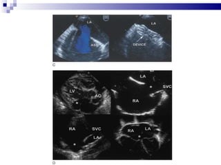
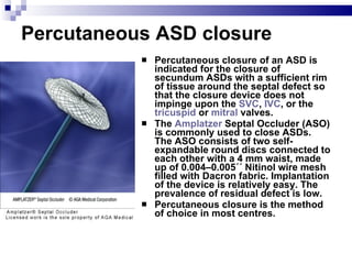
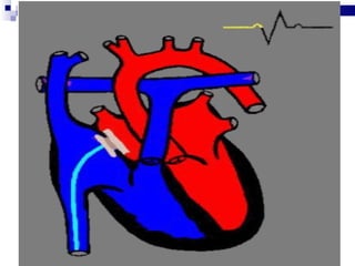
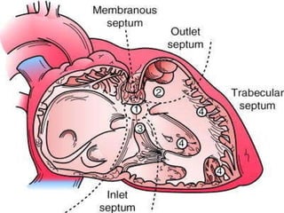
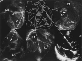
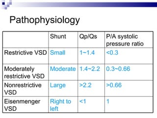
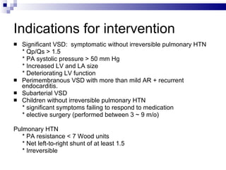
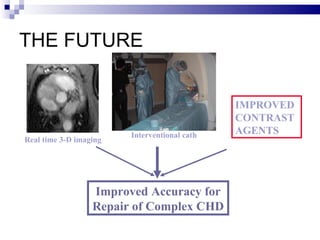
Non Surgical intervention in ASD and VSD
ASD
 
LA RA RUPV RV
LA LA RA RA Fig 9
ASD  An atrialseptal defect (ASD) is a hole in the wall between the two upper chambers of your heart.  The condition is present from birth (congenital).  Smaller atrialseptal defects may close on their own during infancy or early childhood. Large and long-standing atrialseptal defects can damage your heart and lungs. An adult who has had an undetected atrialseptal defect for decades may have a shortened life span from heart failure or high blood pressure in the lungs. Intervention is usually necessary to repair atrialseptal defects to prevent complications.
Symptoms of Atrial Septal Defect (ASD)  Many babies born with atrialseptal defects do not have signs or symptoms. In adults, signs or symptoms may not develop until age 30 or later. Your doctor may first uncover an atrialseptal defect during a regular checkup while listening to your heart using a stethoscope. Hearing a heart murmur may signal a hole in your heart.  Atrialseptal defects are often found when an ultrasound exam of the heart (echocardiogram) is done for another reason. Signs and symptoms of atrialseptal defects develop once damage occurs to the heart and lungs. Infants with larger atrialseptal defects may have poor appetites and not grow as they should. Adults and infants may have signs of heart failure or arrhythmias.
Causes of Atrial Septal Defect (ASD) Doctors know that heart defects present at birth (congenital) arise from errors early in the heart development, but there is often no clear cause. Genetics and environmental factors may play a role. An atrialseptal defect allows freshly oxygenated blood to flow from the left upper chamber of the heart (left atrium) into the right upper chamber of the heart (right atrium). There it mixes with deoxygenated blood and is pumped to the lungs, even though it is already refreshed with oxygen.  If the atrialseptal defect is large, this extra blood volume can overfill the lungs and overwork the heart. If not treated, the right side of the heart eventually enlarges and weakens. In some cases, the blood pressure in your lungs increases as well, leading to pulmonary hypertension.
When to seek medical advice for Atrial Septal Defect (ASD) Contact your doctor if you or your child has any of these signs or symptoms:- Poor appetite  Failure to gain weight (infants)  Bluish discoloration of the skin  Shortness of breath  Easy tiring  Swelling of legs, feet or abdomen  Heart palpitations or skipped beats These could be indications of heart failure or another complication of an atrialseptal defect.
Morphology 4 Types of ASD:  * ostium primum * ostium secundum * sinus venosus * coronary sinus defects  ↑ Left to right shunt:  * left ventricular compliance↓  * left atrial pressure  ↑
 
Clinical feature Large ASD (Qp/Qs > 2): CHF, pulmonary HTN or failure to thrive  Undetected ASD with a significant shunt (Qp/Qs > 1.5): symptoms ↑with aging 80% spontaneous closure occur < 1 y/o
Indications for intervention Asymptomatic children: Right heart dilation + a significant ASD (>5 mm)  without spontaneous closure  Significant ASD (Qp/Qs > 1.5)  ASD associated with RV volume overload To prevent paradoxical emboli in stroke patients  Pulmonary HTN:  * Resistance < 8.0 Wood units/m2  * Net left-to-right shunt of at least 1.5 * Pulmonary artery reactive to vasodilator (e.g., O2 or NO) * Lung biopsy revealed pulmonary arterial changes are potentially  reversible
Intervention Device closure  * For secundum ASD with stretched diameter < 36 mm + adequate rims  * Exception: 1) Anomalous pulmonary venous connection 2) Proximity to the AV valves / coronary sinus / systemic  venous drainage Surgery * For sinus venosus or ostium primum defects or with secundum defects with unsuitable anatomy  * primary suture closure or using a pericardial or synthetic patch
 
Percutaneous ASD closure Percutaneous closure of an ASD is indicated for the closure of secundum ASDs with a sufficient rim of tissue around the septal defect so that the closure device does not impinge upon the  SVC ,  IVC , or the  tricuspid  or  mitral  valves.  The  Amplatzer  Septal Occluder (ASO) is commonly used to close ASDs. The ASO consists of two self-expandable round discs connected to each other with a 4 mm waist, made up of 0.004–0.005´´ Nitinol wire mesh filled with Dacron fabric. Implantation of the device is relatively easy. The prevalence of residual defect is low.  Percutaneous closure is the method of choice in most centres.
 
VSD
Morphology 4 components of Septum:  Membranous, inlet, trabecular, outlet (conal, infundibular) part  3 Types of VSD * Muscular VSD  * Membranous VSD  * Doubly committed subarterial VSD (juxta-arterial/supracristal/ outlet / conal defects)
 
 
Pathophysiology Shunt Qp/Qs P/A systolic pressure ratio Restrictive VSD Small 1~1.4 <0.3 Moderately restrictive VSD Moderate 1.4~2.2 0.3~0.66 Nonrestrictive VSD Large >2.2 >0.66 Eisenmenger VSD Right to left <1 1
Indications for intervention Significant VSD:  symptomatic without irreversible pulmonary HTN * Qp/Qs > 1.5 * PA systolic pressure > 50 mm Hg * Increased LV and LA size * Deteriorating LV function Perimembranous VSD with more than mild AR + recurrent endocarditis. Subarterial VSD Children without irreversible pulmonary HTN * significant symptoms failing to respond to medication * elective surgery (performed between 3 ~ 9 m/o) Pulmonary HTN  * PA resistance < 7 Wood units * Net left-to-right shunt of at least 1.5 * Irreversible
Intervention Surgery: direct suture or with a patch  * Single-stage closure: large defect, CHF s/s, failure to thrive  * Perimembranous and muscular defects + normal PAP + no s/s  delayed op up to 1 year or more * Patient >10 y/o with a small defect (Qp:Qs < 1.5; normal PAP)     controversial Device closure: * Trabecular VSDs have proven more amenable  * Perimembranous VSDs is technically more challenging
Coarc Stent Repair
 
THE FUTURE Improved Accuracy for Repair of Complex CHD Real time 3-D imaging Interventional cath IMPROVED CONTRAST AGENTS
We Care Core Values “ We have a very simple business model that keeps you as the centre.” Having the industry’s most elaborate and exclusive Patient Care and Clinical Coordination teams stationed at each of our hospitals, we provide you the smoothest and seamless care ever imagined.
Thank You

ASD and VSD Closure

  • 1.
  • 2.
  • 3.
  • 4.
  • 5.
    LA LA RARA Fig 9
  • 6.
    ASD Anatrialseptal defect (ASD) is a hole in the wall between the two upper chambers of your heart. The condition is present from birth (congenital). Smaller atrialseptal defects may close on their own during infancy or early childhood. Large and long-standing atrialseptal defects can damage your heart and lungs. An adult who has had an undetected atrialseptal defect for decades may have a shortened life span from heart failure or high blood pressure in the lungs. Intervention is usually necessary to repair atrialseptal defects to prevent complications.
  • 7.
    Symptoms of AtrialSeptal Defect (ASD) Many babies born with atrialseptal defects do not have signs or symptoms. In adults, signs or symptoms may not develop until age 30 or later. Your doctor may first uncover an atrialseptal defect during a regular checkup while listening to your heart using a stethoscope. Hearing a heart murmur may signal a hole in your heart. Atrialseptal defects are often found when an ultrasound exam of the heart (echocardiogram) is done for another reason. Signs and symptoms of atrialseptal defects develop once damage occurs to the heart and lungs. Infants with larger atrialseptal defects may have poor appetites and not grow as they should. Adults and infants may have signs of heart failure or arrhythmias.
  • 8.
    Causes of AtrialSeptal Defect (ASD) Doctors know that heart defects present at birth (congenital) arise from errors early in the heart development, but there is often no clear cause. Genetics and environmental factors may play a role. An atrialseptal defect allows freshly oxygenated blood to flow from the left upper chamber of the heart (left atrium) into the right upper chamber of the heart (right atrium). There it mixes with deoxygenated blood and is pumped to the lungs, even though it is already refreshed with oxygen. If the atrialseptal defect is large, this extra blood volume can overfill the lungs and overwork the heart. If not treated, the right side of the heart eventually enlarges and weakens. In some cases, the blood pressure in your lungs increases as well, leading to pulmonary hypertension.
  • 9.
    When to seekmedical advice for Atrial Septal Defect (ASD) Contact your doctor if you or your child has any of these signs or symptoms:- Poor appetite Failure to gain weight (infants) Bluish discoloration of the skin Shortness of breath Easy tiring Swelling of legs, feet or abdomen Heart palpitations or skipped beats These could be indications of heart failure or another complication of an atrialseptal defect.
  • 10.
    Morphology 4 Typesof ASD: * ostium primum * ostium secundum * sinus venosus * coronary sinus defects ↑ Left to right shunt: * left ventricular compliance↓ * left atrial pressure ↑
  • 11.
  • 12.
    Clinical feature LargeASD (Qp/Qs > 2): CHF, pulmonary HTN or failure to thrive Undetected ASD with a significant shunt (Qp/Qs > 1.5): symptoms ↑with aging 80% spontaneous closure occur < 1 y/o
  • 13.
    Indications for interventionAsymptomatic children: Right heart dilation + a significant ASD (>5 mm) without spontaneous closure Significant ASD (Qp/Qs > 1.5) ASD associated with RV volume overload To prevent paradoxical emboli in stroke patients Pulmonary HTN: * Resistance < 8.0 Wood units/m2 * Net left-to-right shunt of at least 1.5 * Pulmonary artery reactive to vasodilator (e.g., O2 or NO) * Lung biopsy revealed pulmonary arterial changes are potentially reversible
  • 14.
    Intervention Device closure * For secundum ASD with stretched diameter < 36 mm + adequate rims * Exception: 1) Anomalous pulmonary venous connection 2) Proximity to the AV valves / coronary sinus / systemic venous drainage Surgery * For sinus venosus or ostium primum defects or with secundum defects with unsuitable anatomy * primary suture closure or using a pericardial or synthetic patch
  • 15.
  • 16.
    Percutaneous ASD closurePercutaneous closure of an ASD is indicated for the closure of secundum ASDs with a sufficient rim of tissue around the septal defect so that the closure device does not impinge upon the SVC , IVC , or the tricuspid or mitral valves. The Amplatzer Septal Occluder (ASO) is commonly used to close ASDs. The ASO consists of two self-expandable round discs connected to each other with a 4 mm waist, made up of 0.004–0.005´´ Nitinol wire mesh filled with Dacron fabric. Implantation of the device is relatively easy. The prevalence of residual defect is low. Percutaneous closure is the method of choice in most centres.
  • 17.
  • 18.
  • 19.
    Morphology 4 componentsof Septum: Membranous, inlet, trabecular, outlet (conal, infundibular) part 3 Types of VSD * Muscular VSD * Membranous VSD * Doubly committed subarterial VSD (juxta-arterial/supracristal/ outlet / conal defects)
  • 20.
  • 21.
  • 22.
    Pathophysiology Shunt Qp/QsP/A systolic pressure ratio Restrictive VSD Small 1~1.4 <0.3 Moderately restrictive VSD Moderate 1.4~2.2 0.3~0.66 Nonrestrictive VSD Large >2.2 >0.66 Eisenmenger VSD Right to left <1 1
  • 23.
    Indications for interventionSignificant VSD: symptomatic without irreversible pulmonary HTN * Qp/Qs > 1.5 * PA systolic pressure > 50 mm Hg * Increased LV and LA size * Deteriorating LV function Perimembranous VSD with more than mild AR + recurrent endocarditis. Subarterial VSD Children without irreversible pulmonary HTN * significant symptoms failing to respond to medication * elective surgery (performed between 3 ~ 9 m/o) Pulmonary HTN * PA resistance < 7 Wood units * Net left-to-right shunt of at least 1.5 * Irreversible
  • 24.
    Intervention Surgery: directsuture or with a patch * Single-stage closure: large defect, CHF s/s, failure to thrive * Perimembranous and muscular defects + normal PAP + no s/s  delayed op up to 1 year or more * Patient >10 y/o with a small defect (Qp:Qs < 1.5; normal PAP)  controversial Device closure: * Trabecular VSDs have proven more amenable * Perimembranous VSDs is technically more challenging
  • 25.
  • 26.
  • 27.
    THE FUTURE ImprovedAccuracy for Repair of Complex CHD Real time 3-D imaging Interventional cath IMPROVED CONTRAST AGENTS
  • 28.
    We Care CoreValues “ We have a very simple business model that keeps you as the centre.” Having the industry’s most elaborate and exclusive Patient Care and Clinical Coordination teams stationed at each of our hospitals, we provide you the smoothest and seamless care ever imagined.
  • 29.