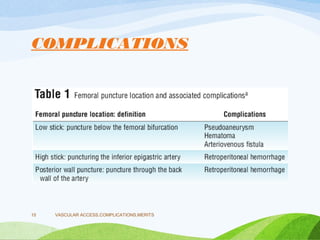
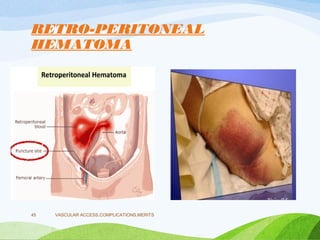
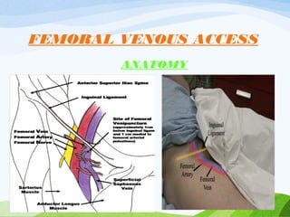
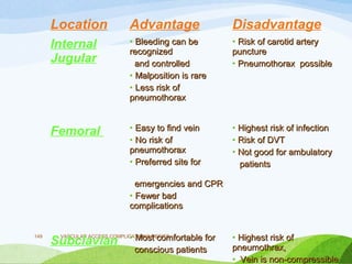
Vascular Access during
Cardiac Catheterization
VASCULAR
ACCESS,COMPLICATIONS,MERITS
1
Dr Vikash M,DM(SR).
NIMS,Hyderabad,India
vikasmedep@yahoo.co.in
VASCULAR ACCESS,COMPLICATIONS,MERITS
2
Profile
VASCULAR ACCESS
ARTERIAL VENOUS
VASCULAR
ACCESS,COMPLICATIONS,MERITS
3
Retrograde/antegrade.
ARTERIAL
FEMORAL RADIAL BRACHIAL ULNAR
VASCULAR
ACCESS,COMPLICATIONS,MERITS
4
Venous Accesss
VENOUS
FEMORAL IJV SUBCLAVIAN
VASCULAR
ACCESS,COMPLICATIONS,MERITS
5
TOPIC OVERLAY
• SITE
• COMPLICATIONS
• ADVANTAGES
• DIS-ADVANTAGES
• COMPARISON
• HEMOSTASIS
VASCULAR ACCESS,COMPLICATIONS,MERITS
6
FEMORAL ACCESS -
ANATOMY
VASCULAR ACCESS,COMPLICATIONS,MERITS
7
ARTERIAL ACCESS
• FEMORAL ARTERIAL ACCESS
• Most commonly used access for PCI
• SITE OF PUNCTURE
• Common femoral artery
• 2 cm below the inguinal ligament.
• Inguinal ligament runs from the anterior superior
iliac spine to the pubic tubercle
VASCULAR ACCESS,COMPLICATIONS,MERITS
8
• Some operators rely on the location of the inguinal
skin crease to position the skin nicks
• The position of the skin crease itself can be
misleading in obese patients
• Localization of the skin nick by
fluoroscopy
• Should show the nick to overlie the inferior border
of the femoral head
VASCULAR ACCESS,COMPLICATIONS,MERITS
9
COMPLICATIONS
• VASCULAR
• Hematoma
• Pseudo-aneurysm
• A-V fistula
• Retropertonial
hemorrhage
• Thrombosis
• NON VASCULAR
• Infections
VASCULAR ACCESS,COMPLICATIONS,MERITS
10
VASCULAR ACCESS,COMPLICATIONS,MERITS
11Nasser TK, Mohler ER 3rd, Wilensky RL, Hathaway DR. Peripheral
vascular complications following coronary interventional procedures.
Clin Cardiol.1995;18:609–614.
PROCEDURAL RISK
STRATIFICATION
• Low Risk:(<1% Complication Rate)
• Diagnostic Angiographic Procedures
• Moderate Risk: (1% to 3% Complication Rate)
• Routine Percutaneous Intervention
• High Risk (>3% Complication Rate)
• Primary PCI for acute MI, prolonged multivessel PCI , or
procedures that require larger sheath sizes (eg,>8F)
VASCULAR ACCESS,COMPLICATIONS,MERITS
13
RISK FACTORS
• Modifiable
• Site of puncture
• Number of attempts
• Size of sheath
• Sheath removal
• Medications
• Non modifiable
• Age
• Gender
• BMI
• Associated disorders -
CKD
VASCULAR ACCESS,COMPLICATIONS,MERITS
14
COMPLICATIONS
VASCULAR ACCESS,COMPLICATIONS,MERITS
15
• NUMBER OF ATTEMPTS
• Best – 1 attempt
• Better – 2 attempts
• Complications - > 2 attempts
• Shift to other side / site.
• SHEATH SIZE
• Greater the size more chances of complications
• Grossman and colleagues found that PCIs performed with 7F
and 8F sheath compared with 6F were associated with more
vascular compliactions
VASCULAR ACCESS,COMPLICATIONS,MERITS
16
• SHEATH REMOVAL
• Time
• Compression
• Adequate compression just proximal to the site of skin
puncture for at least 30 min is ideal.
• MEDICATIONS
• Anti platelets – oral , IV
• Anti coagulants.
VASCULAR ACCESS,COMPLICATIONS,MERITS
17
NON-MODIFIABLE
• AGE – elderly > younger
• SEX – female > male.
• BMI – high > low > normal
• # Delhaye et al – 6% high, 5.1% low, 2.0%
normal
• # Delhaye C, Wakabayashi K, Maluenda G, et al. Body mass index and
bleeding complications after percutaneous coronary ,AmHeart
J.2010;159:1139-1146.
VASCULAR ACCESS,COMPLICATIONS,MERITS
18
• ASSOCIATED CONDITIONS
• HYPERTENSION.
• Manoukian et al, patients with a higher systolic BP (140
vs 120 mm Hg;P= .02) were significantly more likely to
have complications than were patients with lower blood
pressures *
• CKD
• *Manoukian SV, Feit F, Mehran R, et al. Impact of major bleeding on 30-day
mortality and clinical outcomes in patients with acute coronary syndromes: an
analysis from the ACUITY Trial. J Am Coll Cardiol.2007;49:1362-1368
VASCULAR ACCESS,COMPLICATIONS,MERITS
19
• HEMATOMA
• Definition
• Collection of blood in the soft tissue
• Incidence
• Most common vascular complication
• 5- 20 %
• Clinical features
• Pain, swelling, induration
VASCULAR ACCESS,COMPLICATIONS,MERITS
20
VASCULAR ACCESS,COMPLICATIONS,MERITS
21
VASCULAR ACCESS,COMPLICATIONS,MERITS
22
VASCULAR ACCESS,COMPLICATIONS,MERITS
23
Rao SV, O'Grady K,. Impact of bleeding severity on clinical outcomes among patients with acute coronary syndromes. Am J
Cardiol. 2005;96:1200–1206
PSEUDO-ANEURYSM
• Definition
• A contained rupture; with disruption of all 3 layers of
the arterial wall.
• Occur when an arterial puncture site does not
adequately seal.
• Pulsatile blood tracks into the perivascular space
and is contained by the perivascular structures,
which then take on the appearance of a sac.
VASCULAR ACCESS,COMPLICATIONS,MERITS
24
VASCULAR ACCESS,COMPLICATIONS,MERITS
25
• One of the common vascular complications of cardiac and
peripheral angiographic procedures.
• The incidence after diagnostic catheterization ranges from
0.05% to 2%.
• When coronary or peripheral intervention is performed, the
incidence increases to 2% to 6%.*
• *Hessel SJ, Adams DF, Abrams HL. Complications of angiography. Radiology. 1981; 138: 273–281.
VASCULAR ACCESS,COMPLICATIONS,MERITS
26
VASCULAR ACCESS,COMPLICATIONS,MERITS
27
• DIAGNOSIS
• CLINICAL
• Pain and swelling at puncture site.
• Swelling from a large aneurysm may also lead to compression
of nerves and vessels with associated neuropathy, venous
thrombosis, claudication, or, rarely, critical limb ischemia.
• Local ischemia of the skin may lead to necrosis and infection.
• On physical examination, there may be a palpable pulsatile
mass or the presence of a bruit.
VASCULAR ACCESS,COMPLICATIONS,MERITS
28
• However, it should be noted that none of these physical
findings may be present.
• Pain that is disproportionate to that expected
after a PCI should undergo an doppler to
exclude pseudoaneurysm regardless of the
presence of a bruit.
VASCULAR ACCESS,COMPLICATIONS,MERITS
29
• IMAGING
• Duplex ultrasound
• The sensitivity is 94% with a specificity of 97%.
• Echolucent sac that expands and contracts with cardiac
contraction .
• On color Doppler, there is a swirling flow pattern with
turbulence in the chamber(s), there may be 1 or more
chambers.
• A tract connects the chamber to the feeding vessel.
• When a pulsed wave Doppler is placed within the track, a “to-
and-fro” signal is obtained
VASCULAR ACCESS,COMPLICATIONS,MERITS
30
VASCULAR ACCESS,COMPLICATIONS,MERITS
31
• TREATMENT
• Until the early 1990s, the only treatment available
was surgery.
• Since that time, USG compression, USG guided
thrombin injection, FemStop compression devices,
coil insertion, fibrin, adhesives, or balloon occlusion
have been used with variable success.
VASCULAR ACCESS,COMPLICATIONS,MERITS
32
• USG guided compression
• In 1991, Fellmeth and associates introduced a safe and
noninvasive method to treat PSA.
• Success rate of 75% to 98%.
• The ultrasound transducer is positioned and pressure is
applied to compress the chamber and tract while flow in the
native artery is allowed.
• Direct ultrasound visualization confirms cessation of flow.
• Compression is usually held for cycles of 10 minutes
VASCULAR ACCESS,COMPLICATIONS,MERITS
33
• The vertical angle created by the device does not
allow selective compression of the chamber and
tract.
• Nonselective compression leads to longer
compression times, more discomfort to the patient,
and a lower success rate, in addition to an increase
in complications such as DVT
• Body habitus, size, depth, and number of
chambers, as well as concurrent anticoagulation
may limit the success
VASCULAR ACCESS,COMPLICATIONS,MERITS
34
• In patients on anticoagulation, the success is 30% to 73%.
• In 100 cases of pseudoaneurysm, was successful in 94
patients (94%), which included 30 (86%) of 35 patients who
received anticoagulation and 64 (98%) of 65 patients who
were not on anticoagulation.*
• Katzenschlager R, Ugurluoglu A,. Incidence of pseudoaneurysm after diagnostic and therapeutic angiography.Radiology.
1995;195:463–466
VASCULAR ACCESS,COMPLICATIONS,MERITS
35
• DISADVANTAGES
• Long time - average
compression time to
achieve occlusion was 33
min with a range of 10 to
120 min*
• Painful
• Position
• Operator
• *Cox GS, Young JR, Gray BR, Grubb MW,
Hertzer NR. Ultrasound-guided compression
repair of postcatheterization
pseudoaneurysms:results of treatment in one
hundred cases.J Vasc Surg. 1994;19:683–686
• COMPLICATIONS
• Vasovagal reactions,
• Rupture,
• Skin necrosis, and
• DVT
VASCULAR ACCESS,COMPLICATIONS,MERITS
36
• Ultrasound-Guided Thrombin Injection
• The principle - thrombin is important in the conversion of
fibrinogen to fibrin.
• Thus a fibrin clot is formed instantaneously (even in the
presence of antiplatelet therapy or anticoagulation therapy.
• Success ranges from 91% to 100%*
• *Cope C, Zeit R. Coagulation of aneurysms by direct percutaneous thrombin injection. Am J
Roentgenol. 1986;147:383–387.
VASCULAR ACCESS,COMPLICATIONS,MERITS
37
• Complications
• DVT (if the thrombin is inadvertently injected into the vein),
• Pulmonary embolism
• Thrombosis of the artery.
• Allergic reactions and anaphylaxis.
• PARA ANEURYSMAL SALINE INJECTION
VASCULAR ACCESS,COMPLICATIONS,MERITS
38
VASCULAR ACCESS,COMPLICATIONS,MERITS
39
---
---
• ENDOLUMINAL MANAGEMET
• serves to exclude a pseudoaneurysm from the circulation
• Depends on the size of the pseudoaneurysmal neck and the
expendability of the donor artery .
• 2 broad categories: embolization and stent
• The width of the neck relative to the diameter of the donor
artery is the determining factor.
• A vital donor artery may be embolized in certain emergent
situations (eg, rupture with active bleeding); however, distal
blood flow must then be restored by means of a surgical
bypass procedure
VASCULAR ACCESS,COMPLICATIONS,MERITS
40
• COIL CLOSURE
• If the neck is narrow,
• made of either stainless steel or platinum.
• Polyester fibers are incorporated the coil to increase its
thrombogenicity
• Disadvantage
• Potential for recanalization.
• COVERED STENT
• Indications Large neck & larger artery
• Contraindication – mycotic aneurysm
VASCULAR ACCESS,COMPLICATIONS,MERITS
41
SURGERY
VASCULAR ACCESS,COMPLICATIONS,MERITS
42
• Disadvantages of surgery
• Requires anesthesia
• An incision usually in the groin, an area known to
become infected easily after a surgical procedure.
• Lumsden and colleagues reported a surgical
complication rate of 20% repair.
• Complications included bleeding, infection, neuralgia,
prolonged hospital stay
VASCULAR ACCESS,COMPLICATIONS,MERITS
43
• Prevention
• More complex procedures and more potent antithrombotic
therapy have led to the occurrence of more frequent
aneurysm formation.
• The most important strategies to prevent formation are:
• ● Assure a needle puncture in the proper location achieve
vascular access on the first puncture without access through
the posterior wall.
• ● Appropriate groin compression after sheath removal.
VASCULAR ACCESS,COMPLICATIONS,MERITS
44
RETRO-PERITONEAL
HEMATOMA
VASCULAR ACCESS,COMPLICATIONS,MERITS
45
RETROPERITONEAL
HEMATOMA
• Incidence
• 0.1 – 0.2 %
• CAUSES
• High puncture
• Inadvertent puncture of the posterior wall of the femoral or
iliac artery
• Exacerbated by the fact that patients receive antiplatelets,
anticoagulants
• Removal of catheter without wire
VASCULAR ACCESS,COMPLICATIONS,MERITS
46
• Retroperitoneal Hematoma After Percutaneous
Coronary Intervention: Prevalence, Risk Factors,
Management, Outcomes, and Predictors of
Mortality
• Volume 3, Issue 8, August 2010
VASCULAR ACCESS,COMPLICATIONS,MERITS
47
Retroperitoneal Hematoma After Percutaneous Coronary Intervention: Prevalence, Risk Factors,
Management, Outcomes, and Predictors of Mortality
Volume 3, Issue 8, August 2010 , JACC
• CLINICAL FEATURES
• High index of suspicion
• Very subtle clinical signs of haemorrhage
• Back, lower abdominal or groin discomfort and swelling,
• Pallor, sweating.
• Relative hypotension and mild tachycardia that transiently
improves with administration of fluids
• Unable to mount tachycardia because of beta-blockers, and
these patients usually become hypotensive with no change in
their heart rate
VASCULAR ACCESS,COMPLICATIONS,MERITS
49
• Retroperitoneal haematoma near or within the
iliopsoas muscle presents as femoral neuropathy,
begins with groin pain or leg weakness
• Sudden onset severe pain in the affected groin and
hip
• Iliopsoas spasm often results in the flexion and
external rotation of the hip, attempt to extend the
hip results in severe pain.
VASCULAR ACCESS,COMPLICATIONS,MERITS
50
• DIAGNOSIS
• CBP – fall in Hb
• IMAGING
• Ultrasonography of the abdomen and pelvis may detect
haematoma,.
• Limited by patient's discomfort, body habitus, underlying
bowel gas .
• Free fluid or blood in the retroperitoneum pass into the
abdominal or pelvic cavity
VASCULAR ACCESS,COMPLICATIONS,MERITS
51
• CT SCAN
• Type, site and extent of the fluid collections.
• Active bleeding can be seen as extravasation of contrast
material,
• CT angiography may show the site of the bleed and contrast
outside the vessels.
• MRI
• Useful in patients presenting with femoral neuropathy, as MRI
helps to rule out nerve root compression or spinal problems.
• Shows the site of the bleed.
• ANGIOGRAPHY
• Haemodynamically unstable, view to selective embolisation or
placement of a stent graft is indicated
VASCULAR ACCESS,COMPLICATIONS,MERITS
52
• MANAGEMENT
• Fluid resuscitation, blood transfusion and normalisation
of coagulation factor.
• No specific guidelines to suggest when to intervene with
endovascular or open surgery to stop the bleeding.
• If the patient is haemodynamically stable with no
evidence of on-going bleeding, conservative
management is recommended.
VASCULAR ACCESS,COMPLICATIONS,MERITS
53
• ENDOVASCULAR TREATMENT
• Indications - Panetta et al*
• Hemodynamic instablitiy
• Hemodynamiclly stable- four or more units of blood
transfusion within 24 h, or six or more units within 48 h
• Selective intra-arterial embolisation
• Stent-grafts
• Very few heterogeneous case series on stent-grafts in the
management of retroperitoneal haematoma
• * Panetta T, Sclafani SJ, Goldstein AS et al. Percutaneous transcatheter embolization for massive
bleeding from pelvic fractures. J Trauma 1985; 25: 1021-9
VASCULAR ACCESS,COMPLICATIONS,MERITS
54
• OPEN SURGERY
• Indications
• Unstable despite adequate fluid and blood product
resuscitation,
• Failed embloization / stent
• Abdominal compartment syndrome
VASCULAR ACCESS,COMPLICATIONS,MERITS
55
A-V FISTULA
VASCULAR ACCESS,COMPLICATIONS,MERITS
56
• DEFINITION
• Abnormal connections between the arterial and venous
system that bypass the normal anatomic capillary beds
• RISK FACTORS
• Female Hypertension
• Anticoagulation , Low or multiple punctures
• Obesity Advanced age.
VASCULAR ACCESS,COMPLICATIONS,MERITS
57
• Low groin puncture –
• Likely to access SFA just distal to the CFA bifurcation.
• The profunda femoris vein passes between the SFA and the
profunda femoris artery
• Punctures to the proximal SFA are particularly vulnerable to
causing AVF because the needle tip frequently punctures the
underlying profunda vein.
• Sheath placement –
• Dilation of the tract between an artery and vein reduces the
likelihood that the communication will close.
• The larger the sheath size, the greater the risk for AVF
VASCULAR ACCESS,COMPLICATIONS,MERITS
58
• INCIDENCE
• 0.I to 1 %*
• CLINICAL FEATURES
• Initially silent.
• Two days to several months
• Abnormal sensation in the groin, fatigue, new onset or
worsened lower extremity ischemia.
• *Glaser RL, McKellar D, Scher KS. Arteriovenous fistulas after cardiac catheterization. Arch Surg 1989; 124:1313.
VASCULAR ACCESS,COMPLICATIONS,MERITS
59
• Palpation and auscultation of the affected vessel
demonstrates a machinery-like murmur, bruit, hematoma
or pulsatile mass.
• The patient may exhibit lower extremity edema
• CONSEQUENCES
• DVT, nerve compression and new onset or worsened
varicose veins
• The most significant condition related to AVF is high-
output heart failure
VASCULAR ACCESS,COMPLICATIONS,MERITS
60
• DIAGNOSIS
• Duplex ultrasonography
• Current diagnostic test of choice
• High frequency, low resistance flow
• is typical ,with a mosaic color pattern.
• Often the specific artery and vein involved can be identified
• CT ANGIO
• Picks up the defect
• CONVENTIONAL ANGIO
• Appears as a blush with rapid filling of the adjacent deep vein
VASCULAR ACCESS,COMPLICATIONS,MERITS
61
• TREATMENT
• Most small asymptomatic AVFs thrombose spontaneously and thus
should be observed
• INDICATIONS:
• Clinical symptoms related to the AVF
• Steal syndrome causing claudication or distal limb ischemia
• Significant edema or venous insufficiency due to venous
hypertension
• Heart failure due to a high-flow fistula
• Progressive enlargement under ultrasound surveillance
• Iatrogenic AVFs that do not seal spontaneously
VASCULAR ACCESS,COMPLICATIONS,MERITS
62
• Ultrasound-guided compression
• Compression of sufficient force to abolish flow through the fistula
without unduly reducing distal perfusion
• Painful
• Failure is frequent because the fistula track is too short or the AV
fistula is too large
• Chronic AVFs (>2 to 3 weeks) rarely respond to compression.
• Ongoing anticoagulation also decreases success rates of UGC.
• Endovascular repair
• Covered stent placement or embolization techniques
• Surgery
VASCULAR ACCESS,COMPLICATIONS,MERITS
63
VASCULAR ACCESS,COMPLICATIONS,MERITS
64
VASCULAR ACCESS,COMPLICATIONS,MERITS
65
• Incidence
• 0.5 – 1%
• Diagnosis
• Doppler studies
• Peripheral angiogram
• Treatment
• Small – spontaneous lysis
• Large, limb threatening – thrombolysis / thrombectomy
VASCULAR ACCESS,COMPLICATIONS,MERITS
66
• INFECTIONS
• Incidence <1%,
• Bacterial infections occurred in 0.11% at a median of 1.7 days after the procedure*
• CLINICAL FEATURES
• Pain, erythema, swelling at puncture site
• Purulent discharge
• Fever
• *Munoz P, Blanco JR, Rdoriguez-Creixems M, et al. Blood stream infections after invasive nonsurgical cardiology procedures. Arch Intern Med 2001;161:2110–2115
VASCULAR ACCESS,COMPLICATIONS,MERITS
67
• Causes
• Improper shaving
• Improper scrubbing
• TREATMENT
• Antibiotics
• PREVENTION
• Appropriate shaving / scrubbing.
• Using sterile drapes.
VASCULAR ACCESS,COMPLICATIONS,MERITS
68
• FEMORAL NEUROPATHY
• Incidence
• 0.1 – 0.3%
• Mechanism
• Compression of the femoral nerve during puncture or by
hematoma
• Clinical features
• Tingling, numbness, weakness,
• Treatment
• Usually self remitting
VASCULAR ACCESS,COMPLICATIONS,MERITS
69
RADIAL ACCESS
VASCULAR ACCESS,COMPLICATIONS,MERITS
70
VASCULAR ACCESS,COMPLICATIONS,MERITS
71
VASCULAR ACCESS,COMPLICATIONS,MERITS
72
PRE -REQUISITES
VASCULAR ACCESS,COMPLICATIONS,MERITS
73
• Diagnostic Accuracy
• Ruengsakulrach et al.compared the Modified Allen’s Test with
Doppler and found the Modified Allen’s Test to have a
sensitivity of 100% and specificity of 97%.
• Glavin and Jones compared the Modified Allen’s Test with
Doppler a sensitivity of 87% to correctly diagnose the
presence of ulnar artery blood flow and a negative predictive
value of only 0.18; i.e., 80% of all abnormal Modified Allen’s
Test results in their study were incorrect.
• The diagnostic accuracy of the Modified Allen’s Test,
compared with ultrasound, was only 80%, with a sensitivity of
76% and a specificity of 82%
VASCULAR ACCESS,COMPLICATIONS,MERITS
74
BARBEAU TEST
VASCULAR ACCESS,COMPLICATIONS,MERITS
75
COMPLICATIONS
VASCULAR ACCESS,COMPLICATIONS,MERITS
76
• COMPLICATIONS
• PROCEDURAL
• Vaso vagal reaction
• Spasm
• Perforation / Dissection.
• POST PROCEDURE
• Occlusion
• Compartment Syndrome
• Pseudoaneurysm
•
VASCULAR ACCESS,COMPLICATIONS,MERITS
77
• VASOVAGAL REACTIONS
• Due to pain, anxiety
• PREVENTION
• Preprocedural sedation, analgesia, and adequate
local infiltration anesthesia decreases pain, anxiety,
and associated vagal output
VASCULAR ACCESS,COMPLICATIONS,MERITS
78
• SPASM
• Induced by the introduction of a sheath or catheter
• Mechanism
• Prominent medial layer that is largely dominated by alpha-1
receptors.
• Increased levels of catecholamines cause spasm
• Risk factors
• Female young age small artery
• Anxiety Unsuccessful guide wire passage
• Multiple catheter exchanges, prolonged procedure
VASCULAR ACCESS,COMPLICATIONS,MERITS
79
• Prevention
• Adequate vasodilatory cocktail containing
• NTG 100 – 200 mcg + 2.5 mg verapamil, + 40 U/Kg heparin
max 5000 u
• Hydrophilic catheters
• Smaller sheaths
• TREATMENT
• Additional doses of CCB, NTG,
• More analgesia / sedation
• Warm compress
VASCULAR ACCESS,COMPLICATIONS,MERITS
80
• HEMATOMA
• Rare , Easily compressed against bone
• Grades of hematoma *
• <5 cm (grade I),
• <10 cm (grade II),
• Distal to the elbow (grade III), and
• Proximal to elbow (grade IV).
• Hematomas grade III and IV are not directly related to the
puncture site, but result from wire damage to vessels and
small perforations
• Hamon M, Rasmussen LH, Manoukian SV, et al. Choice of arterial access site and
outcomes in patients with acute coronary syndromes managed with an early invasive
strategy: The ACUITY trial. EuroIntervention 2009;5:115–120
VASCULAR ACCESS,COMPLICATIONS,MERITS
81
• COMPARTMENT SYNDROME
• Limb threatening condition
• Foremarm hematoma compressing the ulnar &
radial artery – ischemia.
• incidence of 0.4%*
• *Tizon-Marcos H, Barbeau GR. Incidence of compartment syndrome of the arm in a large series of
transradial approach. J Interv Cardiol. 2008;21:380-384
VASCULAR ACCESS,COMPLICATIONS,MERITS
82
• Causes
• Unrecognized perforation at a distance from the puncture site,
• Unsuccessful compression at the puncture site, or
• Radial artery laceration induced at sheath insertion
• Prevention
• Early recognition and management of hematoma
• Treatment
• Surgical decompression.
VASCULAR ACCESS,COMPLICATIONS,MERITS
83
• AVULSION
• A sheath entrapped by arterial spasm should never be forcibly
removed because traumatic eversion radial artery may result.
• Prevention
• Repeat intra-arterial vasodilators,
• Additional patient sedation and/or analgesia, and
• Reinsertion of the introducer and guidewire may be
necessary.
• In refractory cases, axillary nerve blocks or general
anesthesia may be required for catheter removal
VASCULAR ACCESS,COMPLICATIONS,MERITS
84
• DISSECTION / PERFORATION
• Angiography of the arm should be performed if there is
difficulty with wire or catheter advancement since failure to
identify the problem may lead to vessel perforation or
dissection.
• Rather than aborting the procedure, it is worth trying to
carefully re-cross them with a soft 0.014 angioplasty wire.
• If this attempt is successful, the catheter will usually seal the
dissection or perforation, an
• Aborting the procedure will leave an unsealed dissection or
perforation that may be difficult to control
VASCULAR ACCESS,COMPLICATIONS,MERITS
85
• RADIAL ARTERY OCCLUSION
• Incidence
• 2% to 10% of patients*
• Risk factors**
• Lack of Heparin therapy
• Large artery-catheter mismatch,
• Female sex,
• Lack of pretreatment with clopidogrel,
• Diabetes, and
• Occlusive hemostasis
• Wu CJ, Lo PH, Chang KC, et al. * Transradial coronary angiography and angioplasty. Cathet Cardiovasc Diagn. 1997;40:159-163.
• **Stella PR, Kiemeneij F, Laarman GJ, Odekerken D,. Incidence and outcome of radial artery occlusion following transradial artery coronary angioplasty.Cathet
Cardiovasc Diagn 2007;40:156–158
VASCULAR ACCESS,COMPLICATIONS,MERITS
86
• Consequences
• Usually benign and asymptomatic due to the dual blood
supply to the hand
• Hand ischemia, gangrene
• Spontaneous recanalizaton appears to occur in 50% of
patients
• Prevention
• Pre-procedural heparin > 5000u, without heparin 60-70%, with
2-6%*
• Immediate sheath removal
• Vascular devices better than manual compression.
• *Spaulding C, Lefevre T, Funck F, et al. Left radial approach for coronary angiography:
results of a prospective study. Cathet Cardiovasc Diagn. 2010;39:365-370.
VASCULAR ACCESS,COMPLICATIONS,MERITS
87
VASCULAR ACCESS,COMPLICATIONS,MERITS
88
VASCULAR ACCESS,COMPLICATIONS,MERITS
90
VASCULAR ACCESS,COMPLICATIONS,MERITS
91
VASCULAR ACCESS,COMPLICATIONS,MERITS
92
VASCULAR ACCESS,COMPLICATIONS,MERITS
93
VASCULAR ACCESS,COMPLICATIONS,MERITS
94
VASCULAR ACCESS,COMPLICATIONS,MERITS
95
2011 ACCF/AHA/SCAI/ESC Guideline for Percutaneous
Coronary Intervention
Class IIa
1. The use of radial artery access can be useful to
decrease access site complications.
• CONDITIONS WHERE
READIAL ACCESS SHOULD BE
PREFERRED
• Absent femoral pulses
• Femoral bruit
• Femoral artery graft surgery
• Extensive inguinal scarring from past
surgery
• Surgery / radiation treatment near
inguinal area
• Extensively tortuous iliac system / lower
abdominal aorta
• Abdominal aortic aneurysm
• Patient request
• CONDITIONS WHERE
READIAL ACCESS SHOULD BE
AVOIDED
• Radial artery being considered for
CABG / AV fistula
• Upper limb atherosclerosis, extreme
tortuosity, Raynaud’s or Burger’s
disease.
• Need for 7F or larger sheath.
VASCULAR ACCESS,COMPLICATIONS,MERITS
96
FEMORAL vs RADIAL
APPROACH
VASCULAR ACCESS,COMPLICATIONS,MERITS
97
VASCULAR ACCESS,COMPLICATIONS,MERITS
98
VASCULAR ACCESS,COMPLICATIONS,MERITS
99
VASCULAR ACCESS,COMPLICATIONS,MERITS
100
VASCULAR ACCESS,COMPLICATIONS,MERITS
101
VASCULAR ACCESS,COMPLICATIONS,MERITS
102
VASCULAR ACCESS,COMPLICATIONS,MERITS
103
VASCULAR ACCESS,COMPLICATIONS,MERITS
104
Primary and Secondary Outcomes
Radial
(n=3507)
%
Femoral
(n=3514)
%
HR
HR 95% CI
95% CI P
P
Primary Outcome
Death, MI, Stroke,
Major Bleed
3.7 4.0 0.92
0.92 0.72-1.17
0.72-1.17 0.50
0.50
Secondary Outcomes
Death, MI, Stroke 3.2 3.2 0.98
0.98 0.77-1.28
0.77-1.28 0.90
0.90
Major Bleeding
0.7 0.9 0.73
0.73 0.43-1.23
0.43-1.23 0.23
0.23
VASCULAR ACCESS,COMPLICATIONS,MERITS
105
Other Outcomes
Radial
(n=3507)
Femoral
(n=3514)
P
P
Access site Cross-over (%) 7.6 2.0 <0.0001
<0.0001
PCI Procedure duration (min) 35 34 0.62
0.62
Fluoroscopy time (min) 9.3 8.0 <0.0001
<0.0001
Persistent pain at access site
>2 weeks (%)
2.6 3.1 0.22
0.22
Patient prefers assigned
access site for next
procedure (%)
90 49 <0.0001
<0.0001
VASCULAR ACCESS,COMPLICATIONS,MERITS
106
VASCULAR ACCESS,COMPLICATIONS,MERITS
107
BRACHIAL ARTERY
ACCESS
• SITE OF PUNCTURE
• Medial aspect of cubital fossa, 2-3 cm above the
elbow crease
• INDICATIONS
• Renal / lower limb artery angioplasty
• COMPLICATIONS
• Hematoma
VASCULAR ACCESS,COMPLICATIONS,MERITS
109
• Hand ischemia
• Due to thrombosis
• Compartment syndrome
• Hematoma extends into forearm
• Median nerve injury
• 0.2 and 1.4%
• Orator’s hand posture
• ACCESS trial – radial vs brachial access
• More complications with brachial approach ( 0.2% vs 2.6% p 0.03 )
VASCULAR ACCESS,COMPLICATIONS,MERITS
110
VASCULAR ACCESS,COMPLICATIONS,MERITS
111
ULNAR ARTERY ACCESS
• SITE
• 2-3 cm above the crease of wrist
• ADVANTAGES
• Preservation of radial artery for CABG
• PREREQUISITE
• Reverse Allen’s test
• COMPLICATIOS
• Same as with radial artery access
• EVIDENCE – PCVI-CUBA trial radial vs ulnar
• Success rate - access 96% vs 93%, PCI – 96% vs 95%,
complication rate 1% vs 1.2 % .
VASCULAR ACCESS,COMPLICATIONS,MERITS
112
HEMOSTASIS
• MANUAL COMPRESSION
• MECHANICAL COMPRESSION
• TOPICAL HEMOSTATIC AIDS
• VASCULAR CLOSURE DEVICES
1. Active
2. Passive .
VASCULAR ACCESS,COMPLICATIONS,MERITS
113
• MANUAL COMPRESSION
• Remains the “gold standard”
• Timing
• Diagnostic procedure - Immediately
• Interventions - 4-6 hrs, ACT < 170 sec
• Site
• 2 cm proximal to skin puncture site
• Duration
• 15 – 30 min, larger sheath, longer time
• 3-4 min compression / french.
• Dis advantage
• Ineffective compression due to fatigue
VASCULAR ACCESS,COMPLICATIONS,MERITS
114
VASCULAR ACCESS,COMPLICATIONS,MERITS
115
FEM-STOP
VASCULAR ACCESS,COMPLICATIONS,MERITS
116
70mmHg while sheath removal
MAP for 15 min
MAP for 15 min
Gradually reduce to 30mmHg over 2 hrs and remove.
CLAMP-EASE
VASCULAR ACCESS,COMPLICATIONS,MERITS
117
METAL PAD
C-ARM
PRESSURE PAD
• Advantages
• More effective compression
• Dis-advantages
• Doesn’t decrease time to hemostasis / ambulation.
• Patient discomfort
VASCULAR ACCESS,COMPLICATIONS,MERITS
118
TOPICAL HEMOSTATIC
AIDS
• A variety of topical patches, pads, bandages, and powders are
available for use to assist with hemostasis with manual
compression.
• Accelerate the clotting process and thus accelerate hemostasis
• Advantages
• Topical agents leave no foreign body behind, and act by
• Accelerating natural hemostasis.
• Topical agents still require manual compression
VASCULAR ACCESS,COMPLICATIONS,MERITS
119
VASCULAR ACCESS,COMPLICATIONS,MERITS
120
VASCULAR CLOSURE
DEVICES
• Introduced in 1995 to decrease vascular
complications and reduce the time to
hemostasis and ambulation.
• CLASSIFICATION
• PASSIVE
• enhance hemostasis with prothrombotic
material or mechanical compression, but do not
achieve prompt hemostasis or shorten the time
to ambulation
• ACTIVE
VASCULAR ACCESS,COMPLICATIONS,MERITS
121
VASCULAR ACCESS,COMPLICATIONS,MERITS
122
ANGIO-SEAL
• Success rate –
• 90 - 97%*
• Advantages
• One of the easiest devices to learn and use. •
• Has a very high initial success rate. •
• The collagen plug in the tract also acts to reduce oozing from the
site.
• The retained components of the device are completely resorbed
• *Applegate RJ, Grabarczyk MA, Little WC et al. Vascular closure devices during percutaneous revascularization.
J Am Coll Cardiol 2002;40:78–83.
VASCULAR ACCESS,COMPLICATIONS,MERITS
124
• Disadvantages
• The intravascular anchor has the potential to further
obstruct a heavily diseased vessel.
• Embolization of the intravascular anchor.
• Repeat access of the same vessel within 90 days of
device deployment should be avoided using the
same puncture site.
• Infection.
VASCULAR ACCESS,COMPLICATIONS,MERITS
125
STAR CLOSE DEVICE
• Success rate
• 87%–97%*
• Advantages
• deploys on the outside of the artery, leaving nothing in the lumen.
• Re-puncture through a deployed Starclose clip performed safely at
any time.
• Disadvantages
• Oozing.
• *Applegate RJ, Grabarczyk MA, Little WC et al. Vascular closure devices during percutaneous revascularization.
J Am Coll Cardiol 2002;40:78–83.
VASCULAR ACCESS,COMPLICATIONS,MERITS
128
• Devices:2011 ACCF/AHA/SCAI Guideline for Percutaneous
Coronary Intervention Recommendations
• Class I
• 1. Patients considered for vascular closure devices should undergo a femoral
angiogram to ensure their anatomic suitability for deployment.
• Class IIa
• 1. The use of vascular closure devices is reasonable for the purposes of
achieving faster hemostasis and earlier ambulation
• Class III: NO BENEFIT
• 1. The routine use of vascular closure devices is not recommended for the
purpose of decreasing vascular complications
VASCULAR ACCESS,COMPLICATIONS,MERITS
134
TR band
VASCULAR ACCESS,COMPLICATIONS,MERITS
136
FEMORAL VENOUS ACCESS
ANATOMY
VASCULAR ACCESS,COMPLICATIONS,MERITS
137
• INDICATIONS
• Right heart study TPI
• IVC filter Venous access
• Puncture site
• Medial to femoral artery
• Needle held at 45 degree angle
• Skin insertion 2 cm below inguinal ligament
• Aim toward umbilicus
VASCULAR ACCESS,COMPLICATIONS,MERITS
138
COMPLICATIONS
Local Hematoma
Retroperitoneal hematoma
Pseudoaneurysm
AV fistula
Femoral neuropathy
Infection
DVT
VASCULAR ACCESS,COMPLICATIONS,MERITS
139
SUBCLAVIAN VENOUS
ACCESS
• INDICATIONS
• PPI leads
• TPI
• IVC filter
• Central venous access
• Chemoport
VASCULAR ACCESS,COMPLICATIONS,MERITS
140
• Positioning
• Right side preferred
• Supine position, head neutral, arm abducted
• Trendelenburg (10-15 degrees)
• Shoulders neutral with mild retraction
• Puncture site
• Junction of middle and medial thirds of clavicle
• At the small tubercle in the medial deltopectoral
groove
• Needle should be parallel to skin
• Aim towards the supraclavicular notch and just under
the clavicle
VASCULAR ACCESS,COMPLICATIONS,MERITS
141
• COMPLICATIONS
• Infection Bleeding Pneumothorax
• Thrombosis Air embolization Brachial plexus
injury
• AVOIDED IN
• Coagulopathy Thrombloysis Chest wall
deformity
VASCULAR ACCESS,COMPLICATIONS,MERITS
143
IJV ACCESS
• INDICATIONS
• TPI
• Central venous line
• Positioning
• Right side preferred
• Trendelenburg position
• Head turned slightly away from side of
venipuncture
VASCULAR ACCESS,COMPLICATIONS,MERITS
144
Needle placement
• Central approach
• Locate the triangle formed by the clavicle and the
sternal and clavicular heads of the SCM muscle
• Place 3 fingers of left hand on carotid artery
• Place needle at 30 to 40 degrees to the skin,
lateral to the carotid artery
• Aim toward the ipsilateral nipple under the medial
border of the lateral head of the SCM muscle
• Vein is 1-1.5 cm deep, avoid deep probing in the
neck
VASCULAR ACCESS,COMPLICATIONS,MERITS
145
COMPLICATIONS
• Infection Bleeding – airway compression
• Thrombosis Air embolization Pneumothorax
• AVOIDED IN
• Trendelenburg tilt is not possible – pulmonary
edema
• Child < 1 yr who cannot be sedated / paralysed
VASCULAR ACCESS,COMPLICATIONS,MERITS
147
COMPLICATIONS
VASCULAR ACCESS,COMPLICATIONS,MERITS
149
Location Advantage Disadvantage
Internal
Jugular
• Bleeding can be
Bleeding can be
recognized
recognized
and controlled
and controlled
• Malposition is rare
Malposition is rare
• Less risk of
Less risk of
pneumothorax
pneumothorax
• Risk of carotid artery
Risk of carotid artery
puncture
puncture
• Pneumothorax possible
Pneumothorax possible
Femoral • Easy to find vein
Easy to find vein
• No risk of
No risk of
pneumothorax
pneumothorax
• Preferred site for
Preferred site for
emergencies and CPR
emergencies and CPR
• Fewer bad
Fewer bad
complications
complications
• Highest risk of infection
Highest risk of infection
• Risk of DVT
Risk of DVT
• Not good for ambulatory
Not good for ambulatory
patients
patients
Subclavian • Most comfortable for
Most comfortable for
conscious patients
conscious patients
• Highest risk of
Highest risk of
pneumothrax,
pneumothrax,
• Vein is non-compressible
Vein is non-compressible
Thank You.
VASCULAR ACCESS,COMPLICATIONS,MERITS
150

vascularaccessincardiaccatheterization-130517040431-phpapp02 (1).pdf

  • 1.
    Vascular Access during CardiacCatheterization VASCULAR ACCESS,COMPLICATIONS,MERITS 1 Dr Vikash M,DM(SR). NIMS,Hyderabad,India vikasmedep@yahoo.co.in
  • 2.
  • 3.
  • 4.
    Retrograde/antegrade. ARTERIAL FEMORAL RADIAL BRACHIALULNAR VASCULAR ACCESS,COMPLICATIONS,MERITS 4
  • 5.
    Venous Accesss VENOUS FEMORAL IJVSUBCLAVIAN VASCULAR ACCESS,COMPLICATIONS,MERITS 5
  • 6.
    TOPIC OVERLAY • SITE •COMPLICATIONS • ADVANTAGES • DIS-ADVANTAGES • COMPARISON • HEMOSTASIS VASCULAR ACCESS,COMPLICATIONS,MERITS 6
  • 7.
    FEMORAL ACCESS - ANATOMY VASCULARACCESS,COMPLICATIONS,MERITS 7
  • 8.
    ARTERIAL ACCESS • FEMORALARTERIAL ACCESS • Most commonly used access for PCI • SITE OF PUNCTURE • Common femoral artery • 2 cm below the inguinal ligament. • Inguinal ligament runs from the anterior superior iliac spine to the pubic tubercle VASCULAR ACCESS,COMPLICATIONS,MERITS 8
  • 9.
    • Some operatorsrely on the location of the inguinal skin crease to position the skin nicks • The position of the skin crease itself can be misleading in obese patients • Localization of the skin nick by fluoroscopy • Should show the nick to overlie the inferior border of the femoral head VASCULAR ACCESS,COMPLICATIONS,MERITS 9
  • 10.
    COMPLICATIONS • VASCULAR • Hematoma •Pseudo-aneurysm • A-V fistula • Retropertonial hemorrhage • Thrombosis • NON VASCULAR • Infections VASCULAR ACCESS,COMPLICATIONS,MERITS 10
  • 11.
    VASCULAR ACCESS,COMPLICATIONS,MERITS 11Nasser TK,Mohler ER 3rd, Wilensky RL, Hathaway DR. Peripheral vascular complications following coronary interventional procedures. Clin Cardiol.1995;18:609–614.
  • 13.
    PROCEDURAL RISK STRATIFICATION • LowRisk:(<1% Complication Rate) • Diagnostic Angiographic Procedures • Moderate Risk: (1% to 3% Complication Rate) • Routine Percutaneous Intervention • High Risk (>3% Complication Rate) • Primary PCI for acute MI, prolonged multivessel PCI , or procedures that require larger sheath sizes (eg,>8F) VASCULAR ACCESS,COMPLICATIONS,MERITS 13
  • 14.
    RISK FACTORS • Modifiable •Site of puncture • Number of attempts • Size of sheath • Sheath removal • Medications • Non modifiable • Age • Gender • BMI • Associated disorders - CKD VASCULAR ACCESS,COMPLICATIONS,MERITS 14
  • 15.
  • 16.
    • NUMBER OFATTEMPTS • Best – 1 attempt • Better – 2 attempts • Complications - > 2 attempts • Shift to other side / site. • SHEATH SIZE • Greater the size more chances of complications • Grossman and colleagues found that PCIs performed with 7F and 8F sheath compared with 6F were associated with more vascular compliactions VASCULAR ACCESS,COMPLICATIONS,MERITS 16
  • 17.
    • SHEATH REMOVAL •Time • Compression • Adequate compression just proximal to the site of skin puncture for at least 30 min is ideal. • MEDICATIONS • Anti platelets – oral , IV • Anti coagulants. VASCULAR ACCESS,COMPLICATIONS,MERITS 17
  • 18.
    NON-MODIFIABLE • AGE –elderly > younger • SEX – female > male. • BMI – high > low > normal • # Delhaye et al – 6% high, 5.1% low, 2.0% normal • # Delhaye C, Wakabayashi K, Maluenda G, et al. Body mass index and bleeding complications after percutaneous coronary ,AmHeart J.2010;159:1139-1146. VASCULAR ACCESS,COMPLICATIONS,MERITS 18
  • 19.
    • ASSOCIATED CONDITIONS •HYPERTENSION. • Manoukian et al, patients with a higher systolic BP (140 vs 120 mm Hg;P= .02) were significantly more likely to have complications than were patients with lower blood pressures * • CKD • *Manoukian SV, Feit F, Mehran R, et al. Impact of major bleeding on 30-day mortality and clinical outcomes in patients with acute coronary syndromes: an analysis from the ACUITY Trial. J Am Coll Cardiol.2007;49:1362-1368 VASCULAR ACCESS,COMPLICATIONS,MERITS 19
  • 20.
    • HEMATOMA • Definition •Collection of blood in the soft tissue • Incidence • Most common vascular complication • 5- 20 % • Clinical features • Pain, swelling, induration VASCULAR ACCESS,COMPLICATIONS,MERITS 20
  • 21.
  • 22.
  • 23.
    VASCULAR ACCESS,COMPLICATIONS,MERITS 23 Rao SV,O'Grady K,. Impact of bleeding severity on clinical outcomes among patients with acute coronary syndromes. Am J Cardiol. 2005;96:1200–1206
  • 24.
    PSEUDO-ANEURYSM • Definition • Acontained rupture; with disruption of all 3 layers of the arterial wall. • Occur when an arterial puncture site does not adequately seal. • Pulsatile blood tracks into the perivascular space and is contained by the perivascular structures, which then take on the appearance of a sac. VASCULAR ACCESS,COMPLICATIONS,MERITS 24
  • 25.
  • 26.
    • One ofthe common vascular complications of cardiac and peripheral angiographic procedures. • The incidence after diagnostic catheterization ranges from 0.05% to 2%. • When coronary or peripheral intervention is performed, the incidence increases to 2% to 6%.* • *Hessel SJ, Adams DF, Abrams HL. Complications of angiography. Radiology. 1981; 138: 273–281. VASCULAR ACCESS,COMPLICATIONS,MERITS 26
  • 27.
  • 28.
    • DIAGNOSIS • CLINICAL •Pain and swelling at puncture site. • Swelling from a large aneurysm may also lead to compression of nerves and vessels with associated neuropathy, venous thrombosis, claudication, or, rarely, critical limb ischemia. • Local ischemia of the skin may lead to necrosis and infection. • On physical examination, there may be a palpable pulsatile mass or the presence of a bruit. VASCULAR ACCESS,COMPLICATIONS,MERITS 28
  • 29.
    • However, itshould be noted that none of these physical findings may be present. • Pain that is disproportionate to that expected after a PCI should undergo an doppler to exclude pseudoaneurysm regardless of the presence of a bruit. VASCULAR ACCESS,COMPLICATIONS,MERITS 29
  • 30.
    • IMAGING • Duplexultrasound • The sensitivity is 94% with a specificity of 97%. • Echolucent sac that expands and contracts with cardiac contraction . • On color Doppler, there is a swirling flow pattern with turbulence in the chamber(s), there may be 1 or more chambers. • A tract connects the chamber to the feeding vessel. • When a pulsed wave Doppler is placed within the track, a “to- and-fro” signal is obtained VASCULAR ACCESS,COMPLICATIONS,MERITS 30
  • 31.
  • 32.
    • TREATMENT • Untilthe early 1990s, the only treatment available was surgery. • Since that time, USG compression, USG guided thrombin injection, FemStop compression devices, coil insertion, fibrin, adhesives, or balloon occlusion have been used with variable success. VASCULAR ACCESS,COMPLICATIONS,MERITS 32
  • 33.
    • USG guidedcompression • In 1991, Fellmeth and associates introduced a safe and noninvasive method to treat PSA. • Success rate of 75% to 98%. • The ultrasound transducer is positioned and pressure is applied to compress the chamber and tract while flow in the native artery is allowed. • Direct ultrasound visualization confirms cessation of flow. • Compression is usually held for cycles of 10 minutes VASCULAR ACCESS,COMPLICATIONS,MERITS 33
  • 34.
    • The verticalangle created by the device does not allow selective compression of the chamber and tract. • Nonselective compression leads to longer compression times, more discomfort to the patient, and a lower success rate, in addition to an increase in complications such as DVT • Body habitus, size, depth, and number of chambers, as well as concurrent anticoagulation may limit the success VASCULAR ACCESS,COMPLICATIONS,MERITS 34
  • 35.
    • In patientson anticoagulation, the success is 30% to 73%. • In 100 cases of pseudoaneurysm, was successful in 94 patients (94%), which included 30 (86%) of 35 patients who received anticoagulation and 64 (98%) of 65 patients who were not on anticoagulation.* • Katzenschlager R, Ugurluoglu A,. Incidence of pseudoaneurysm after diagnostic and therapeutic angiography.Radiology. 1995;195:463–466 VASCULAR ACCESS,COMPLICATIONS,MERITS 35
  • 36.
    • DISADVANTAGES • Longtime - average compression time to achieve occlusion was 33 min with a range of 10 to 120 min* • Painful • Position • Operator • *Cox GS, Young JR, Gray BR, Grubb MW, Hertzer NR. Ultrasound-guided compression repair of postcatheterization pseudoaneurysms:results of treatment in one hundred cases.J Vasc Surg. 1994;19:683–686 • COMPLICATIONS • Vasovagal reactions, • Rupture, • Skin necrosis, and • DVT VASCULAR ACCESS,COMPLICATIONS,MERITS 36
  • 37.
    • Ultrasound-Guided ThrombinInjection • The principle - thrombin is important in the conversion of fibrinogen to fibrin. • Thus a fibrin clot is formed instantaneously (even in the presence of antiplatelet therapy or anticoagulation therapy. • Success ranges from 91% to 100%* • *Cope C, Zeit R. Coagulation of aneurysms by direct percutaneous thrombin injection. Am J Roentgenol. 1986;147:383–387. VASCULAR ACCESS,COMPLICATIONS,MERITS 37
  • 38.
    • Complications • DVT(if the thrombin is inadvertently injected into the vein), • Pulmonary embolism • Thrombosis of the artery. • Allergic reactions and anaphylaxis. • PARA ANEURYSMAL SALINE INJECTION VASCULAR ACCESS,COMPLICATIONS,MERITS 38
  • 39.
  • 40.
    • ENDOLUMINAL MANAGEMET •serves to exclude a pseudoaneurysm from the circulation • Depends on the size of the pseudoaneurysmal neck and the expendability of the donor artery . • 2 broad categories: embolization and stent • The width of the neck relative to the diameter of the donor artery is the determining factor. • A vital donor artery may be embolized in certain emergent situations (eg, rupture with active bleeding); however, distal blood flow must then be restored by means of a surgical bypass procedure VASCULAR ACCESS,COMPLICATIONS,MERITS 40
  • 41.
    • COIL CLOSURE •If the neck is narrow, • made of either stainless steel or platinum. • Polyester fibers are incorporated the coil to increase its thrombogenicity • Disadvantage • Potential for recanalization. • COVERED STENT • Indications Large neck & larger artery • Contraindication – mycotic aneurysm VASCULAR ACCESS,COMPLICATIONS,MERITS 41
  • 42.
  • 43.
    • Disadvantages ofsurgery • Requires anesthesia • An incision usually in the groin, an area known to become infected easily after a surgical procedure. • Lumsden and colleagues reported a surgical complication rate of 20% repair. • Complications included bleeding, infection, neuralgia, prolonged hospital stay VASCULAR ACCESS,COMPLICATIONS,MERITS 43
  • 44.
    • Prevention • Morecomplex procedures and more potent antithrombotic therapy have led to the occurrence of more frequent aneurysm formation. • The most important strategies to prevent formation are: • ● Assure a needle puncture in the proper location achieve vascular access on the first puncture without access through the posterior wall. • ● Appropriate groin compression after sheath removal. VASCULAR ACCESS,COMPLICATIONS,MERITS 44
  • 45.
  • 46.
    RETROPERITONEAL HEMATOMA • Incidence • 0.1– 0.2 % • CAUSES • High puncture • Inadvertent puncture of the posterior wall of the femoral or iliac artery • Exacerbated by the fact that patients receive antiplatelets, anticoagulants • Removal of catheter without wire VASCULAR ACCESS,COMPLICATIONS,MERITS 46
  • 47.
    • Retroperitoneal HematomaAfter Percutaneous Coronary Intervention: Prevalence, Risk Factors, Management, Outcomes, and Predictors of Mortality • Volume 3, Issue 8, August 2010 VASCULAR ACCESS,COMPLICATIONS,MERITS 47 Retroperitoneal Hematoma After Percutaneous Coronary Intervention: Prevalence, Risk Factors, Management, Outcomes, and Predictors of Mortality Volume 3, Issue 8, August 2010 , JACC
  • 48.
    • CLINICAL FEATURES •High index of suspicion • Very subtle clinical signs of haemorrhage • Back, lower abdominal or groin discomfort and swelling, • Pallor, sweating. • Relative hypotension and mild tachycardia that transiently improves with administration of fluids • Unable to mount tachycardia because of beta-blockers, and these patients usually become hypotensive with no change in their heart rate VASCULAR ACCESS,COMPLICATIONS,MERITS 49
  • 49.
    • Retroperitoneal haematomanear or within the iliopsoas muscle presents as femoral neuropathy, begins with groin pain or leg weakness • Sudden onset severe pain in the affected groin and hip • Iliopsoas spasm often results in the flexion and external rotation of the hip, attempt to extend the hip results in severe pain. VASCULAR ACCESS,COMPLICATIONS,MERITS 50
  • 50.
    • DIAGNOSIS • CBP– fall in Hb • IMAGING • Ultrasonography of the abdomen and pelvis may detect haematoma,. • Limited by patient's discomfort, body habitus, underlying bowel gas . • Free fluid or blood in the retroperitoneum pass into the abdominal or pelvic cavity VASCULAR ACCESS,COMPLICATIONS,MERITS 51
  • 51.
    • CT SCAN •Type, site and extent of the fluid collections. • Active bleeding can be seen as extravasation of contrast material, • CT angiography may show the site of the bleed and contrast outside the vessels. • MRI • Useful in patients presenting with femoral neuropathy, as MRI helps to rule out nerve root compression or spinal problems. • Shows the site of the bleed. • ANGIOGRAPHY • Haemodynamically unstable, view to selective embolisation or placement of a stent graft is indicated VASCULAR ACCESS,COMPLICATIONS,MERITS 52
  • 52.
    • MANAGEMENT • Fluidresuscitation, blood transfusion and normalisation of coagulation factor. • No specific guidelines to suggest when to intervene with endovascular or open surgery to stop the bleeding. • If the patient is haemodynamically stable with no evidence of on-going bleeding, conservative management is recommended. VASCULAR ACCESS,COMPLICATIONS,MERITS 53
  • 53.
    • ENDOVASCULAR TREATMENT •Indications - Panetta et al* • Hemodynamic instablitiy • Hemodynamiclly stable- four or more units of blood transfusion within 24 h, or six or more units within 48 h • Selective intra-arterial embolisation • Stent-grafts • Very few heterogeneous case series on stent-grafts in the management of retroperitoneal haematoma • * Panetta T, Sclafani SJ, Goldstein AS et al. Percutaneous transcatheter embolization for massive bleeding from pelvic fractures. J Trauma 1985; 25: 1021-9 VASCULAR ACCESS,COMPLICATIONS,MERITS 54
  • 54.
    • OPEN SURGERY •Indications • Unstable despite adequate fluid and blood product resuscitation, • Failed embloization / stent • Abdominal compartment syndrome VASCULAR ACCESS,COMPLICATIONS,MERITS 55
  • 55.
  • 56.
    • DEFINITION • Abnormalconnections between the arterial and venous system that bypass the normal anatomic capillary beds • RISK FACTORS • Female Hypertension • Anticoagulation , Low or multiple punctures • Obesity Advanced age. VASCULAR ACCESS,COMPLICATIONS,MERITS 57
  • 57.
    • Low groinpuncture – • Likely to access SFA just distal to the CFA bifurcation. • The profunda femoris vein passes between the SFA and the profunda femoris artery • Punctures to the proximal SFA are particularly vulnerable to causing AVF because the needle tip frequently punctures the underlying profunda vein. • Sheath placement – • Dilation of the tract between an artery and vein reduces the likelihood that the communication will close. • The larger the sheath size, the greater the risk for AVF VASCULAR ACCESS,COMPLICATIONS,MERITS 58
  • 58.
    • INCIDENCE • 0.Ito 1 %* • CLINICAL FEATURES • Initially silent. • Two days to several months • Abnormal sensation in the groin, fatigue, new onset or worsened lower extremity ischemia. • *Glaser RL, McKellar D, Scher KS. Arteriovenous fistulas after cardiac catheterization. Arch Surg 1989; 124:1313. VASCULAR ACCESS,COMPLICATIONS,MERITS 59
  • 59.
    • Palpation andauscultation of the affected vessel demonstrates a machinery-like murmur, bruit, hematoma or pulsatile mass. • The patient may exhibit lower extremity edema • CONSEQUENCES • DVT, nerve compression and new onset or worsened varicose veins • The most significant condition related to AVF is high- output heart failure VASCULAR ACCESS,COMPLICATIONS,MERITS 60
  • 60.
    • DIAGNOSIS • Duplexultrasonography • Current diagnostic test of choice • High frequency, low resistance flow • is typical ,with a mosaic color pattern. • Often the specific artery and vein involved can be identified • CT ANGIO • Picks up the defect • CONVENTIONAL ANGIO • Appears as a blush with rapid filling of the adjacent deep vein VASCULAR ACCESS,COMPLICATIONS,MERITS 61
  • 61.
    • TREATMENT • Mostsmall asymptomatic AVFs thrombose spontaneously and thus should be observed • INDICATIONS: • Clinical symptoms related to the AVF • Steal syndrome causing claudication or distal limb ischemia • Significant edema or venous insufficiency due to venous hypertension • Heart failure due to a high-flow fistula • Progressive enlargement under ultrasound surveillance • Iatrogenic AVFs that do not seal spontaneously VASCULAR ACCESS,COMPLICATIONS,MERITS 62
  • 62.
    • Ultrasound-guided compression •Compression of sufficient force to abolish flow through the fistula without unduly reducing distal perfusion • Painful • Failure is frequent because the fistula track is too short or the AV fistula is too large • Chronic AVFs (>2 to 3 weeks) rarely respond to compression. • Ongoing anticoagulation also decreases success rates of UGC. • Endovascular repair • Covered stent placement or embolization techniques • Surgery VASCULAR ACCESS,COMPLICATIONS,MERITS 63
  • 63.
  • 64.
  • 65.
    • Incidence • 0.5– 1% • Diagnosis • Doppler studies • Peripheral angiogram • Treatment • Small – spontaneous lysis • Large, limb threatening – thrombolysis / thrombectomy VASCULAR ACCESS,COMPLICATIONS,MERITS 66
  • 66.
    • INFECTIONS • Incidence<1%, • Bacterial infections occurred in 0.11% at a median of 1.7 days after the procedure* • CLINICAL FEATURES • Pain, erythema, swelling at puncture site • Purulent discharge • Fever • *Munoz P, Blanco JR, Rdoriguez-Creixems M, et al. Blood stream infections after invasive nonsurgical cardiology procedures. Arch Intern Med 2001;161:2110–2115 VASCULAR ACCESS,COMPLICATIONS,MERITS 67
  • 67.
    • Causes • Impropershaving • Improper scrubbing • TREATMENT • Antibiotics • PREVENTION • Appropriate shaving / scrubbing. • Using sterile drapes. VASCULAR ACCESS,COMPLICATIONS,MERITS 68
  • 68.
    • FEMORAL NEUROPATHY •Incidence • 0.1 – 0.3% • Mechanism • Compression of the femoral nerve during puncture or by hematoma • Clinical features • Tingling, numbness, weakness, • Treatment • Usually self remitting VASCULAR ACCESS,COMPLICATIONS,MERITS 69
  • 69.
  • 70.
  • 71.
  • 72.
  • 73.
    • Diagnostic Accuracy •Ruengsakulrach et al.compared the Modified Allen’s Test with Doppler and found the Modified Allen’s Test to have a sensitivity of 100% and specificity of 97%. • Glavin and Jones compared the Modified Allen’s Test with Doppler a sensitivity of 87% to correctly diagnose the presence of ulnar artery blood flow and a negative predictive value of only 0.18; i.e., 80% of all abnormal Modified Allen’s Test results in their study were incorrect. • The diagnostic accuracy of the Modified Allen’s Test, compared with ultrasound, was only 80%, with a sensitivity of 76% and a specificity of 82% VASCULAR ACCESS,COMPLICATIONS,MERITS 74
  • 74.
  • 75.
  • 76.
    • COMPLICATIONS • PROCEDURAL •Vaso vagal reaction • Spasm • Perforation / Dissection. • POST PROCEDURE • Occlusion • Compartment Syndrome • Pseudoaneurysm • VASCULAR ACCESS,COMPLICATIONS,MERITS 77
  • 77.
    • VASOVAGAL REACTIONS •Due to pain, anxiety • PREVENTION • Preprocedural sedation, analgesia, and adequate local infiltration anesthesia decreases pain, anxiety, and associated vagal output VASCULAR ACCESS,COMPLICATIONS,MERITS 78
  • 78.
    • SPASM • Inducedby the introduction of a sheath or catheter • Mechanism • Prominent medial layer that is largely dominated by alpha-1 receptors. • Increased levels of catecholamines cause spasm • Risk factors • Female young age small artery • Anxiety Unsuccessful guide wire passage • Multiple catheter exchanges, prolonged procedure VASCULAR ACCESS,COMPLICATIONS,MERITS 79
  • 79.
    • Prevention • Adequatevasodilatory cocktail containing • NTG 100 – 200 mcg + 2.5 mg verapamil, + 40 U/Kg heparin max 5000 u • Hydrophilic catheters • Smaller sheaths • TREATMENT • Additional doses of CCB, NTG, • More analgesia / sedation • Warm compress VASCULAR ACCESS,COMPLICATIONS,MERITS 80
  • 80.
    • HEMATOMA • Rare, Easily compressed against bone • Grades of hematoma * • <5 cm (grade I), • <10 cm (grade II), • Distal to the elbow (grade III), and • Proximal to elbow (grade IV). • Hematomas grade III and IV are not directly related to the puncture site, but result from wire damage to vessels and small perforations • Hamon M, Rasmussen LH, Manoukian SV, et al. Choice of arterial access site and outcomes in patients with acute coronary syndromes managed with an early invasive strategy: The ACUITY trial. EuroIntervention 2009;5:115–120 VASCULAR ACCESS,COMPLICATIONS,MERITS 81
  • 81.
    • COMPARTMENT SYNDROME •Limb threatening condition • Foremarm hematoma compressing the ulnar & radial artery – ischemia. • incidence of 0.4%* • *Tizon-Marcos H, Barbeau GR. Incidence of compartment syndrome of the arm in a large series of transradial approach. J Interv Cardiol. 2008;21:380-384 VASCULAR ACCESS,COMPLICATIONS,MERITS 82
  • 82.
    • Causes • Unrecognizedperforation at a distance from the puncture site, • Unsuccessful compression at the puncture site, or • Radial artery laceration induced at sheath insertion • Prevention • Early recognition and management of hematoma • Treatment • Surgical decompression. VASCULAR ACCESS,COMPLICATIONS,MERITS 83
  • 83.
    • AVULSION • Asheath entrapped by arterial spasm should never be forcibly removed because traumatic eversion radial artery may result. • Prevention • Repeat intra-arterial vasodilators, • Additional patient sedation and/or analgesia, and • Reinsertion of the introducer and guidewire may be necessary. • In refractory cases, axillary nerve blocks or general anesthesia may be required for catheter removal VASCULAR ACCESS,COMPLICATIONS,MERITS 84
  • 84.
    • DISSECTION /PERFORATION • Angiography of the arm should be performed if there is difficulty with wire or catheter advancement since failure to identify the problem may lead to vessel perforation or dissection. • Rather than aborting the procedure, it is worth trying to carefully re-cross them with a soft 0.014 angioplasty wire. • If this attempt is successful, the catheter will usually seal the dissection or perforation, an • Aborting the procedure will leave an unsealed dissection or perforation that may be difficult to control VASCULAR ACCESS,COMPLICATIONS,MERITS 85
  • 85.
    • RADIAL ARTERYOCCLUSION • Incidence • 2% to 10% of patients* • Risk factors** • Lack of Heparin therapy • Large artery-catheter mismatch, • Female sex, • Lack of pretreatment with clopidogrel, • Diabetes, and • Occlusive hemostasis • Wu CJ, Lo PH, Chang KC, et al. * Transradial coronary angiography and angioplasty. Cathet Cardiovasc Diagn. 1997;40:159-163. • **Stella PR, Kiemeneij F, Laarman GJ, Odekerken D,. Incidence and outcome of radial artery occlusion following transradial artery coronary angioplasty.Cathet Cardiovasc Diagn 2007;40:156–158 VASCULAR ACCESS,COMPLICATIONS,MERITS 86
  • 86.
    • Consequences • Usuallybenign and asymptomatic due to the dual blood supply to the hand • Hand ischemia, gangrene • Spontaneous recanalizaton appears to occur in 50% of patients • Prevention • Pre-procedural heparin > 5000u, without heparin 60-70%, with 2-6%* • Immediate sheath removal • Vascular devices better than manual compression. • *Spaulding C, Lefevre T, Funck F, et al. Left radial approach for coronary angiography: results of a prospective study. Cathet Cardiovasc Diagn. 2010;39:365-370. VASCULAR ACCESS,COMPLICATIONS,MERITS 87
  • 87.
  • 89.
  • 90.
  • 91.
  • 92.
  • 93.
  • 94.
    VASCULAR ACCESS,COMPLICATIONS,MERITS 95 2011 ACCF/AHA/SCAI/ESCGuideline for Percutaneous Coronary Intervention Class IIa 1. The use of radial artery access can be useful to decrease access site complications.
  • 95.
    • CONDITIONS WHERE READIALACCESS SHOULD BE PREFERRED • Absent femoral pulses • Femoral bruit • Femoral artery graft surgery • Extensive inguinal scarring from past surgery • Surgery / radiation treatment near inguinal area • Extensively tortuous iliac system / lower abdominal aorta • Abdominal aortic aneurysm • Patient request • CONDITIONS WHERE READIAL ACCESS SHOULD BE AVOIDED • Radial artery being considered for CABG / AV fistula • Upper limb atherosclerosis, extreme tortuosity, Raynaud’s or Burger’s disease. • Need for 7F or larger sheath. VASCULAR ACCESS,COMPLICATIONS,MERITS 96
  • 96.
    FEMORAL vs RADIAL APPROACH VASCULARACCESS,COMPLICATIONS,MERITS 97
  • 97.
  • 98.
  • 99.
  • 100.
  • 101.
  • 102.
  • 103.
    VASCULAR ACCESS,COMPLICATIONS,MERITS 104 Primary andSecondary Outcomes Radial (n=3507) % Femoral (n=3514) % HR HR 95% CI 95% CI P P Primary Outcome Death, MI, Stroke, Major Bleed 3.7 4.0 0.92 0.92 0.72-1.17 0.72-1.17 0.50 0.50 Secondary Outcomes Death, MI, Stroke 3.2 3.2 0.98 0.98 0.77-1.28 0.77-1.28 0.90 0.90 Major Bleeding 0.7 0.9 0.73 0.73 0.43-1.23 0.43-1.23 0.23 0.23
  • 104.
    VASCULAR ACCESS,COMPLICATIONS,MERITS 105 Other Outcomes Radial (n=3507) Femoral (n=3514) P P Accesssite Cross-over (%) 7.6 2.0 <0.0001 <0.0001 PCI Procedure duration (min) 35 34 0.62 0.62 Fluoroscopy time (min) 9.3 8.0 <0.0001 <0.0001 Persistent pain at access site >2 weeks (%) 2.6 3.1 0.22 0.22 Patient prefers assigned access site for next procedure (%) 90 49 <0.0001 <0.0001
  • 105.
  • 106.
  • 107.
    BRACHIAL ARTERY ACCESS • SITEOF PUNCTURE • Medial aspect of cubital fossa, 2-3 cm above the elbow crease • INDICATIONS • Renal / lower limb artery angioplasty • COMPLICATIONS • Hematoma VASCULAR ACCESS,COMPLICATIONS,MERITS 109
  • 108.
    • Hand ischemia •Due to thrombosis • Compartment syndrome • Hematoma extends into forearm • Median nerve injury • 0.2 and 1.4% • Orator’s hand posture • ACCESS trial – radial vs brachial access • More complications with brachial approach ( 0.2% vs 2.6% p 0.03 ) VASCULAR ACCESS,COMPLICATIONS,MERITS 110
  • 109.
  • 110.
    ULNAR ARTERY ACCESS •SITE • 2-3 cm above the crease of wrist • ADVANTAGES • Preservation of radial artery for CABG • PREREQUISITE • Reverse Allen’s test • COMPLICATIOS • Same as with radial artery access • EVIDENCE – PCVI-CUBA trial radial vs ulnar • Success rate - access 96% vs 93%, PCI – 96% vs 95%, complication rate 1% vs 1.2 % . VASCULAR ACCESS,COMPLICATIONS,MERITS 112
  • 111.
    HEMOSTASIS • MANUAL COMPRESSION •MECHANICAL COMPRESSION • TOPICAL HEMOSTATIC AIDS • VASCULAR CLOSURE DEVICES 1. Active 2. Passive . VASCULAR ACCESS,COMPLICATIONS,MERITS 113
  • 112.
    • MANUAL COMPRESSION •Remains the “gold standard” • Timing • Diagnostic procedure - Immediately • Interventions - 4-6 hrs, ACT < 170 sec • Site • 2 cm proximal to skin puncture site • Duration • 15 – 30 min, larger sheath, longer time • 3-4 min compression / french. • Dis advantage • Ineffective compression due to fatigue VASCULAR ACCESS,COMPLICATIONS,MERITS 114
  • 113.
  • 114.
    FEM-STOP VASCULAR ACCESS,COMPLICATIONS,MERITS 116 70mmHg whilesheath removal 70mmHg while sheath removal MAP for 15 min MAP for 15 min Gradually reduce to 30mmHg over 2 hrs and remove. Gradually reduce to 30mmHg over 2 hrs and remove.
  • 115.
  • 116.
    • Advantages • Moreeffective compression • Dis-advantages • Doesn’t decrease time to hemostasis / ambulation. • Patient discomfort VASCULAR ACCESS,COMPLICATIONS,MERITS 118
  • 117.
    TOPICAL HEMOSTATIC AIDS • Avariety of topical patches, pads, bandages, and powders are available for use to assist with hemostasis with manual compression. • Accelerate the clotting process and thus accelerate hemostasis • Advantages • Topical agents leave no foreign body behind, and act by • Accelerating natural hemostasis. • Topical agents still require manual compression VASCULAR ACCESS,COMPLICATIONS,MERITS 119
  • 118.
  • 119.
    VASCULAR CLOSURE DEVICES • Introducedin 1995 to decrease vascular complications and reduce the time to hemostasis and ambulation. • CLASSIFICATION • PASSIVE • enhance hemostasis with prothrombotic material or mechanical compression, but do not achieve prompt hemostasis or shorten the time to ambulation • ACTIVE VASCULAR ACCESS,COMPLICATIONS,MERITS 121
  • 120.
  • 121.
  • 122.
    • Success rate– • 90 - 97%* • Advantages • One of the easiest devices to learn and use. • • Has a very high initial success rate. • • The collagen plug in the tract also acts to reduce oozing from the site. • The retained components of the device are completely resorbed • *Applegate RJ, Grabarczyk MA, Little WC et al. Vascular closure devices during percutaneous revascularization. J Am Coll Cardiol 2002;40:78–83. VASCULAR ACCESS,COMPLICATIONS,MERITS 124
  • 123.
    • Disadvantages • Theintravascular anchor has the potential to further obstruct a heavily diseased vessel. • Embolization of the intravascular anchor. • Repeat access of the same vessel within 90 days of device deployment should be avoided using the same puncture site. • Infection. VASCULAR ACCESS,COMPLICATIONS,MERITS 125
  • 124.
  • 126.
    • Success rate •87%–97%* • Advantages • deploys on the outside of the artery, leaving nothing in the lumen. • Re-puncture through a deployed Starclose clip performed safely at any time. • Disadvantages • Oozing. • *Applegate RJ, Grabarczyk MA, Little WC et al. Vascular closure devices during percutaneous revascularization. J Am Coll Cardiol 2002;40:78–83. VASCULAR ACCESS,COMPLICATIONS,MERITS 128
  • 127.
    • Devices:2011 ACCF/AHA/SCAIGuideline for Percutaneous Coronary Intervention Recommendations • Class I • 1. Patients considered for vascular closure devices should undergo a femoral angiogram to ensure their anatomic suitability for deployment. • Class IIa • 1. The use of vascular closure devices is reasonable for the purposes of achieving faster hemostasis and earlier ambulation • Class III: NO BENEFIT • 1. The routine use of vascular closure devices is not recommended for the purpose of decreasing vascular complications
  • 128.
  • 129.
  • 130.
    FEMORAL VENOUS ACCESS ANATOMY VASCULARACCESS,COMPLICATIONS,MERITS 137
  • 131.
    • INDICATIONS • Rightheart study TPI • IVC filter Venous access • Puncture site • Medial to femoral artery • Needle held at 45 degree angle • Skin insertion 2 cm below inguinal ligament • Aim toward umbilicus VASCULAR ACCESS,COMPLICATIONS,MERITS 138
  • 132.
    COMPLICATIONS Local Hematoma Retroperitoneal hematoma Pseudoaneurysm AVfistula Femoral neuropathy Infection DVT VASCULAR ACCESS,COMPLICATIONS,MERITS 139
  • 133.
    SUBCLAVIAN VENOUS ACCESS • INDICATIONS •PPI leads • TPI • IVC filter • Central venous access • Chemoport VASCULAR ACCESS,COMPLICATIONS,MERITS 140
  • 134.
    • Positioning • Rightside preferred • Supine position, head neutral, arm abducted • Trendelenburg (10-15 degrees) • Shoulders neutral with mild retraction • Puncture site • Junction of middle and medial thirds of clavicle • At the small tubercle in the medial deltopectoral groove • Needle should be parallel to skin • Aim towards the supraclavicular notch and just under the clavicle VASCULAR ACCESS,COMPLICATIONS,MERITS 141
  • 136.
    • COMPLICATIONS • InfectionBleeding Pneumothorax • Thrombosis Air embolization Brachial plexus injury • AVOIDED IN • Coagulopathy Thrombloysis Chest wall deformity VASCULAR ACCESS,COMPLICATIONS,MERITS 143
  • 137.
    IJV ACCESS • INDICATIONS •TPI • Central venous line • Positioning • Right side preferred • Trendelenburg position • Head turned slightly away from side of venipuncture VASCULAR ACCESS,COMPLICATIONS,MERITS 144
  • 138.
    Needle placement • Centralapproach • Locate the triangle formed by the clavicle and the sternal and clavicular heads of the SCM muscle • Place 3 fingers of left hand on carotid artery • Place needle at 30 to 40 degrees to the skin, lateral to the carotid artery • Aim toward the ipsilateral nipple under the medial border of the lateral head of the SCM muscle • Vein is 1-1.5 cm deep, avoid deep probing in the neck VASCULAR ACCESS,COMPLICATIONS,MERITS 145
  • 140.
    COMPLICATIONS • Infection Bleeding– airway compression • Thrombosis Air embolization Pneumothorax • AVOIDED IN • Trendelenburg tilt is not possible – pulmonary edema • Child < 1 yr who cannot be sedated / paralysed VASCULAR ACCESS,COMPLICATIONS,MERITS 147
  • 141.
  • 142.
    VASCULAR ACCESS,COMPLICATIONS,MERITS 149 Location AdvantageDisadvantage Internal Jugular • Bleeding can be Bleeding can be recognized recognized and controlled and controlled • Malposition is rare Malposition is rare • Less risk of Less risk of pneumothorax pneumothorax • Risk of carotid artery Risk of carotid artery puncture puncture • Pneumothorax possible Pneumothorax possible Femoral • Easy to find vein Easy to find vein • No risk of No risk of pneumothorax pneumothorax • Preferred site for Preferred site for emergencies and CPR emergencies and CPR • Fewer bad Fewer bad complications complications • Highest risk of infection Highest risk of infection • Risk of DVT Risk of DVT • Not good for ambulatory Not good for ambulatory patients patients Subclavian • Most comfortable for Most comfortable for conscious patients conscious patients • Highest risk of Highest risk of pneumothrax, pneumothrax, • Vein is non-compressible Vein is non-compressible
  • 143.