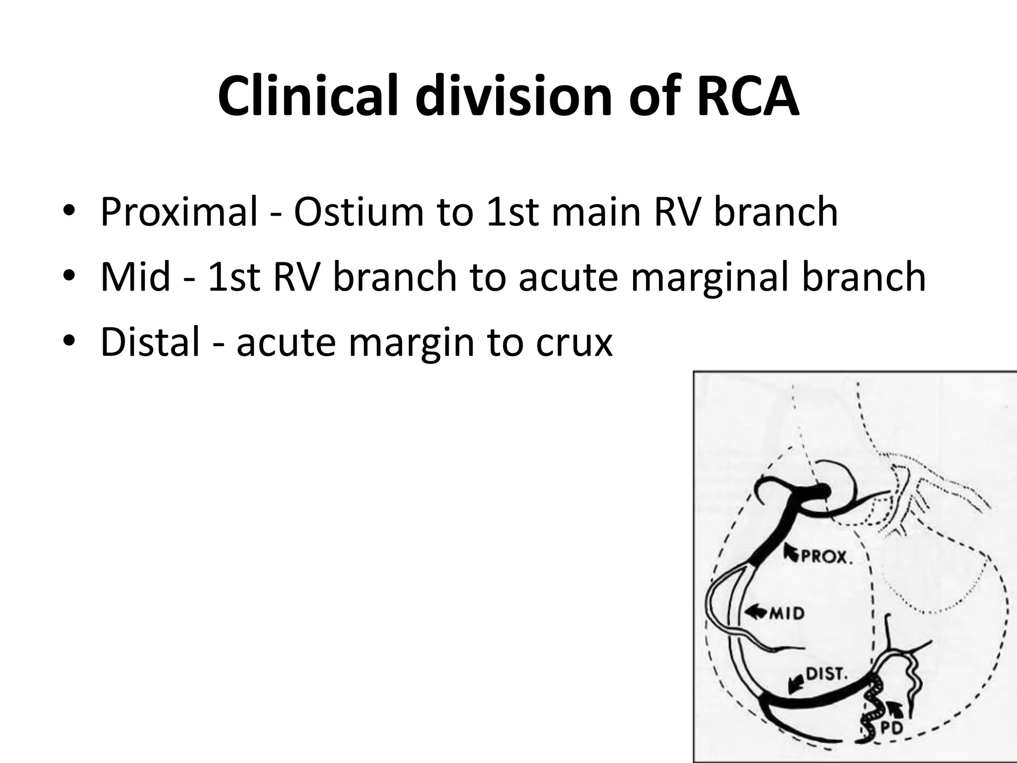
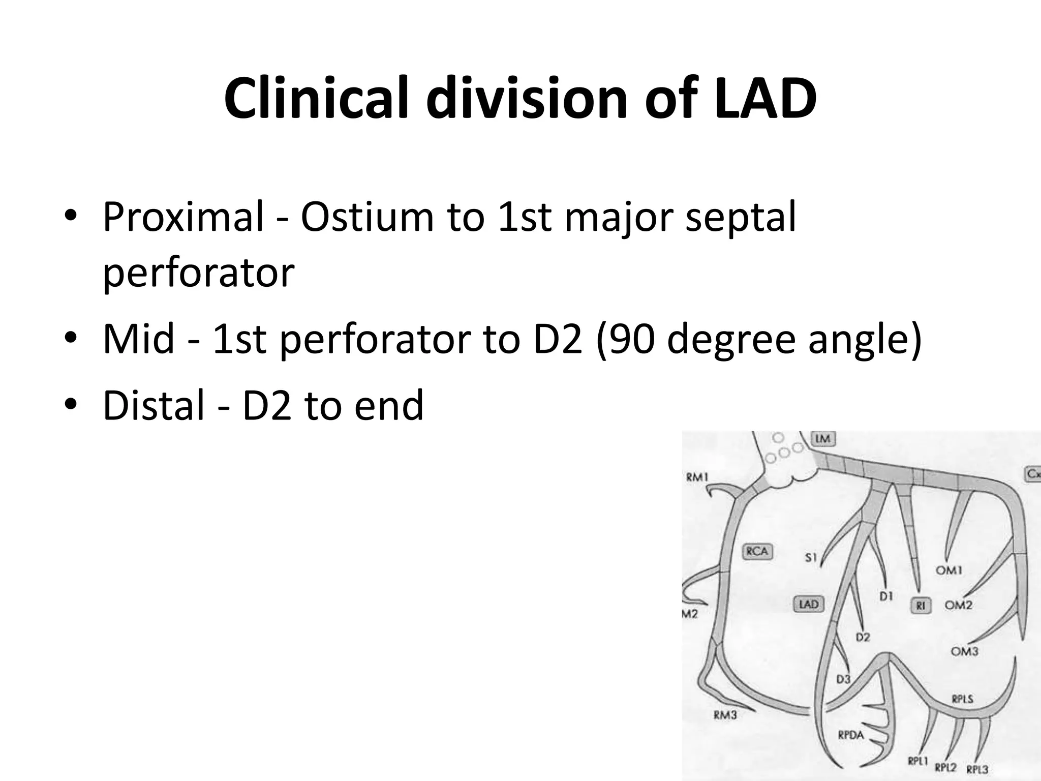
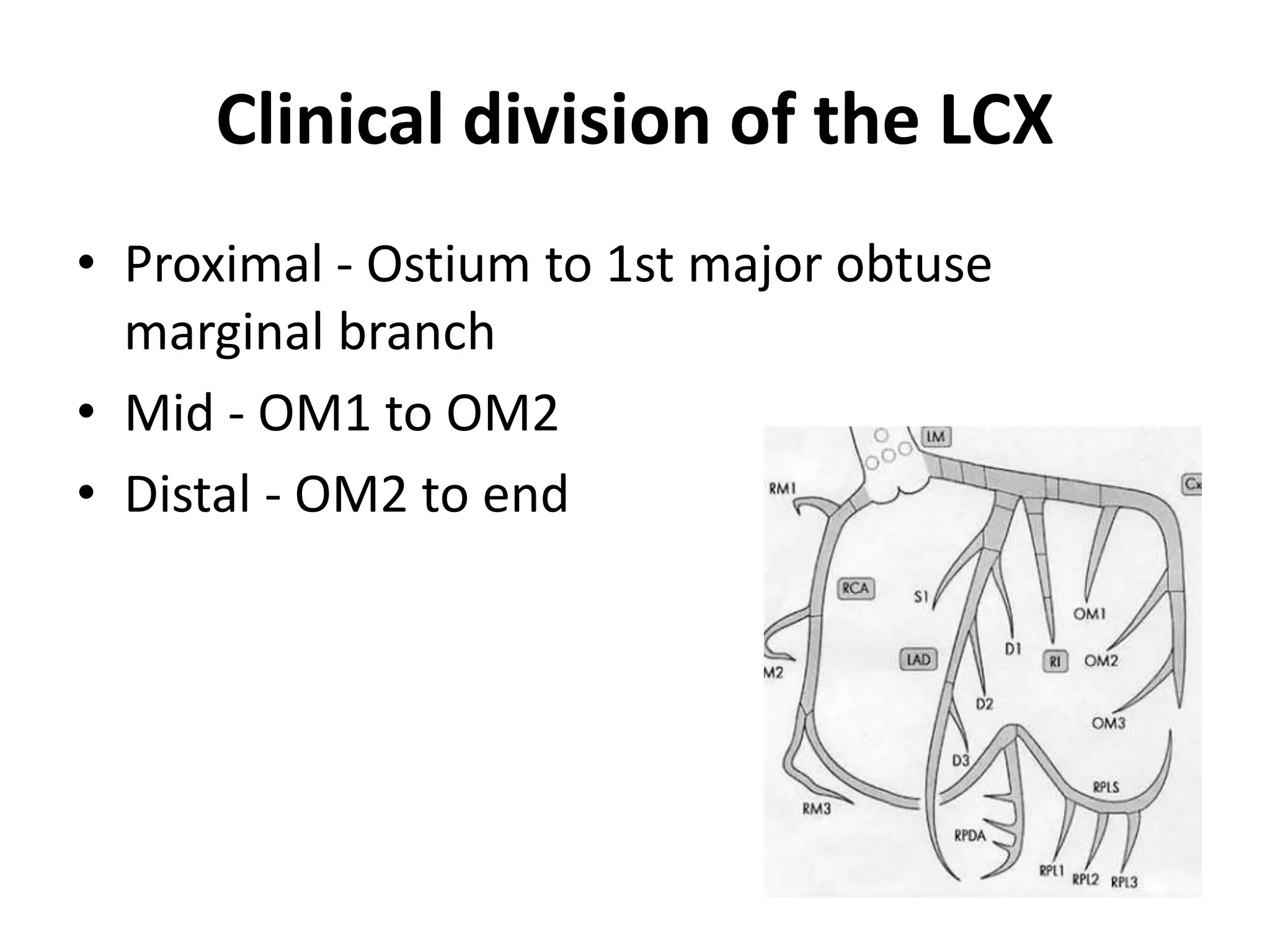
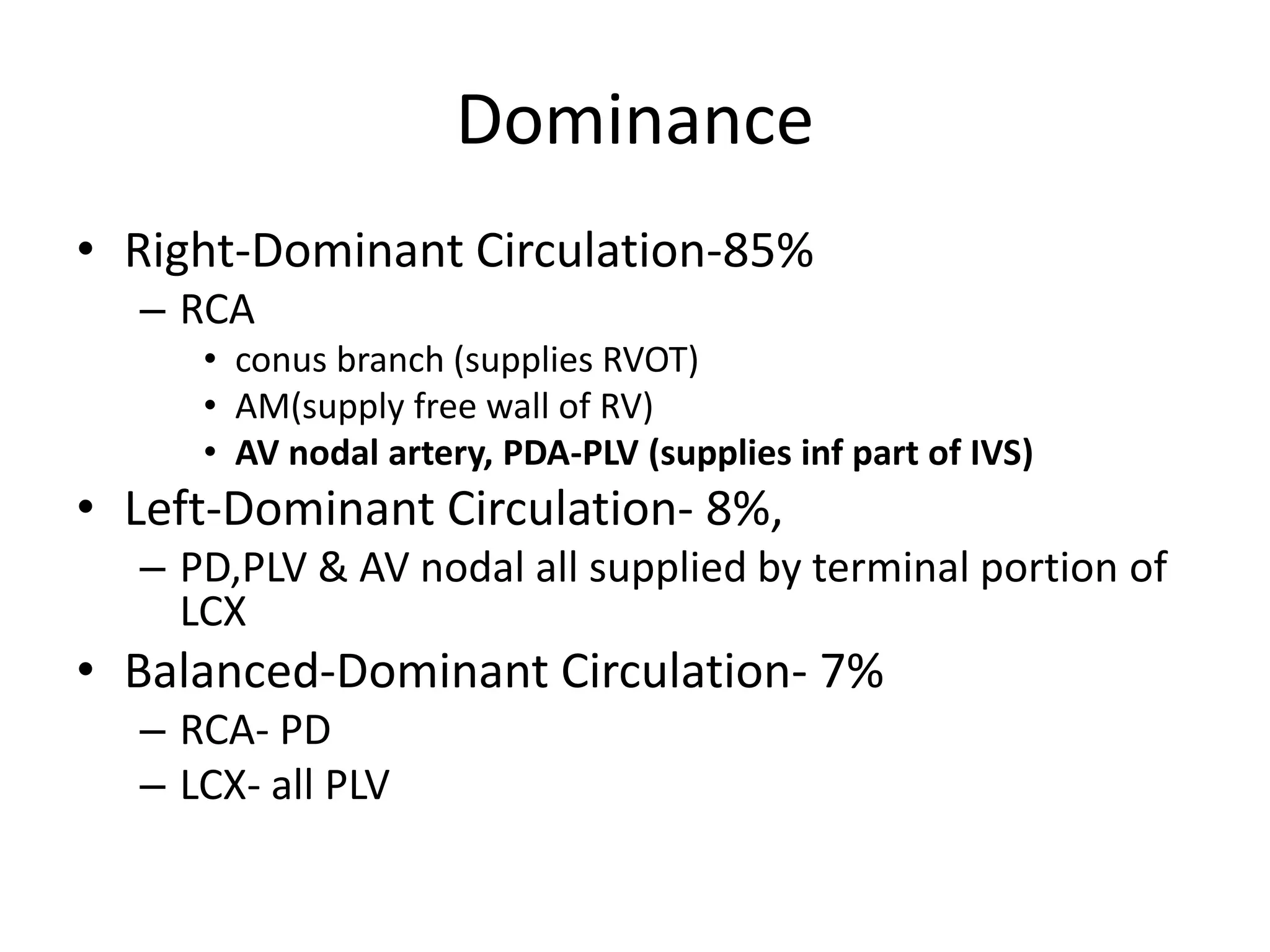
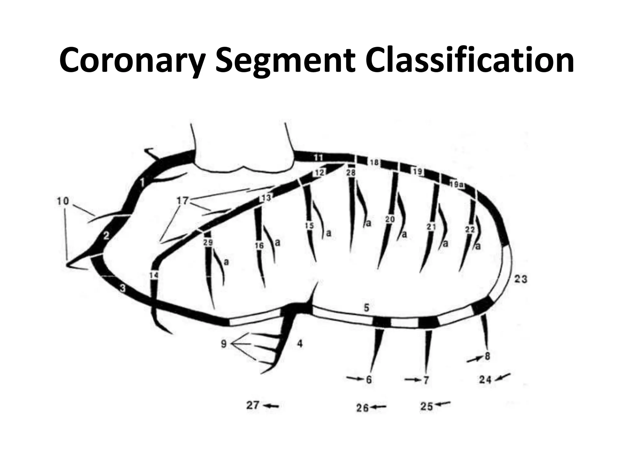
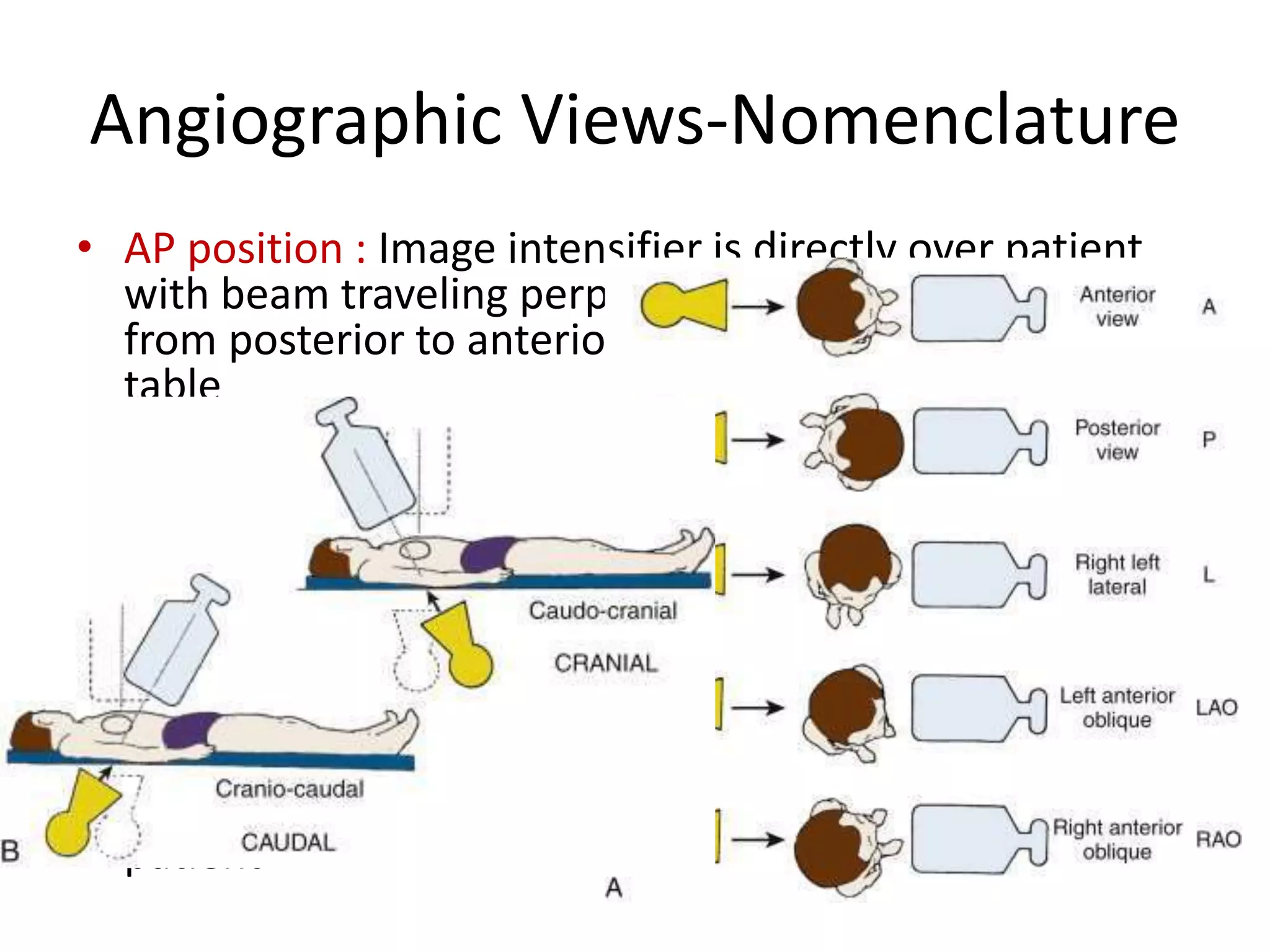
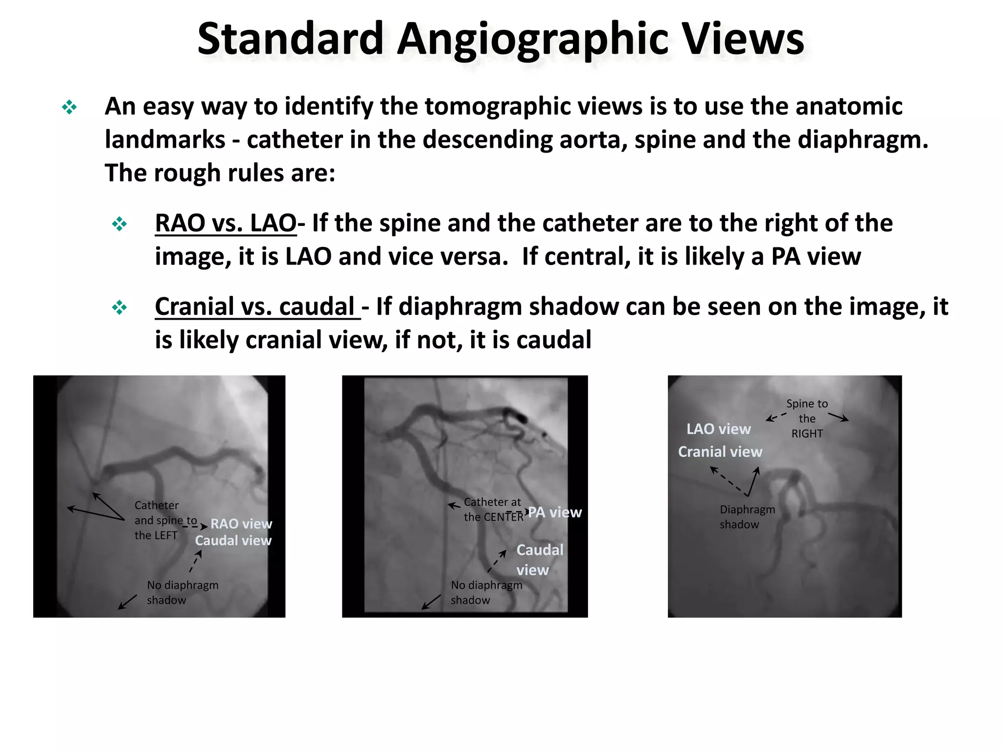
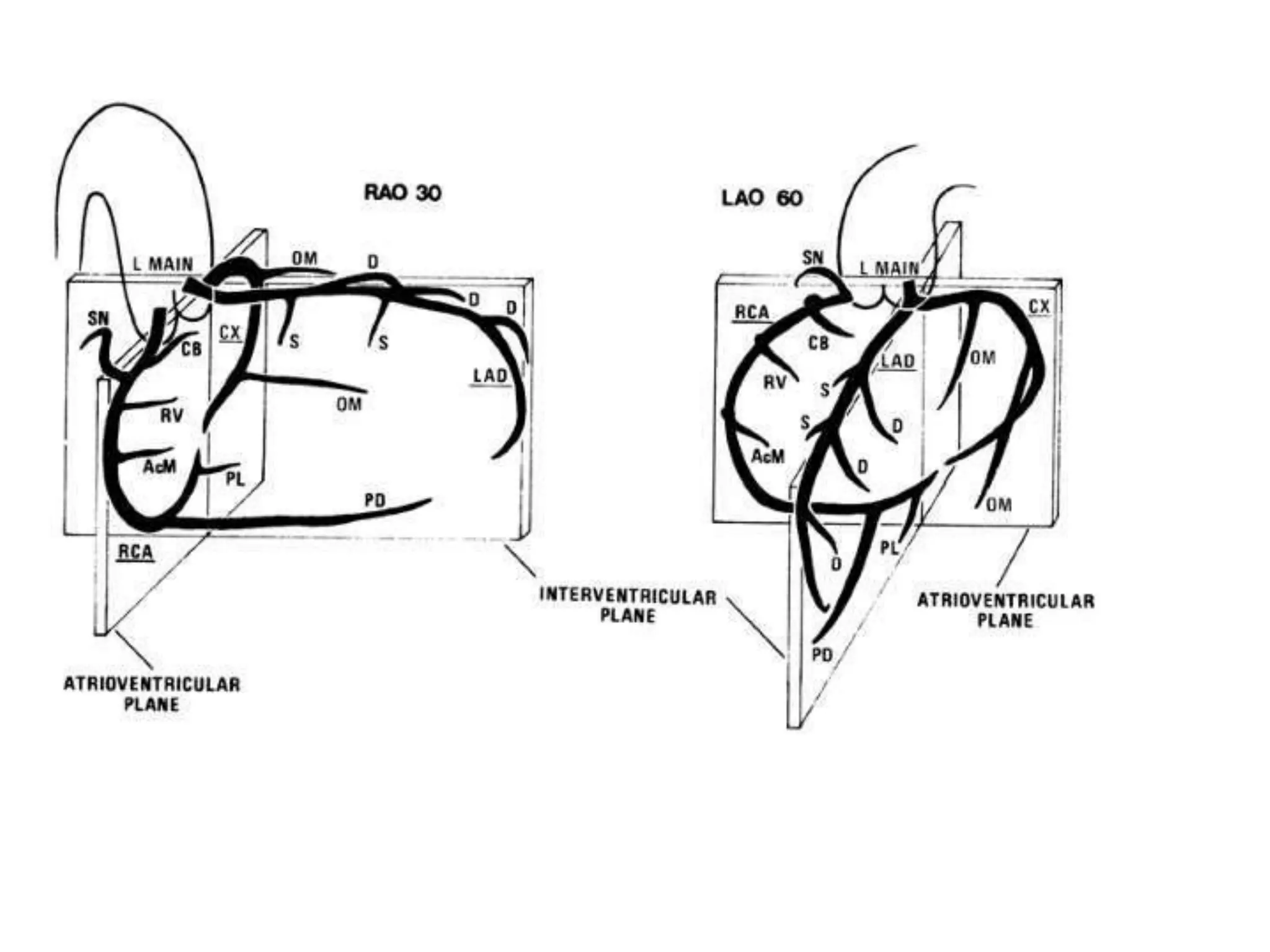
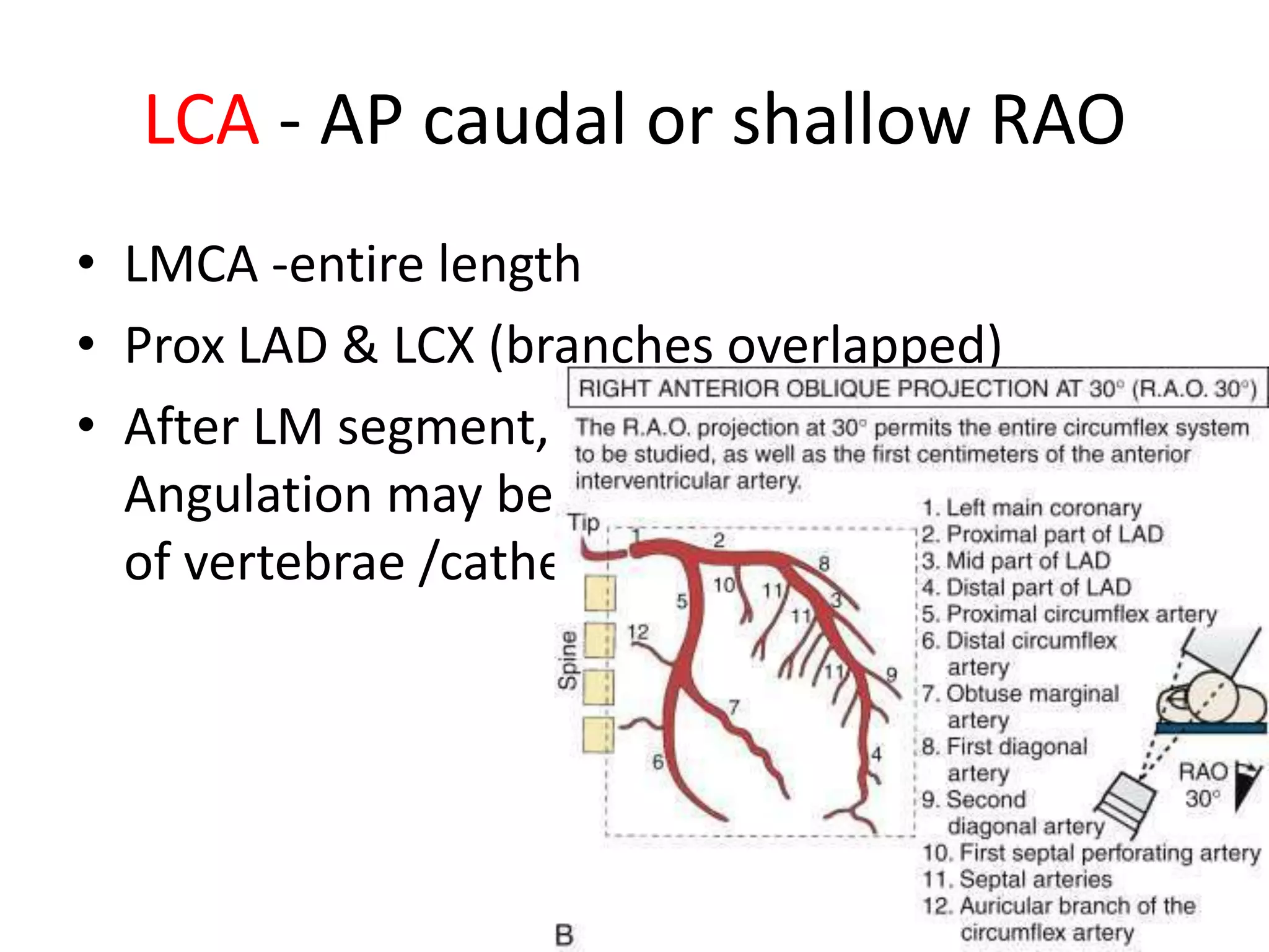
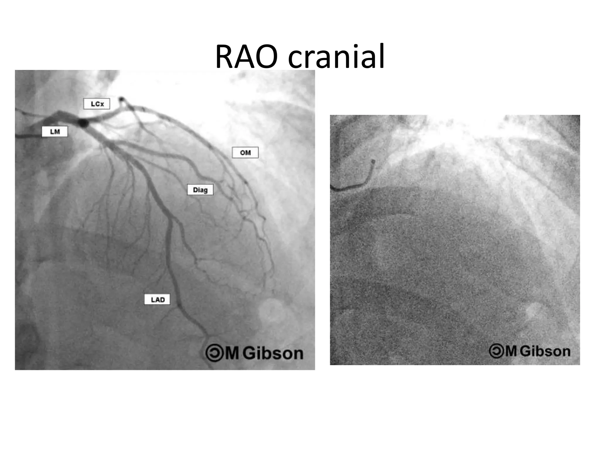
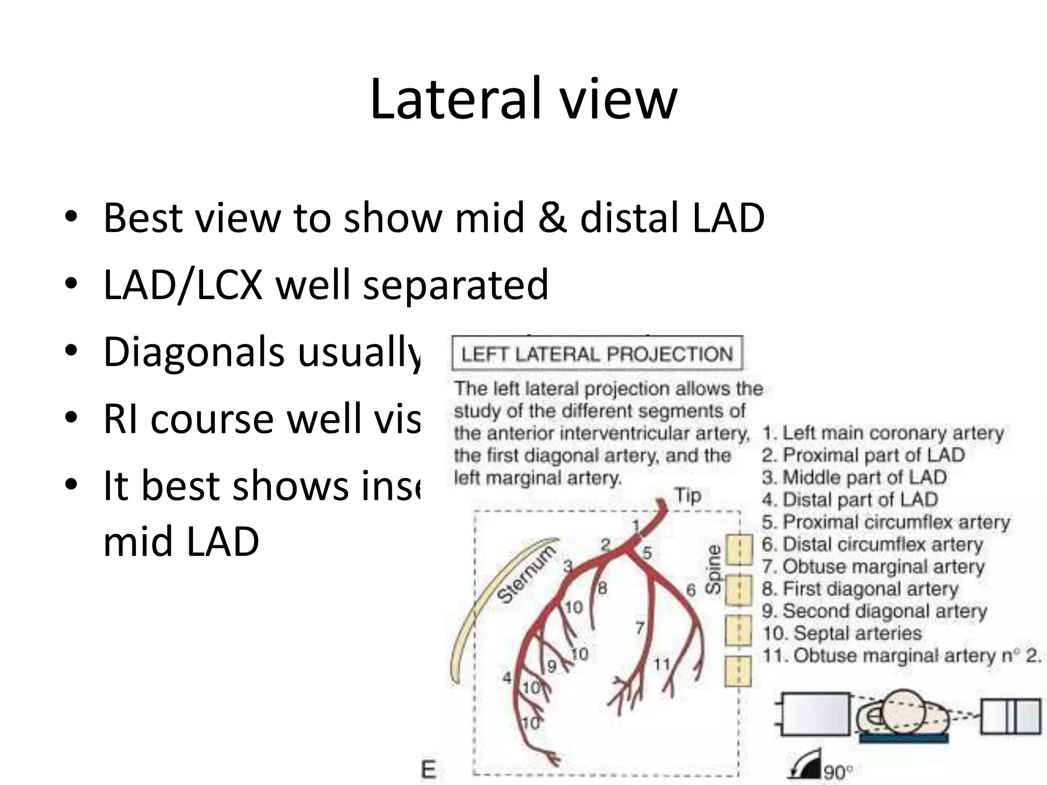
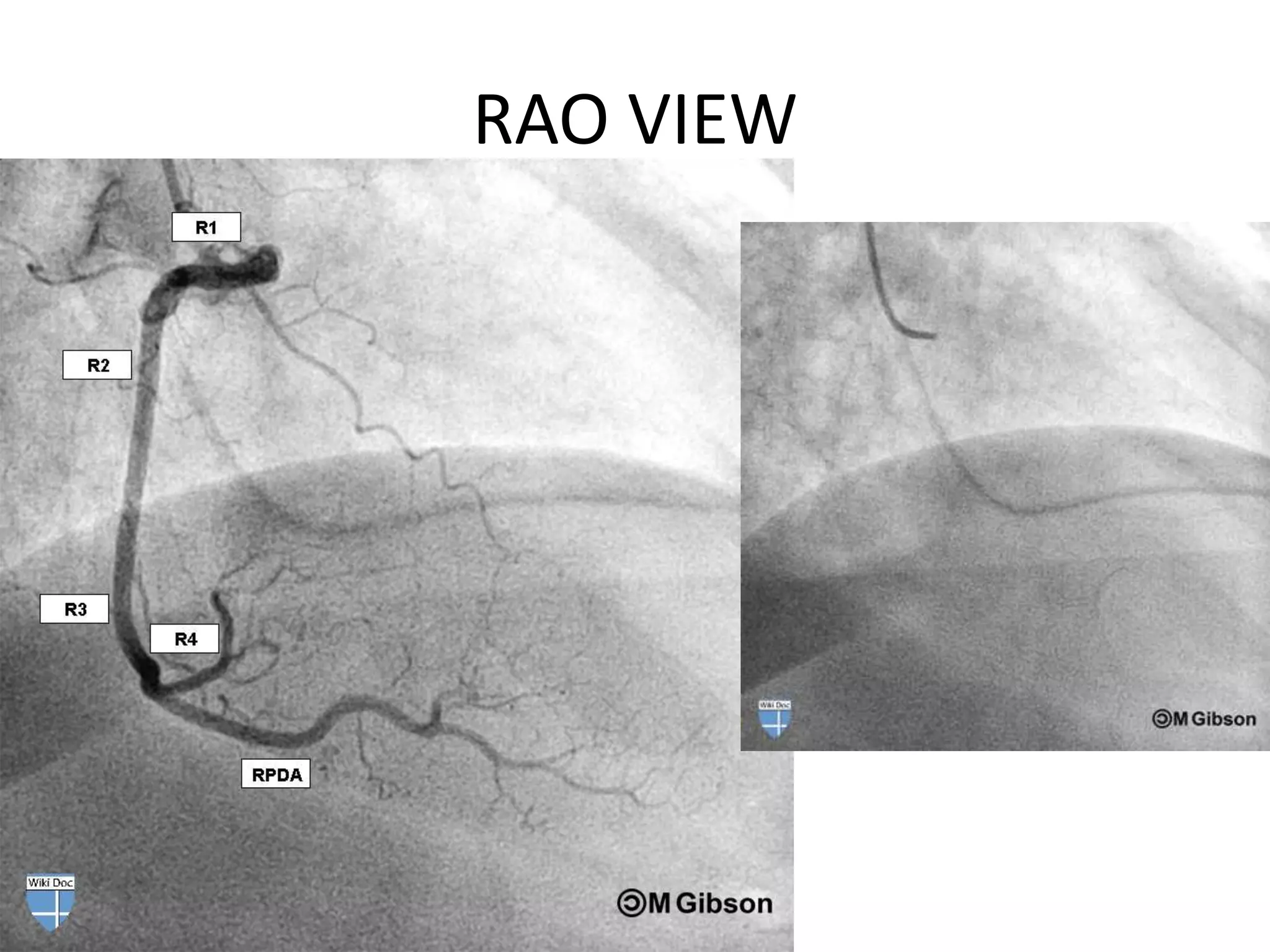
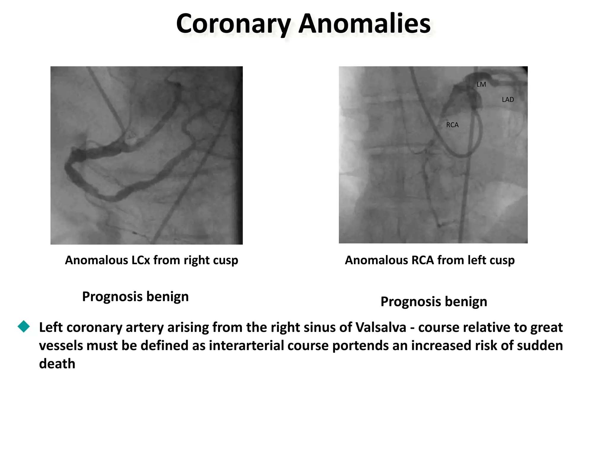
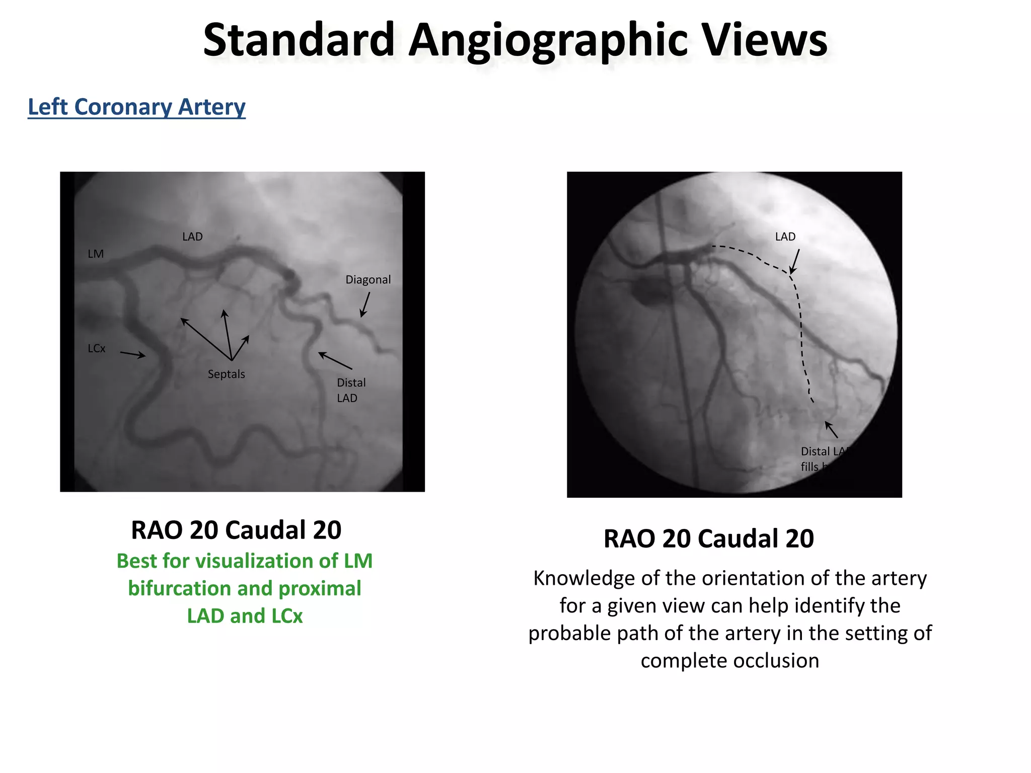
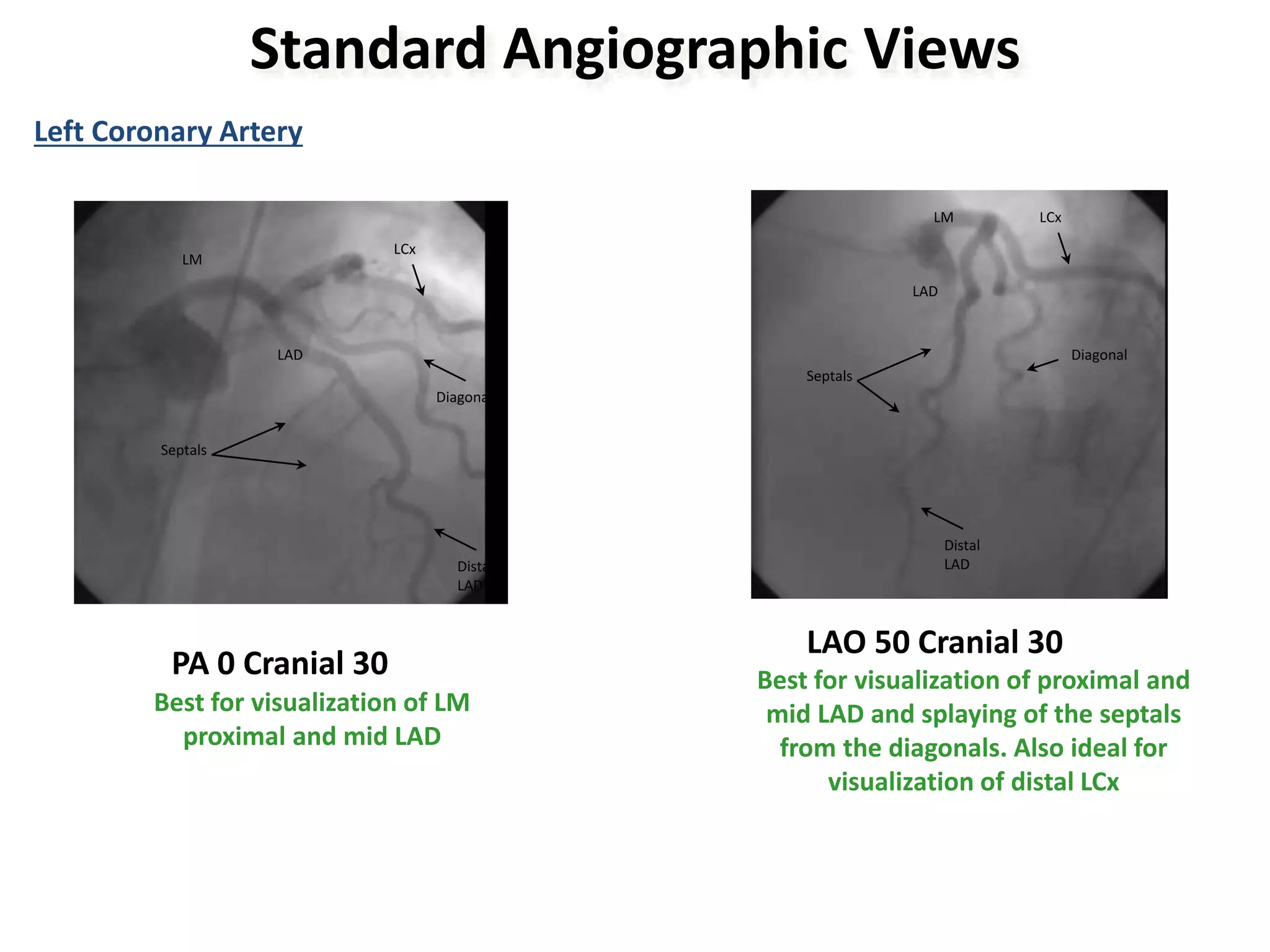
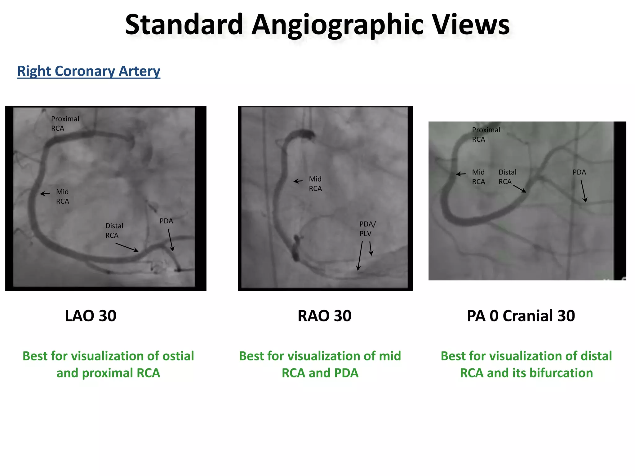
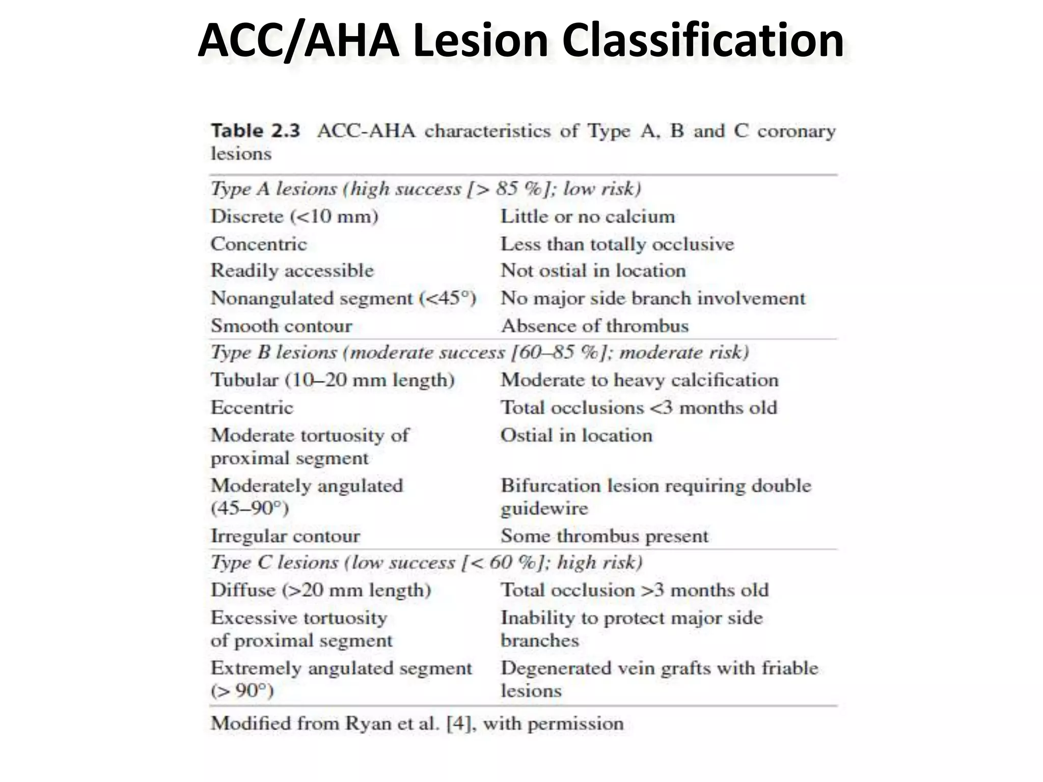
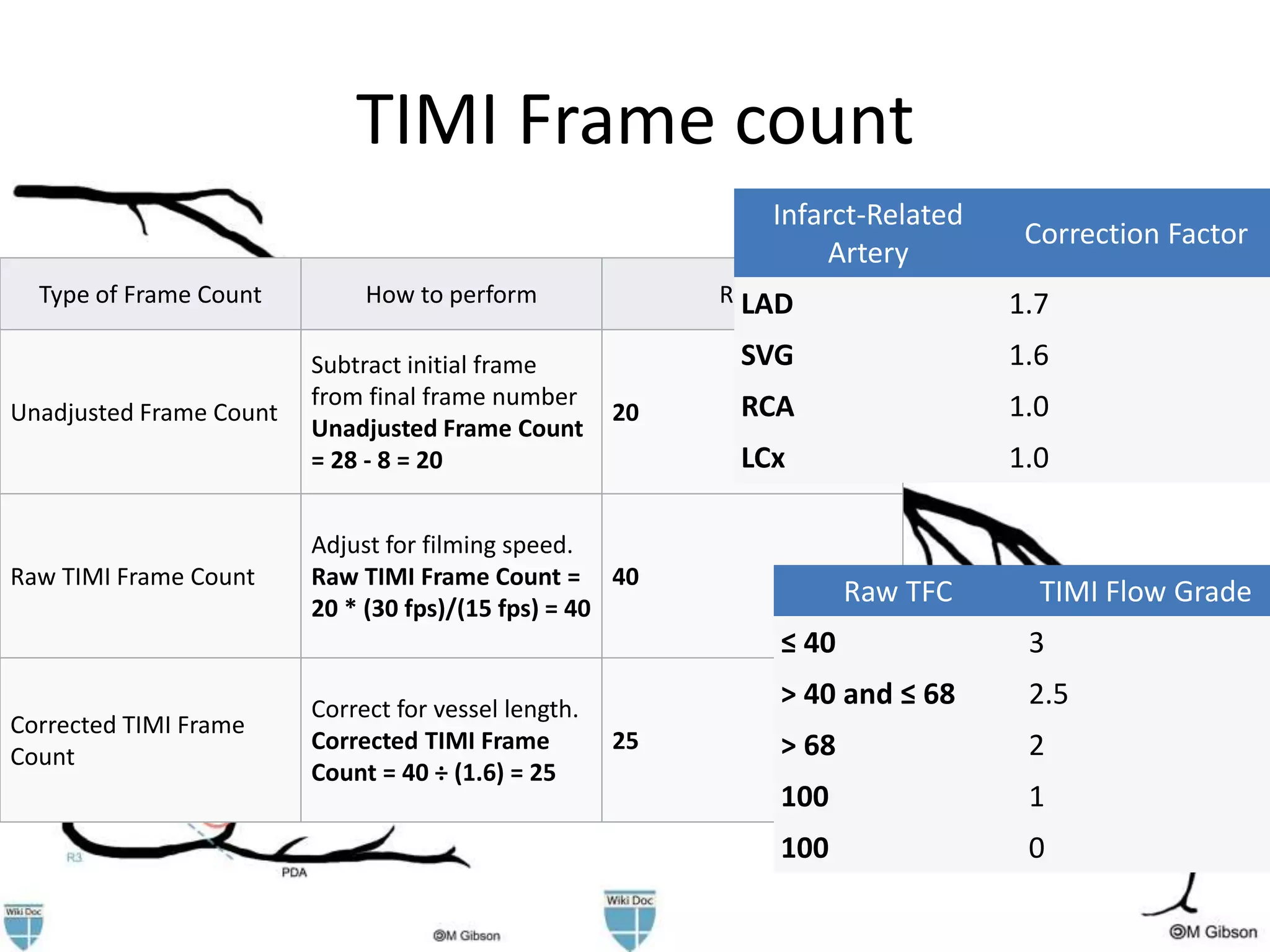
Coronary angiography and
angiographic views
-Dr. Rohit Walse
Intervention Fellow,
SCTIMST, Kerala
ANATOMY OF CORONARIES
Clinical division of RCA
• Proximal - Ostium to 1st main RV branch
• Mid - 1st RV branch to acute marginal branch
• Distal - acute margin to crux
Clinical division of LAD
• Proximal - Ostium to 1st major septal
perforator
• Mid - 1st perforator to D2 (90 degree angle)
• Distal - D2 to end
Clinical division of the LCX
• Proximal - Ostium to 1st major obtuse
marginal branch
• Mid - OM1 to OM2
• Distal - OM2 to end
Normal calibre
• LMCA: 4.5 ± 0.5 mm
• LAD: 3.7 ± 0.4 mm
• LCX : 3.5 ± 0.5 mm ( 4.2 mm if dominant)
• RCA: 3.9 ± 0.6 mm ( 2.8 mm if non-dominant)
• LCA ostium ~ 4mm
• RCA ostium~ 3.2mm
Dominance
• Right-Dominant Circulation-85%
– RCA
• conus branch (supplies RVOT)
• AM(supply free wall of RV)
• AV nodal artery, PDA-PLV (supplies inf part of IVS)
• Left-Dominant Circulation- 8%,
– PD,PLV & AV nodal all supplied by terminal portion of
LCX
• Balanced-Dominant Circulation- 7%
– RCA- PD
– LCX- all PLV
What is significant CAD?
• >50% - significant disease
• >2 mm vessel only considered for
revscularisation
• >70% considered hemodynamically significant
ds
• >50% is considered in LMCA
Coronary Segment Classification
Angiographic Views-Nomenclature
• AP position : Image intensifier is directly over patient
with beam traveling perpendicularly back to front (i.e.,
from posterior to anterior) to patient lying flat on x-ray
table
• RAO position : Image intensifier is on right side of
patient. A, anterior; O, oblique
• LAO position: Image intensifier is on left side of patient
• Lt Lateral position :Image intensifier rotated 90 deg
parallel to floor
• Cranial :Image intensifier is tilted toward head of
patient
• Caudal : Image intensifier is tilted toward feet of
patient
Standard Angiographic Views
 An easy way to identify the tomographic views is to use the anatomic
landmarks - catheter in the descending aorta, spine and the diaphragm.
The rough rules are:
 RAO vs. LAO- If the spine and the catheter are to the right of the
image, it is LAO and vice versa. If central, it is likely a PA view
 Cranial vs. caudal - If diaphragm shadow can be seen on the image, it
is likely cranial view, if not, it is caudal
Catheter
and spine to
the LEFT
RAO view
No diaphragm
shadow
Caudal view
Catheter at
the CENTER PA view
No diaphragm
shadow
Caudal
view
Spine to
the
RIGHT
LAO view
Diaphragm
shadow
Cranial view
LCA - AP caudal or shallow RAO
• LMCA -entire length
• Prox LAD & LCX (branches overlapped)
• After LM segment, slight RAO or LAO
Angulation may be necessary to clear density
of vertebrae /catheter shaft
LAO-cranial view
• LMCA (slightly foreshortened)
• LAD-Septal & diagonal are separated clearly
• LCX/OM: foreshortened/ overlapped
• PD/PL of left-dominant circulation are displayed clearly
• Deep inspiration helpful
• Cranial angulation permits view of LAD/LCX bifurcation
• LAO-cranial angulation that is too steep or inspiration
that is too shallow produces considerable overlapping
with diaphragm and liver, degrading the image
LAO Cranial
RAO-caudal
• LMCA bifurcation
• Origin & course of LCX/OM, RI & prox LAD
seen clearly
• One of the best for visualization of LCX
• LAD beyond proximal segment obscured
• Apical LAD displayed clearly
RAO caudal
RAO-cranial
• Used for origins of diagonals along mid /distal
LAD
• Diagonals bifurcations well visualized
• Diagonals projected upward
• Prox LAD/LCX usually overlapped
RAO cranial
LAO-caudal (“spider” view)
• LMCA (foreshortened) & LMCA bifurcation
• Prox & mid LCX with origins of OM
LAO caudal
Lateral view
• Best view to show mid & distal LAD
• LAD/LCX well separated
• Diagonals usually overlapped
• RI course well visualized
• It best shows insertions of bypass grafts into
mid LAD
RCA --- LAO-cranial
• Origin of RCA
• Entire length of mid RCA
• PDA bifurcation (crux)
• Cranial angulation tilts PDA down to see vessel
contour / reduce foreshortening
• Deep inspiration is necessary to clear
diaphragm
LAO cranial
RCA - RAO view
• Shows mid RCA & length of PDA / PL
• Septals coursing upward from PDA, supplying
occluded LAD artery via collaterals, may be
clearly identified
• PL are overlapped, may need addition of
cranial view
RAO VIEW
AP cranial
• Shows origin of RCA
• Mid segment foreshortened
• Best view for PD/PL of dominant RCA system
and size of collateralized LAD
AP cranial
Lateral view
• Shows RCA origin (especially in pt with more
anteriorly oriented orifices) and mid RCA
• PDA and PL are foreshortened
Saphenous vein graft views
• RCA graft—LAO cranial, RAO, and AP cranial
• LAD graft (or internal mammary artery)—
lateral,RAO cranial, LAO cranial, and AP
(lateral view is especially useful to visualize
anastomosis to LAD)
• LCX (and obtuse marginal branches) grafts—
LAO caudal and RAO caudal
• Diagonal graft—LAO cranial and RAO cranial
DUAL OSTIA
Coronary Anomalies
Increased risk of sudden death
Anomalous LCA from right sinus -
Inter-arterial Course
Anomalous LCA from right sinus -
Retro-aortic course
Prognosis benign
AORTA
PULMONARY
ARTERY
AORTA
PULMONARY
ARTERY
RCA RCA
LM
LM
LAD LAD
LCX
LCX
Coronary Anomalies
Prognosis benign
Anomalous LCx from right cusp Anomalous RCA from left cusp
LM
RCA
LAD
Prognosis benign
 Left coronary artery arising from the right sinus of Valsalva - course relative to great
vessels must be defined as interarterial course portends an increased risk of sudden
death
Coronary Aneursym
 Coronary Aneurysm: Vessel diameter >
1.5x neighboring segment
 Incidence: 0.15%-4.9%; very rare in LMCA
 Etiology: mainly atherosclerosis; other
causes include Kawasaki’s, PCI,
inflammatory disease, trauma, connective
tissue disease
Standard Angiographic Views
Left Coronary Artery
RAO 20 Caudal 20
LM
LAD
Diagonal
Septals
Distal
LAD
LCx
RAO 20 Caudal 20
Knowledge of the orientation of the artery
for a given view can help identify the
probable path of the artery in the setting of
complete occlusion
Distal LAD
fills by
collaterals
LAD
Best for visualization of LM
bifurcation and proximal
LAD and LCx
Standard Angiographic Views
Left Coronary Artery
LAO 50 Cranial 30
LM
LAD
Diagonal
Septals
Distal
LAD
LCx
PA 0 Cranial 30
LM
LAD
Diagonal
Septals
Distal
LAD
LCx
Best for visualization of LM
proximal and mid LAD
Best for visualization of proximal and
mid LAD and splaying of the septals
from the diagonals. Also ideal for
visualization of distal LCx
Standard Angiographic Views
Left Coronary Artery
PA0 Caudal 30
LM
LAD
Diagonal
Septals
Distal
LAD
LCx
LAO 50 Caudal 30
OM
LM
LAD
Diagonal
Distal
LAD
LCx
OM
‘Spider’ view
Best for visualization of LM
bifurcation and proximal LAD
and LCx
Best for visualization of LM
bifurcation, proximal LAD and LCx and
OM
Standard Angiographic Views
Right Coronary Artery
LAO 30
Proximal
RCA
PDA
Distal
RCA
Mid
RCA
RAO 30
Mid
RCA
PDA/
PLV
PA 0 Cranial 30
Proximal
RCA
PDA
Distal
RCA
Mid
RCA
Best for visualization of ostial
and proximal RCA
Best for visualization of mid
RCA and PDA
Best for visualization of distal
RCA and its bifurcation
Angiogram-Interpretation
 A systematic interpretation of a coronary angiogram would involve:
 Evaluation of the extent and severity of coronary calcification just
prior to or soon after contrast opacification
 Lesion quantification in at least 2 orthogonal views:
 Severity
 Calcification
 Presence of ulceration/thrombus
 Degree of tortuosity
 ACC/AHA lesion classification
 Reference vessel size
 Grading TIMI flow
 Grading TIMI myocardial perfusion blush grade
 Identifying and quantifying coronary collaterals
ACC/AHA Lesion Classification
Other Definitions
 Lesion length: Measured “shoulder-to-shoulder” in an unforeshortened view
Discrete Lesion length < 10 mm
Tubular Lesion length 10–20 mm
Diffuse Lesion length ≥ 20 mm
 Lesion angulation: Vessel angle formed by the centerline through the lumen proximal to
the stenosis and extending beyond it and a second centerline in the straight portion of the
artery distal to the stenosis
Moderate: Lesion angulation ≥ 45 degrees
Severe: Lesion angulation ≥ 90 degrees
 Calcification: Readily apparent densities noted within the apparent vascular wall at the
site of the stenosis
Moderate: Densities noted only with cardiac motion prior to contrast injection
Severe: Radiopacities noted without cardiac motion prior to contrast injection
TIMI Frame count
Type of Frame Count How to perform Result
Unadjusted Frame Count
Subtract initial frame
from final frame number
Unadjusted Frame Count
= 28 - 8 = 20
20
Raw TIMI Frame Count
Adjust for filming speed.
Raw TIMI Frame Count =
20 * (30 fps)/(15 fps) = 40
40
Corrected TIMI Frame
Count
Correct for vessel length.
Corrected TIMI Frame
Count = 40 ÷ (1.6) = 25
25
Raw TFC TIMI Flow Grade
≤ 40 3
> 40 and ≤ 68 2.5
> 68 2
100 1
100 0
Infarct-Related
Artery
Correction Factor
LAD 1.7
SVG 1.6
RCA 1.0
LCx 1.0
Clinical syntax scoring
Rentrop collateral circulation
Thank You!

coronaryangiography-220330142539 (1).pdf

  • 1.
    Coronary angiography and angiographicviews -Dr. Rohit Walse Intervention Fellow, SCTIMST, Kerala
  • 3.
  • 4.
    Clinical division ofRCA • Proximal - Ostium to 1st main RV branch • Mid - 1st RV branch to acute marginal branch • Distal - acute margin to crux
  • 5.
    Clinical division ofLAD • Proximal - Ostium to 1st major septal perforator • Mid - 1st perforator to D2 (90 degree angle) • Distal - D2 to end
  • 6.
    Clinical division ofthe LCX • Proximal - Ostium to 1st major obtuse marginal branch • Mid - OM1 to OM2 • Distal - OM2 to end
  • 7.
    Normal calibre • LMCA:4.5 ± 0.5 mm • LAD: 3.7 ± 0.4 mm • LCX : 3.5 ± 0.5 mm ( 4.2 mm if dominant) • RCA: 3.9 ± 0.6 mm ( 2.8 mm if non-dominant) • LCA ostium ~ 4mm • RCA ostium~ 3.2mm
  • 8.
    Dominance • Right-Dominant Circulation-85% –RCA • conus branch (supplies RVOT) • AM(supply free wall of RV) • AV nodal artery, PDA-PLV (supplies inf part of IVS) • Left-Dominant Circulation- 8%, – PD,PLV & AV nodal all supplied by terminal portion of LCX • Balanced-Dominant Circulation- 7% – RCA- PD – LCX- all PLV
  • 9.
    What is significantCAD? • >50% - significant disease • >2 mm vessel only considered for revscularisation • >70% considered hemodynamically significant ds • >50% is considered in LMCA
  • 10.
  • 11.
    Angiographic Views-Nomenclature • APposition : Image intensifier is directly over patient with beam traveling perpendicularly back to front (i.e., from posterior to anterior) to patient lying flat on x-ray table • RAO position : Image intensifier is on right side of patient. A, anterior; O, oblique • LAO position: Image intensifier is on left side of patient • Lt Lateral position :Image intensifier rotated 90 deg parallel to floor • Cranial :Image intensifier is tilted toward head of patient • Caudal : Image intensifier is tilted toward feet of patient
  • 13.
    Standard Angiographic Views An easy way to identify the tomographic views is to use the anatomic landmarks - catheter in the descending aorta, spine and the diaphragm. The rough rules are:  RAO vs. LAO- If the spine and the catheter are to the right of the image, it is LAO and vice versa. If central, it is likely a PA view  Cranial vs. caudal - If diaphragm shadow can be seen on the image, it is likely cranial view, if not, it is caudal Catheter and spine to the LEFT RAO view No diaphragm shadow Caudal view Catheter at the CENTER PA view No diaphragm shadow Caudal view Spine to the RIGHT LAO view Diaphragm shadow Cranial view
  • 15.
    LCA - APcaudal or shallow RAO • LMCA -entire length • Prox LAD & LCX (branches overlapped) • After LM segment, slight RAO or LAO Angulation may be necessary to clear density of vertebrae /catheter shaft
  • 16.
    LAO-cranial view • LMCA(slightly foreshortened) • LAD-Septal & diagonal are separated clearly • LCX/OM: foreshortened/ overlapped • PD/PL of left-dominant circulation are displayed clearly • Deep inspiration helpful • Cranial angulation permits view of LAD/LCX bifurcation • LAO-cranial angulation that is too steep or inspiration that is too shallow produces considerable overlapping with diaphragm and liver, degrading the image
  • 17.
  • 18.
    RAO-caudal • LMCA bifurcation •Origin & course of LCX/OM, RI & prox LAD seen clearly • One of the best for visualization of LCX • LAD beyond proximal segment obscured • Apical LAD displayed clearly
  • 19.
  • 20.
    RAO-cranial • Used fororigins of diagonals along mid /distal LAD • Diagonals bifurcations well visualized • Diagonals projected upward • Prox LAD/LCX usually overlapped
  • 21.
  • 22.
    LAO-caudal (“spider” view) •LMCA (foreshortened) & LMCA bifurcation • Prox & mid LCX with origins of OM
  • 23.
  • 24.
    Lateral view • Bestview to show mid & distal LAD • LAD/LCX well separated • Diagonals usually overlapped • RI course well visualized • It best shows insertions of bypass grafts into mid LAD
  • 25.
    RCA --- LAO-cranial •Origin of RCA • Entire length of mid RCA • PDA bifurcation (crux) • Cranial angulation tilts PDA down to see vessel contour / reduce foreshortening • Deep inspiration is necessary to clear diaphragm
  • 26.
  • 27.
    RCA - RAOview • Shows mid RCA & length of PDA / PL • Septals coursing upward from PDA, supplying occluded LAD artery via collaterals, may be clearly identified • PL are overlapped, may need addition of cranial view
  • 28.
  • 29.
    AP cranial • Showsorigin of RCA • Mid segment foreshortened • Best view for PD/PL of dominant RCA system and size of collateralized LAD
  • 30.
  • 31.
    Lateral view • ShowsRCA origin (especially in pt with more anteriorly oriented orifices) and mid RCA • PDA and PL are foreshortened
  • 32.
    Saphenous vein graftviews • RCA graft—LAO cranial, RAO, and AP cranial • LAD graft (or internal mammary artery)— lateral,RAO cranial, LAO cranial, and AP (lateral view is especially useful to visualize anastomosis to LAD) • LCX (and obtuse marginal branches) grafts— LAO caudal and RAO caudal • Diagonal graft—LAO cranial and RAO cranial
  • 34.
  • 35.
    Coronary Anomalies Increased riskof sudden death Anomalous LCA from right sinus - Inter-arterial Course Anomalous LCA from right sinus - Retro-aortic course Prognosis benign AORTA PULMONARY ARTERY AORTA PULMONARY ARTERY RCA RCA LM LM LAD LAD LCX LCX
  • 36.
    Coronary Anomalies Prognosis benign AnomalousLCx from right cusp Anomalous RCA from left cusp LM RCA LAD Prognosis benign  Left coronary artery arising from the right sinus of Valsalva - course relative to great vessels must be defined as interarterial course portends an increased risk of sudden death
  • 37.
    Coronary Aneursym  CoronaryAneurysm: Vessel diameter > 1.5x neighboring segment  Incidence: 0.15%-4.9%; very rare in LMCA  Etiology: mainly atherosclerosis; other causes include Kawasaki’s, PCI, inflammatory disease, trauma, connective tissue disease
  • 38.
    Standard Angiographic Views LeftCoronary Artery RAO 20 Caudal 20 LM LAD Diagonal Septals Distal LAD LCx RAO 20 Caudal 20 Knowledge of the orientation of the artery for a given view can help identify the probable path of the artery in the setting of complete occlusion Distal LAD fills by collaterals LAD Best for visualization of LM bifurcation and proximal LAD and LCx
  • 39.
    Standard Angiographic Views LeftCoronary Artery LAO 50 Cranial 30 LM LAD Diagonal Septals Distal LAD LCx PA 0 Cranial 30 LM LAD Diagonal Septals Distal LAD LCx Best for visualization of LM proximal and mid LAD Best for visualization of proximal and mid LAD and splaying of the septals from the diagonals. Also ideal for visualization of distal LCx
  • 40.
    Standard Angiographic Views LeftCoronary Artery PA0 Caudal 30 LM LAD Diagonal Septals Distal LAD LCx LAO 50 Caudal 30 OM LM LAD Diagonal Distal LAD LCx OM ‘Spider’ view Best for visualization of LM bifurcation and proximal LAD and LCx Best for visualization of LM bifurcation, proximal LAD and LCx and OM
  • 41.
    Standard Angiographic Views RightCoronary Artery LAO 30 Proximal RCA PDA Distal RCA Mid RCA RAO 30 Mid RCA PDA/ PLV PA 0 Cranial 30 Proximal RCA PDA Distal RCA Mid RCA Best for visualization of ostial and proximal RCA Best for visualization of mid RCA and PDA Best for visualization of distal RCA and its bifurcation
  • 42.
    Angiogram-Interpretation  A systematicinterpretation of a coronary angiogram would involve:  Evaluation of the extent and severity of coronary calcification just prior to or soon after contrast opacification  Lesion quantification in at least 2 orthogonal views:  Severity  Calcification  Presence of ulceration/thrombus  Degree of tortuosity  ACC/AHA lesion classification  Reference vessel size  Grading TIMI flow  Grading TIMI myocardial perfusion blush grade  Identifying and quantifying coronary collaterals
  • 43.
  • 45.
    Other Definitions  Lesionlength: Measured “shoulder-to-shoulder” in an unforeshortened view Discrete Lesion length < 10 mm Tubular Lesion length 10–20 mm Diffuse Lesion length ≥ 20 mm  Lesion angulation: Vessel angle formed by the centerline through the lumen proximal to the stenosis and extending beyond it and a second centerline in the straight portion of the artery distal to the stenosis Moderate: Lesion angulation ≥ 45 degrees Severe: Lesion angulation ≥ 90 degrees  Calcification: Readily apparent densities noted within the apparent vascular wall at the site of the stenosis Moderate: Densities noted only with cardiac motion prior to contrast injection Severe: Radiopacities noted without cardiac motion prior to contrast injection
  • 48.
    TIMI Frame count Typeof Frame Count How to perform Result Unadjusted Frame Count Subtract initial frame from final frame number Unadjusted Frame Count = 28 - 8 = 20 20 Raw TIMI Frame Count Adjust for filming speed. Raw TIMI Frame Count = 20 * (30 fps)/(15 fps) = 40 40 Corrected TIMI Frame Count Correct for vessel length. Corrected TIMI Frame Count = 40 ÷ (1.6) = 25 25 Raw TFC TIMI Flow Grade ≤ 40 3 > 40 and ≤ 68 2.5 > 68 2 100 1 100 0 Infarct-Related Artery Correction Factor LAD 1.7 SVG 1.6 RCA 1.0 LCx 1.0
  • 49.
  • 51.
  • 53.