Stereotactic biopsy is a neurosurgical procedure that uses CT or MRI guidance to take tissue samples from tumor sites in order to determine the type, grade, molecular biology, and growth pattern of suspected tumors or infections. The procedure takes about 3 hours to perform under local anesthesia and allows for deep-seated or multiple lesions to be biopsied safely.
This document discusses the use of high-intensity focused ultrasound (HIFU) to treat localized prostate cancer. It explains that HIFU uses focused ultrasound beams to heat and destroy cancerous prostate cells in a non-invasive procedure. The document outlines the indications and contraindications for HIFU, as well as the preparation, technique, post-procedure care, advantages, and disadvantages of using HIFU to treat prostate cancer.
This document discusses best practices for transradial access procedures. It emphasizes that transradial access can be safer and more convenient than femoral access. Key points include selecting appropriate patients, using the proper access technique and hardware, and achieving appropriate hemostasis. The document provides guidance on patient selection, radial artery assessment, equipment, access techniques including ultrasound-guided puncture, and tips for managing complications to help ensure safe and successful transradial procedures.
Mechanical and bioprosthetic heart valves have evolved significantly since the first prosthetic valve implantation in 1952. Modern bileaflet mechanical valves provide improved central blood flow compared to older caged ball designs. Tissue valves like porcine and pericardial valves do not require lifelong anticoagulation but have limited durability. Prosthetic heart valves are prone to complications like thrombosis, structural deterioration, endocarditis, and paravalvular leak. Careful monitoring and treatment is needed to optimize outcomes.
Basics of Interventional Radiology and Vascular Interventions RVRoshan Valentine
Brief overview of the general principles of interventional radiology, DSA, vascular interventions, catheters, guidewires, patient management, complications
Current applications of interventional radiology 97Arun Jagannathan
Interventional radiologists are physicians who specialize in minimally invasive targeted treatments using image guidance. They undergo 4 years of undergraduate education, 4 years of medical school, 5 years of residency training, and 1 year of fellowship training. Interventional radiology pioneered modern medicine by inventing angioplasty and stenting to treat blocked arteries. Some key milestones include the first angioplasty in 1964, development of embolization therapy in the 1960s, and invention of the catheter-delivered stent in the late 1960s and early 1970s. Interventional radiologists now perform a variety of minimally invasive procedures to treat cancer, vascular diseases, trauma, and other conditions.
An Overview of Filter-Protected Carotid Artery Stentinggailms
This document provides an overview of filter-protected carotid artery stenting. It discusses carotid artery disease and treatment options like carotid endarterectomy and carotid artery stenting. Embolic protection filters are used during carotid artery stenting to prevent plaque and debris from entering the bloodstream and causing strokes. The document summarizes various embolic protection devices and filter designs. It also reviews several in vitro studies that evaluate the capture efficiency and performance of different filter devices using particle models and benchtop flow loops. Overall, the document presents background information on carotid artery disease and stenting and evaluates the performance of embolic protection filters through in vitro testing.
This is a much less visited and often less spoken of topic about MRI Imaging... Herein we present a compilation of the various aspects of MRI Safety regarding both the patient, precautions and any contraindications to better the understanding of magnetic resonance imaging.
Interventional Radiology : Devices and Embolic Agents that a Resident NEEDS T...Saurabh Joshi
Interventional Radiology is full of various devices and materials. The general radiology resident needs to know these in order to impress the examiner. This file also contains information on various embolic agents.
The implementation of MDCT in urological imaging has solved much of the diagnostic dilemma. Thanks to its multiplanar capabilities and post processing techniques.
This document outlines the protocol for performing CT angiography (CTA) from the cerebral arteries to the lower limbs. It discusses indications for CTA including aneurysms, stenosis, dissections, and more. The preparation, positioning, and scanning protocols are provided for CTA of the head to lower limbs as well as the subclavian arteries. Pediatric protocols are also summarized. The document concludes with examples of CTA findings and references.
This document provides an overview of key concepts in radiation protection for diagnostic radiology, including:
- Medical exposure involves exposing patients for diagnosis or treatment, following principles of justification and optimization.
- Justification involves assessing if a procedure does more good than harm at the individual, generic, and general levels.
- Optimization aims to keep patient doses as low as reasonably achievable given image quality needs.
- Guidance levels indicate typical dose levels and help identify unusually high exposures requiring review. They are not dose limits.
DIGITAL SUBTRACTION ANGIOGRAPHY IN CEREBROVASCULAR DISEASE AND PERSPECTIVE.pptxNeurologyKota
This document discusses digital subtraction angiography (DSA) for diagnosing cerebrovascular diseases. It begins by introducing DSA as the gold standard technique for visualizing blood vessels through fluoroscopy and contrast injection. It then covers the indications for DSA including vascular diseases and tumors. The document provides details on the technique, approach for ischemic infarction cases, signs of acute ischemia, and examples of non-atherosclerotic vasculopathies that can be identified with DSA such as dissection, moyamoya disease, and fibromuscular dysplasia.
Radiation Protection in Diagnostic and Interventional Radiology, MDIRT Nchanj...Nchanji Nkeh Keneth
The document provides an overview of radiation protection in diagnostic radiology from materials from the International Atomic Energy Agency (IAEA). It discusses key topics including the definition of medical exposure, justification of medical exposures, optimization of protection, diagnostic reference levels, and effective doses. Examples of diagnostic reference levels are provided for various radiographic and CT examinations for adults as well as for mammography. The document emphasizes that diagnostic reference levels are not dose limits but are intended to provide guidance and indicate levels where doses should be reviewed.
This document discusses the basics of PET imaging including positron emission, fluorine-18 production and decay, FDG synthesis, tumor physiology and uptake, detection methods, and scanning techniques. It also covers applications of PET imaging in cardiac imaging like assessing myocardial viability and perfusion, as well as atherosclerotic plaque characterization and inflammation.
Cardiac MRI provides detailed images of the heart and blood vessels without using radiation. It has advanced significantly since its introduction in the 1980s. Developments in pulse sequences have improved image quality and allowed for faster acquisition times. Techniques like T2-weighted imaging and delayed enhancement MRI can identify heart muscle damage. Perfusion MRI evaluates blood flow and identifies areas with reduced flow. Further advances may integrate sequences, allow free breathing scans, and expand MRI's clinical and research roles in cardiology.
Type B aortic dissection occurs when blood flows within the aortic wall separating the layers of the media. Computed tomography angiography is useful for establishing the diagnosis by showing two distinct lumens separated by an intimal flap, and for determining the location of the primary intimal tear and extent of dissection. Initial medical management aims to control blood pressure and pain. Intervention may be indicated for complications such as persistent pain, expansion of the dissection, or impaired blood flow to organs. Both open surgical repair and endovascular techniques can be used to treat type B dissection, with endovascular options becoming more common due to lower risks.
This document discusses abdominal aortic aneurysms (AAAs) and their endovascular repair (EVAR). It defines AAAs as a dilatation of the abdominal aorta over 3cm in diameter. EVAR involves inserting a folded graft through the femoral artery which expands to exclude the aneurysm sac from blood flow and pressure. The benefits of EVAR over open repair include lower peri-operative mortality and complications. Proper patient assessment including vascular anatomy and medical comorbidities is important for determining candidacy for EVAR. The procedure involves deploying graft components in the aorta and iliac arteries under imaging guidance. Post-operative surveillance with imaging is needed to monitor for complications like endoleaks.
Endovascular repair of thoracic and abdominal aortic aneurysmsApollo Hospitals
Endovascular repair of thoracic and abdominal aortic aneurysms has significantly reduced mortality and morbidity compared to open surgery. It involves placing stent grafts using catheters to exclude aneurysms from blood flow. Proper patient selection based on aneurysm anatomy and vessel access is important for success. Follow up imaging is needed to monitor for complications like endoleaks. Mid-term results show endovascular repair provides good outcomes with 85% survival at 18-24 months for thoracic aneurysms. It has emerged as an alternative to open surgical repair for properly selected abdominal aortic aneurysm cases.
The document contains questions and answers about various topics related to CT scans. It includes definitions and explanations of ring artifacts, HRCT techniques, image reconstruction methods, CT numbers, scintillation detectors, pixels, radiation profile width in CT collimators, CT number, resolution types, mass attenuation coefficient, parallel multi-hole collimators, low dose CT scans, CT guided biopsies, and CT artifacts. The document consists of questions from several students on technical aspects of computed tomography imaging.
Evar in ruptured aaa + fast track 9.7.61Mai Parachy
This document discusses endovascular repair (EVAR) and open surgical repair for ruptured abdominal aortic aneurysms (rAAA). It provides definitions of terminology used and outlines the clinical features, initial management strategies, and operative strategies for rAAA repair. Specifically, it summarizes evidence from studies comparing outcomes of EVAR versus open repair for rAAA, finding that EVAR is associated with lower short-term mortality and fewer complications but long-term outcomes are less certain. It also describes the concept of a "fast-track" approach to rAAA focusing on rapid diagnosis, preparation, and treatment to minimize time to exclusion of the rupture site. Key aspects of this approach include multidisciplinary team coordination, equipment preparation, and
Coronary CT angiography is a noninvasive imaging modality used to evaluate coronary artery disease. It has a high sensitivity of 87-99% and specificity of 93-96% for detecting coronary artery stenosis. Coronary CT angiography is most useful in low- to intermediate-risk patients with chest pain to rule out coronary artery disease given its high negative predictive value of 93-100%. Coronary CT angiography involves acquiring images using ionizing radiation as the patient holds their breath and synchronizing the images with the patient's ECG signal.
This document discusses various imaging approaches for evaluating acute chest pain and thoracic abnormalities. It begins by outlining how cardiac CT angiography can be used to assess coronary artery disease and causes of non-cardiac chest pain such as aortic disorders, pulmonary embolism, and other thoracic issues. Examples of CT and MRI images are provided to illustrate different pathologies. The document then focuses on specific conditions like aortic dissection, pulmonary embolism, pneumothorax, and pleural effusions. Imaging findings and diagnostic criteria for each condition are summarized.
Занятие 2. Физико-технические и морфологические основы рентгенодиагностики.
Методы рентгенологического исследования. Преимущества и недостатки методов.
Контрольные вопросы:
1. Устройство и принцип работы рентгеновской трубки.
2. Физические и морфологические основы формирования рентгеновского изображения.
3. Основные методы рентгенологического исследования. Принципы флюорографии,
рентгенографии, рентгеноскопии,.
4. Принципы получения диагностического изображения при линейной томографии.
5. Факторы, определяющие качество рентгеновского изображения (резкость, контрастность,
жесткость).
6. Искусственное контрастирования в рентгенологии и область применения. Контрастные
вещества, используемые в рентгенологической практике. Противопоказания для
применения контрастных веществ, возможные побочные эффекты (симптомы, первая
помощь).
Формула успеха в офтальмологии. М.М.Дронов
лауреат Государственной премии СССР,
доктор мед. наук, профессор, академик МАИ,
Санкт-Петербургский государственный университет. Общество православных врачей СПб www.opvspb.ru
ОТОЖДЕСТВЛЕНИЕ БИОЛОГИЧЕСКИХ ТКАНЕЙ С ПОМОЩЬЮ ТЕЛЕКОММУНИКАЦИОННЫХ МИКРОСИСТЕМITMO University
В статье рассматриваются телекоммуникационные способы и устройства, основанные на функциональных узлах микроскопов, для верификации (отождествления) расположения конца медицинской иглы в биологических тканях в процессе проведения медицинских операций.
Занятие 1. Ионизирующее и неионизирующее излучение на службе лучевой
диагностики. Виды излучений, их применение в медицине.
Контрольные вопросы:
1. Ультразвук: физические основы, использование в медицине.
2. Инфракрасное излучение: определение, спектральная характеристика. Термография:
понятие, возможность использования на современном этапе.
3. Физика магнитного резонанса. Формирование МР-изображения. Применение в медицине.
4. Рентгеновские лучи: основные свойства, их использование в медицине. Принципы и
средства защиты от ионизирующего излучения.
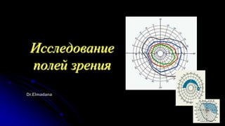
2. Поле зрения
Поле зрения - одна из основных составляющих
зрительного восприятия человека. Состояние поля зрения
обеспечивает ориентацию в пространстве и позволяет дать
функциональную характеристику зрительного анализатора
при выборе профессии, прохождении призывной
комиссии, экспертизе трудоспособности, а также при
научных исследованиях.
3. В литературе можно найти различные определения
понятия «поля зрения».
В американских источниках классическим считается
определение, данное H.M. Traquair (1875-1954): «Поле
зрения – это возвышающийся остров видения в море
тьмы».
S. Duke-Elder в многотомном руководстве по
офтальмологии определяет поле зрения как проекцию в
пространстве всех точек сетчатки, обладающих
зрительной чувствительностью.
4. Поле зрения
- пространство, одновременно воспринимаемое глазом
при неподвижном взоре и фиксированном положении
головы. Оно имеет определенные границы,
соответствующие переходу оптически деятельной
части сетчатки в оптически слепую.
- Более коротко – это видимое пространство,
воспринимаемое глазом при неподвижном взоре.
Измерение поля зрения — неотъемлемая часть любого,
как рутинного, так и специального, обследования
пациента в офтальмологии, поскольку нарушение
периферического зрения может быть ранним, а иногда
и единственным признаком многих глазных болезней.
5. Развитие клинической периметрии.
Альбрехт фон Грефе в 1855 г. первым стал исследовать поле
зрения с клинической целью, выполняя его измерения вначале на
плоскости. Для поиска и регистрации скотом, слепого пятна
великий ученый использовал обычную школьную доску и кусочек
мела.
Однако еще задолго до Грефе существовали представления о
границах поля зрения в норме и при патологии. Так, в трудах
древнегреческого врача Гиппократа (460-377 гг. до н.э.),
древнеримского врача Галена (ок. 130 – ок. 200 гг.) упоминается о
сужении периферических границ поля зрения. В 1553г. Б. Порто
исследовал границы поля зрения на листе белой бумаги, учитывая
яркость освещения экрана, его фон, но допустил ошибку, считая,
что границы поля зрения имеют вид круга.
6. Развитие клинической периметрии.
В 1668 г. Мариотт впервые описал так называемое слепое
пятно.
В 1825 г. Пуркинье первым отметил, что перемещение объекта
по дуге дает равные отрезки, а на плоскости они
увеличиваются к периферии.
В 1857 г. Ауберт и Ферстер создали первый дуговой
периметр,
а в 1889 г. Бьеррум вновь возвратился к кампиметру и, описав
характерную для глаукомы арочную скотому, показал
важность исследования центрального поля зрения (ЦПЗ) на
плоскости при этом заболевании.
7. С этого времени было предложено огромное количество разных
аппаратов для исследования поля зрения и методов его
исследования, от простых до очень сложных. Однако нужно
помнить, что аппаратура сама по себе еще не обусловливает
точности исследования. Результаты периметрии
определяются не конструкцией периметра, а знаниями и
искусством исследующего. Опытный исследователь,
применив самые простые приемы исследования и
приспособления, может получить более полные и точные
данные о состоянии поля зрения пациента, чем менее
опытный исследователь на сложно устроенном приборе.
8. Поле зрения искусственно ограничивается
выступающими частями лица — спинкой носа, верхним
краем глазницы. Кроме того, его границы зависят от
положения глазного яблока в глазнице. Поле зрения имеет
периферические и центральные отделы. Первые
соответствуют отделам сетчатки, более чувствительным к
прерывистым раздражениям, в частности к восприятию
движущихся предметов (периферическое зрение,
осуществляемое палочками). Центральные отделы
находятся в проекции желтого пятна и обеспечивают
центральное зрение, осуществляемое колбочками.
Выделяют также парацентральные отделы поля зрения. В
зависимости от участия в зрении одного или обоих глаз,
различают монокулярное и бинокулярное поле зрения. В
клинической практике обычно исследуют монокулярное
поле зрения.
9. Наиболее точные данные получают при инструментальном исследовании поля
зрения, основанном на фиксации момента появления движущегося или
неподвижного тест-объекта на дуге либо полусфере (кинетическая и
статическая периметрия) или на плоскости (кампиметрия). Периметрию
применяют в основном для изучения периферических отделов поля зрения, при
котором определяют границы поля зрения, выявляют дефекты зрительного
восприятия — скотомы, обусловленные расположенными впереди сетчатки
кровеносными сосудами (ангиоскотомы) или нарушениями зрительной
функции (патологической скотомы).
В норме наиболее широкие границы поля зрения получают при периметрии с
использованием белого тест-объекта, более узкие границы — при
использовании тест-объекта синего цвета, еще более узкие — при
использовании красного тест-объекта, наиболее узкие границы поля зрения —
при исследовании с помощью зеленого тест-объекта.
10. Оценка состояния поля зрения в динамике - критерий
течения заболевания, эффективности лечения; кроме того,
она имеет прогностическое значение. Исследованию поля
зрения придают существенное значение в нейрохирургии и
нейроофтальмологии при топической диагностике
заболеваний головного мозга и проводящих путей, так как
при этом возникают характерные дефекты,
свидетельствующие о повреждении различных участков
зрительного пути.
11. Показаниями к периметрии являются:
1. Глаукома.
2. Заболевания зрительного нерва (неврит, травма, ишемия).
3. Патология сетчатки (дистрофия, кровоизлияния, лучевой ожог,
отслойка,опухоль).
4. Гипертоническая болезнь.
5. Опухоли головного мозга.
6. Черепно-мозговые травмы.
7. Нарушения мозгового кровообращения.
8. Оценка зрения при профилактических осмотрах.
Противопоказания к проведению периметрии:
1. Психические заболевания пациента.
2. Алкогольное или наркотическое опьянение.
12. К основным методам диагностики относятся кинетическая
периметрия и статическая периметрия. Кинетическая
периметрия и ее разновидности (квантитативная,
хронопериметрия, разноэнерготическая периметрия)
основаны на сочетании различных комбинаций размеров и
яркости стимула, что позволяет определить границы
периферического поля зрения и границы скотом, однако не
устанавливает глубину выявленного дефекта.
13. Кинетическая периметрия
Позволяет выявить сужение поля зрения, определить
границы абсолютных скотом в условиях
стандартизированных освещения фона, величины и
яркости объектов
Предложено несколько периметров для исследования поля зрения по методу
кинетической периметрии; при этом тестируемый объект смещается по дуге
периметра от периферии к центру либо наоборот. Границу видения определяют
в момент появления объекта в поле зрения, иногда используют критерии его
исчезновения. Наиболее простым и до последнего времени распространенным
прибором для исследования поля зрения является периметр Ферстера
14. Основным достоинством
периметра Ферстера является
простота в обращении и
дешевизна, а недостатком —
непостоянство освещения дуги
и тестов. На нем трудно
обнаруживать небольшие
скотомы в поле зрения;
пигментные испытательные
объекты быстро пачкаются от
употребления и выходят из
строя.
Периметр настольный ПНР-03
15. На самой периферии сетчатки светоощущения нет,
крайняя периферия её воспринимает только белый свет, а
по мере продвижения к центру появляется ощущение
синего, жёлтого, красного и зелёного. В центральной
части сетчатки различаются все цвета. Таким образом,
поле зрения каждого глаза на белый объект
характеризуется следующими границами: кнаружи (к
виску) – 90, кверху кнаружи – 70, кверху – 50-55, кверху
кнутри – 60, кнутри (к носу) – 55, книзу кнутри – 50,
книзу – 65-70, книзу кнаружи – 90. Возможны небольшие
колебания в пределах 5-10 градусов.
16.
17. Абсолютный или относительный дефект в поле зрения
называется скотомой. Абсолютная скотома представляет
собой полную потерю зрения, при которой даже самый
яркий и крупный объект не воспринимается;
относительная скотома - это зона частичной потери
зрения, в которой некоторые объекты могут быть видны.
Скотома может иметь пологие края, так что ее
абсолютная часть оказывается окруженной
относительной скотомой. Выделяют положительные
скотомы, которые воспринимаются пациентом, и
отрицательные, которые обнаруживаются только при
исследовании.
18. Статическая
периметрия
Принцип статической периметрии — предъявление
светового стимула переменной величины и яркости в
фиксированной точке поля зрения. Методика дает
возможность не только выявить дефекты, но и определить
уровень светочувствительности сетчатки в заранее
обусловленных участках. Среди современных периметров
наиболее широкое распространение получил
автоматизированный анализатор поля зрения Humphrey.
19. Задачи периметрии:
Выявить возможные дефекты поля зрения
Определить характер выявленных изменений
Уточнить соответствие выявленных дефектов поля
изменения на сетчатке
Осуществлять динамическое наблюдение пациентов с
патологическими изменениями сетчатки и ЗН с целью
определения степени прогрессирования.
20. КОМПЬЮТЕРНАЯ ПЕРИМЕТРИЯ
Компьютерные анализаторы поля зрения предназначены в
мерную очередь для статической периметрии, но на большин-
стве из них может проводиться и кинетическая периметрия в
полном объеме или в пределах 60° от точки фиксации.
В современных компьютерных анализаторах поля зрения
приняты стандарты, рекомендованные Международным
периметрическим обществом в 1979 г. Они, в свою очередь,
базируются на стандартах, использованных Гольдманом в его
полусферическом периметре.
21. КОМПЬЮТЕРНАЯ ПЕРИМЕТРИЯ
Яркость поверхности полусферы, служащей фоном для
демонстрации тестовых объектов, должна составлять 31,5
апостильб, яркость тестовых объектов (стимулов) может
колебаться в пределах от 0,08 апостильб до 10 ООО
апостильб, диаметр — от 1 до 5 мм. Однако, в отличие от
периметров докомпьютерной эры, чувствительность сетчатки
в точке исследования оценивается не косвенно — в единицах
яркости различаемого стимула, а в единицах
чувствительности сетчатки, обратных яркости стимула. Такой
единицей чувствительности является Бел, соответствующий
1 логарифмической единице чувствительности. Это значит,
что изменение чувствительности на 1 Бел — это изменение
ее в 10 раз.
22. КОМПЬЮТЕРНАЯ ПЕРИМЕТРИЯ
При периметрии измерения производятся в
десятых долях Бела — децибелах
(сокращенное обозначение дБ). Каждый
децибел — это изменение чувствительности
на 0,1 логарифмическую единицу.
Соотношение между яркостью стимула в
апостильбах и чувствительностью на рисунке.
Если пациент не различает стимул яркостью
10000 апостильб, то прибор регистрирует 0
чувствительность сетчатки в данной точке.
При анализе поля зрения в ходе
статической периметрии изменяется
только яркость стимула, а не его
диаметр.
1000
24. Принципы исследования поля зрения на
компьютерном периметре.
Контроль точности фиксации (это
главное условие достоверности)
Наличие фиксационного объекта
Все исследования по всем
стратегиям по умолчанию
проводятся при размере
тестового объекта III (4 мм2).
Интервалы между отдельными
стимулами в ходе исследования
изменяются автоматически в
довольно широком диапазоне
времени.
25. Стратегии тестирования поля зрения
Во всех современных компьютерных периметрах
используется несколько способов тестирования поля,
которые по принятой на Западе терминологии
называют стратегиями. Основными из них являются:
пороговая стратегия;
скрининговая стратегия;
кинетическое тестирование.
26. Пороговая стратегия
используется для определения порога световой
чувствительности данной точки сетчатки. Порог
определяется минимальной яркостью стимула,
которую еще различает испытуемый. Исследование
порогов имеет первостепенное значение для ранней
диагностики депрессий в зоне Бьеррума и,
следовательно, для ранней диагностики глаукомы.
27. Тестированию по пороговой стратегии могут быть
подвергнуты разные участки сетчатки в зависимости
от цели исследования, так как тестирование каждый
раз всего поля зрения заняло бы слишком много
времени. В связи с этим разработчики предлагают
несколько «наборов», или «решеток». С полным
ассортиментом «наборов» стимулов можно
ознакомиться в инструкции к конкретному прибору. В
связи с тем, что наибольший интерес представляют
пороги чувствительности парацентральной зоны
сетчатки, большинство наборов преследуют именно
эту цель.
28. Использование специальных алгоритмов,
сокращающих время порогового исследования за счет
оптимизации многих его элементов. В периметре
Humphrey это алгоритмы SITA Standard и SITA Fast, в
периметре Octopus – алгоритм TOP. С учетом
значительного (в 3-4 раза) сокращения времени
исследования использование указанных алгоритмов
следует считать оправданным, несмотря на некоторое
снижение точности исследования.
29. Факторы, влияющие на результаты
компьютерной периметрии
Размер зрачка (особенно у пациентов с
катарактой, на фоне миотиков)
Помутнение оптических сред
Отсутствие или неправильная коррекция
аномалий рефракции
Загрязненные контактные линзы
30. Факторы, влияющие на результаты
компьютерной периметрии
Неправильно введенная дата рождения
Блефарохалазис. Птоз
Косой выход ДЗН
Первое в жизни исследование («эффект
обучения»)
31. Ошибки при компьютерной
периметрии
Ложноположительные (false positive)
Ложноотрицательные (false negative)
Достоверность исследования сомнительна, если количество ложноположительных и
ложноотрицательных ответов превышает 20%.
32. Ошибки при компьютерной периметрии
Ложноположительные и ложноотрицательные ответы на
стимулы заведомо высокой и заведомо низкой яркости,
которые компьютер периодически посылает в процессе
исследования. Если пациент не реагирует на стимул
надпороговой яркости, то прибор фиксирует
ложноотрицательный ответ, а если он среагировал на стимул
яркостью заведомо ниже порога — ложноположительный
ответ. Большое количество таких ответов, равно как и
случаев потери фиксации, указывает на низкую
достоверность полученных результатов.
33. Способы проверки достоверности
исследования
Периодическая подача сигналов в зону слепого пятна
для учета частоты ошибочно положительных ответов
пациента, свидетельствующих о нарушении фиксации
взора (метод Хейл-Крокау)
График отклонений взора
Определение количества ложноположительных и
ложноотрицательных ответов на стимулы.
35. 1-5 – информация о пациенте и условиях
проведения теста
6 – показатели достоверности теста
7 – цифровое выражение
светочувствительности
8 – изображение в режиме серо-белой
шкалы
9 – отклонение светочувствительности от
средней возрастной нормы со
статистической оценкой достоверности
(р <0,5%)
10 – отклонение в отдельных участках от
средней индивидуальной
светочувствительности
11 – показатель глаукомного
полуполярного теста
12 – MD и PSD
Основные периметрические индексы
36. Основные периметрические индексы
MD – (mean deviation) среднее отклонение или средний
дефект: общая разница между нормальной
светочувствительностью (с учетом возраста) и
светочувствительностью сетчатки у данного пациента
MD увеличивается при непрозрачности сред, диффузных или
выраженных локальных поражениях. Значения
чувствительности меньше нормальных отмечаются знаком
«-» в периметрах Humphrey (обозначает девиацию) и знаком
«+» в периметрах Octopus (обозначает наличие нарушений)
37. Основные периметрические индексы
PSD или LV (паттерн стандартное отклонение или
вариабельность дефектов): сравнивает результаты в
тестируемых точках между собой и отражает
выраженность очаговых поражений поля зрения
Может быть нормальным в случаях диффузных
поражений и не подходит для наблюдения в динамике
в продвинутые стадии глаукомы.
38. периметрические индексы
Значение «MS» - относится к средней
чувствительности, абсолютное измеренное среднее
значение.
«RF» является коэффициентом надежности
исследования. Производится контроль фиксации и
учет количества ложных положительных ответов.
Наибольшее возможное значение — 1.0, но значение
должно быть не ниже 0,7.
39. периметрические индексы
SF – short term fluctuation (краткосрочные флюктуации, только
Humphrey) – говорит о стабильности (повторяемости)
измерений светочувствительности в точках, которые
проверялись дважды в ходе исследования. SF>7,0 дБ
рассматривается как признак ненадежности полученных
результатов.
CPSD – corrected PSD / CLV – corrected LV –
скорректированные с учетом величины краткосрочных
флюктуаций значения PSD / LV
(При использовании алгоритмов SITA Standard и SITA Fast
индексы CF и CPSD не указываются)
40. периметрические индексы
На периметре Humphrey оценивается вероятность наличия
данной величины индексов в норме. Например, запись «MD -
9.96 dB P<0.5%» указывает, что снижение индекса MD на 9,96
дБ встречается реже, чем в 0,5% (то есть реже, чем у 1 из 200
здоровых лиц).
Суммарные индексы, особенно первые два из них,
используются преимущественно в научных исследованиях, а
также, у отдельных пациентов,– при оценке динамики
изменений. Однако в целом они намного менее
информативны, чем схемы «Pattern deviation» или «Corrected
probability».
41. периметрические индексы
Распечатка периметра Humphrey содержит также результат
GHT –Glaucoma Hemifield Test – Глаукомного теста полуполей
(сравнения верхнего и нижнего полуполей по 5
соответственным участкам) в виде сообщений: GHT within /
outside normal limits (в пределах / за пределами нормы) или
GHT borderline (на пограничном уровне).
42. Скрининговые стратегии
Надпороговые – проводятся при постоянной,
заведомо надпороговой яркости стимула и только
позволяет выявить есть или отсутствует дефект в
поле зрения.
43. Распечатка периметра Octopus включает кривую Bebie Curve,
называемую также Cumulative Defect Curve (кумулятивная
кривая дефектов). На кривой слева направо последовательно
отложена светочувствительность всех точек от наибольшей к
наименьшей. Данная кривая, если она равномерно снижена
относительно кривой нормы, указывает на наличие общего
(диффузного) снижения светочувствительности. При наличии
же локальных дефектов левый край кривой остается на
нормальном уровне, в то время, как правый край резко
отклоняется книзу.
44.
45. Пример ОКТ И КПЗ (макулярный тест 10-2)
пациента с дистрофическими изменениями
47. Пример КПЗ (центральный тест 30-2)
пациента с пигментной абиотрофией
Фото глазного дна и периметрия ребенок 10 лет. Уже
видны пигментные изменения пигментного пигмента
средней периферии сетчатки. Периметрия показывает
концентрическое сужение поля зрения у пациента