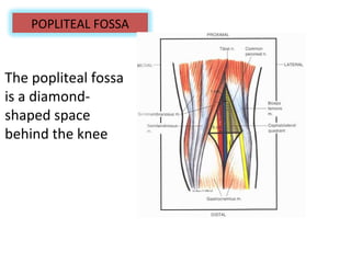
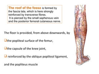
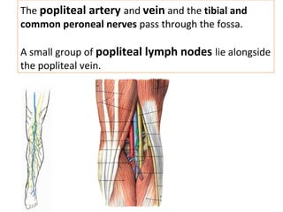
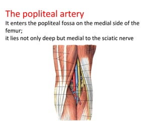
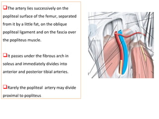
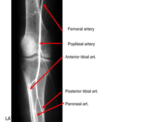
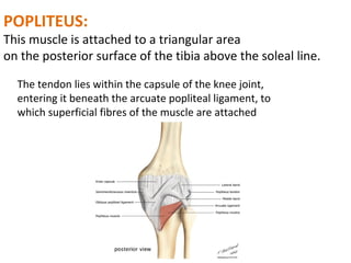
ANATOMY OF POPLITEAL FOSSA
The popliteal fossa
is a diamond-
shaped space
behind the knee
POPLITEAL FOSSA
The adductor hiatus is an opening or
gap formed by the fascia of the adductor
magnus.The adductor hiatus transmits the
femoral artery and vein from the adductor
canal in the thigh to the popliteal fossa
posterior to the knee
The roof of the fossa is formed by
the fascia lata, which is here strongly
reinforced by transverse fibres.
It is pierced by the small saphenous vein
and the posterior femoral cutaneous nerve.
The floor is provided, from above downwards, by
the popliteal surface of the femur,
the capsule of the knee joint,
 reinforced by the oblique popliteal ligament,
and the popliteus muscle
Deeply, the superior boundaries are
formed by the diverging medial and
lateral supracondylar lines of the
femur. The inferior boundary is
formed by the soleal line of the tibia
The popliteal artery and vein and the tibial and
common peroneal nerves pass through the fossa.
A small group of popliteal lymph nodes lie alongside
the popliteal vein.
The popliteal artery
It enters the popliteal fossa on the medial side of the
femur;
it lies not only deep but medial to the sciatic nerve
The artery lies successively on the
popliteal surface of the femur, separated
from it by a little fat, on the oblique
popliteal ligament and on the fascia over
the popliteus muscle.
It passes under the fibrous arch in
soleus and immediately divides into
anterior and posterior tibial arteries.
Rarely the popliteal artery may divide
proximal to popliteus
LATERAL
Femoral artery
Popliteal artery
Anterior tibial art.
Posterior tibial art.
Peroneal art.
CLINICALLY
popliteal pulsation against the
back of the femur, with the
fingertips of both hands pressing
into the centre of the fossa
BRANCHES OF POPLITEAL ARTERY
sural arteries that supply the two
heads of gastrocnemius
The genicular arteries are five in
number
 The middle genicular artery
pierces the oblique popliteal
ligament to supply the cruciate
ligaments
GENICULAR ANASTOMOSIS a network of vessels surrounding
the knee that provides collateral circulation capable of maintaining
blood supply to the leg during full knee flexion
Genicular branches
descending genicular branch of
the femoral artery,
 the descending branch of the
lateral circumflex femoral artery,
the circumflex fibular branch of
the posterior tibial artery
the anterior and posterior
recurrent branches of the anterior
tibial artery
The common peroneal nerve
(common fibular nerve)
runs downwards and laterally, medial to the biceps
tendon, and disappears into the substance of peroneus
longus to lie on the neck of the fibula,
I. The sural communicating nerue
2.The lateral cutaneous nerue of the cal
3.The superior and inferior genicular nerues,
4.The recurrent genicular nerue
The tibial nerve runs vertically
down along the middle of the fossa
and disappears by passing deeply
between the heads of
gastrocnemius
The tibial nerve here has only
one cutaneous branch,the
sural nerve. lt runs vertically
down in the narrow
POPLITEUS:
This muscle is attached to a triangular area
on the posterior surface of the tibia above the soleal line.
The tendon lies within the capsule of the knee joint,
entering it beneath the arcuate popliteal ligament, to
which superficial fibres of the muscle are attached
The femur is rotated laterally
to unlock the joint at the
commencement of flexion by
the popliteus

Popliteal fossa

  • 1.
  • 2.
    The popliteal fossa isa diamond- shaped space behind the knee POPLITEAL FOSSA
  • 3.
    The adductor hiatusis an opening or gap formed by the fascia of the adductor magnus.The adductor hiatus transmits the femoral artery and vein from the adductor canal in the thigh to the popliteal fossa posterior to the knee
  • 4.
    The roof ofthe fossa is formed by the fascia lata, which is here strongly reinforced by transverse fibres. It is pierced by the small saphenous vein and the posterior femoral cutaneous nerve. The floor is provided, from above downwards, by the popliteal surface of the femur, the capsule of the knee joint,  reinforced by the oblique popliteal ligament, and the popliteus muscle
  • 5.
    Deeply, the superiorboundaries are formed by the diverging medial and lateral supracondylar lines of the femur. The inferior boundary is formed by the soleal line of the tibia
  • 6.
    The popliteal arteryand vein and the tibial and common peroneal nerves pass through the fossa. A small group of popliteal lymph nodes lie alongside the popliteal vein.
  • 7.
    The popliteal artery Itenters the popliteal fossa on the medial side of the femur; it lies not only deep but medial to the sciatic nerve
  • 8.
    The artery liessuccessively on the popliteal surface of the femur, separated from it by a little fat, on the oblique popliteal ligament and on the fascia over the popliteus muscle. It passes under the fibrous arch in soleus and immediately divides into anterior and posterior tibial arteries. Rarely the popliteal artery may divide proximal to popliteus
  • 9.
    LATERAL Femoral artery Popliteal artery Anteriortibial art. Posterior tibial art. Peroneal art.
  • 10.
    CLINICALLY popliteal pulsation againstthe back of the femur, with the fingertips of both hands pressing into the centre of the fossa
  • 11.
    BRANCHES OF POPLITEALARTERY sural arteries that supply the two heads of gastrocnemius The genicular arteries are five in number  The middle genicular artery pierces the oblique popliteal ligament to supply the cruciate ligaments
  • 12.
    GENICULAR ANASTOMOSIS anetwork of vessels surrounding the knee that provides collateral circulation capable of maintaining blood supply to the leg during full knee flexion Genicular branches descending genicular branch of the femoral artery,  the descending branch of the lateral circumflex femoral artery, the circumflex fibular branch of the posterior tibial artery the anterior and posterior recurrent branches of the anterior tibial artery
  • 13.
    The common peronealnerve (common fibular nerve) runs downwards and laterally, medial to the biceps tendon, and disappears into the substance of peroneus longus to lie on the neck of the fibula, I. The sural communicating nerue 2.The lateral cutaneous nerue of the cal 3.The superior and inferior genicular nerues, 4.The recurrent genicular nerue
  • 14.
    The tibial nerveruns vertically down along the middle of the fossa and disappears by passing deeply between the heads of gastrocnemius The tibial nerve here has only one cutaneous branch,the sural nerve. lt runs vertically down in the narrow
  • 15.
    POPLITEUS: This muscle isattached to a triangular area on the posterior surface of the tibia above the soleal line. The tendon lies within the capsule of the knee joint, entering it beneath the arcuate popliteal ligament, to which superficial fibres of the muscle are attached
  • 16.
    The femur isrotated laterally to unlock the joint at the commencement of flexion by the popliteus