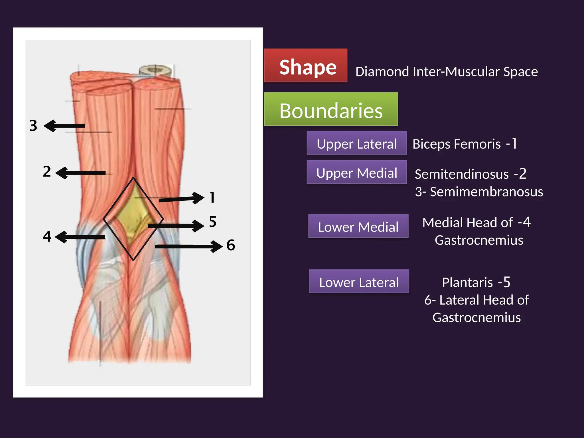
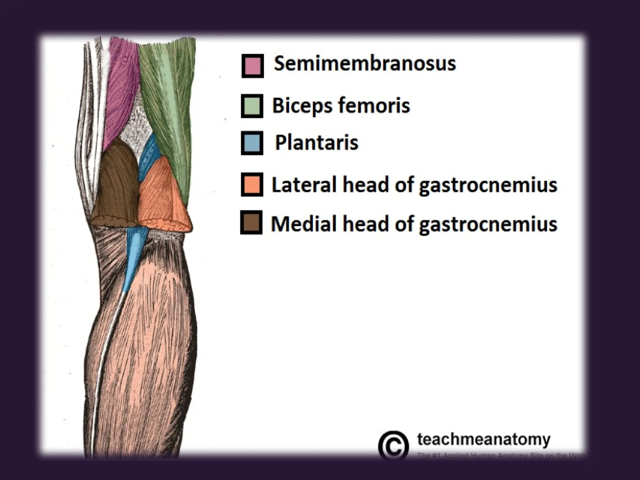
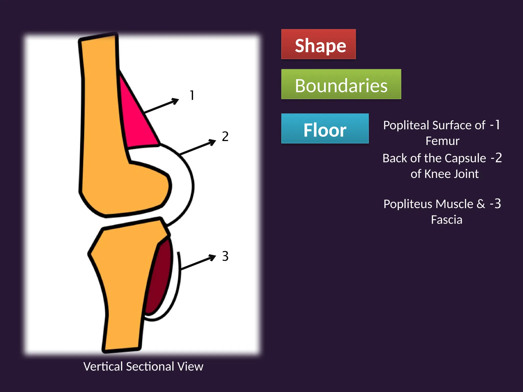
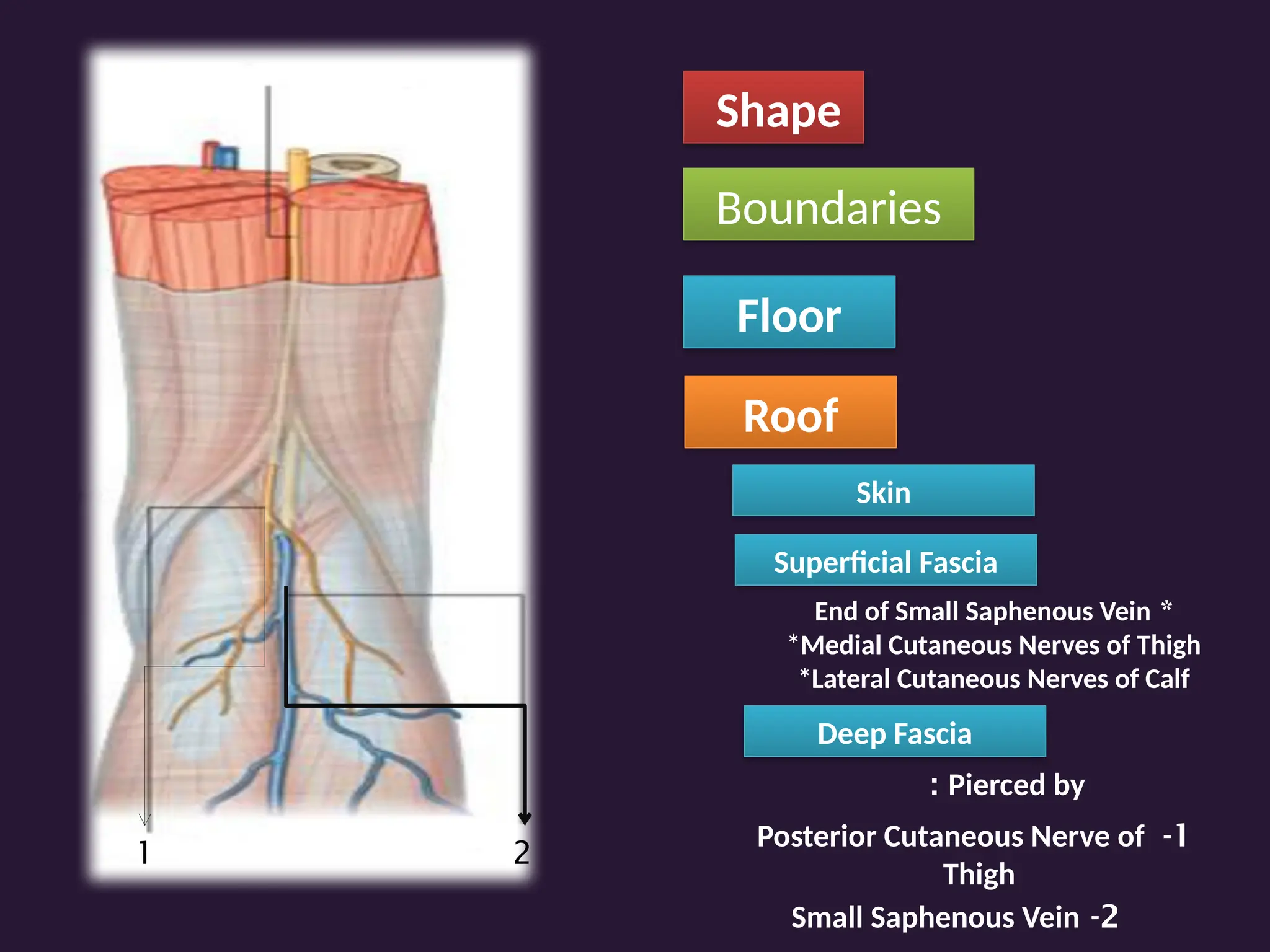
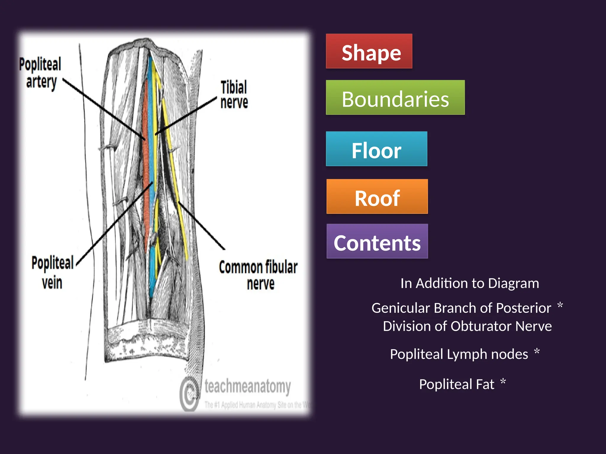
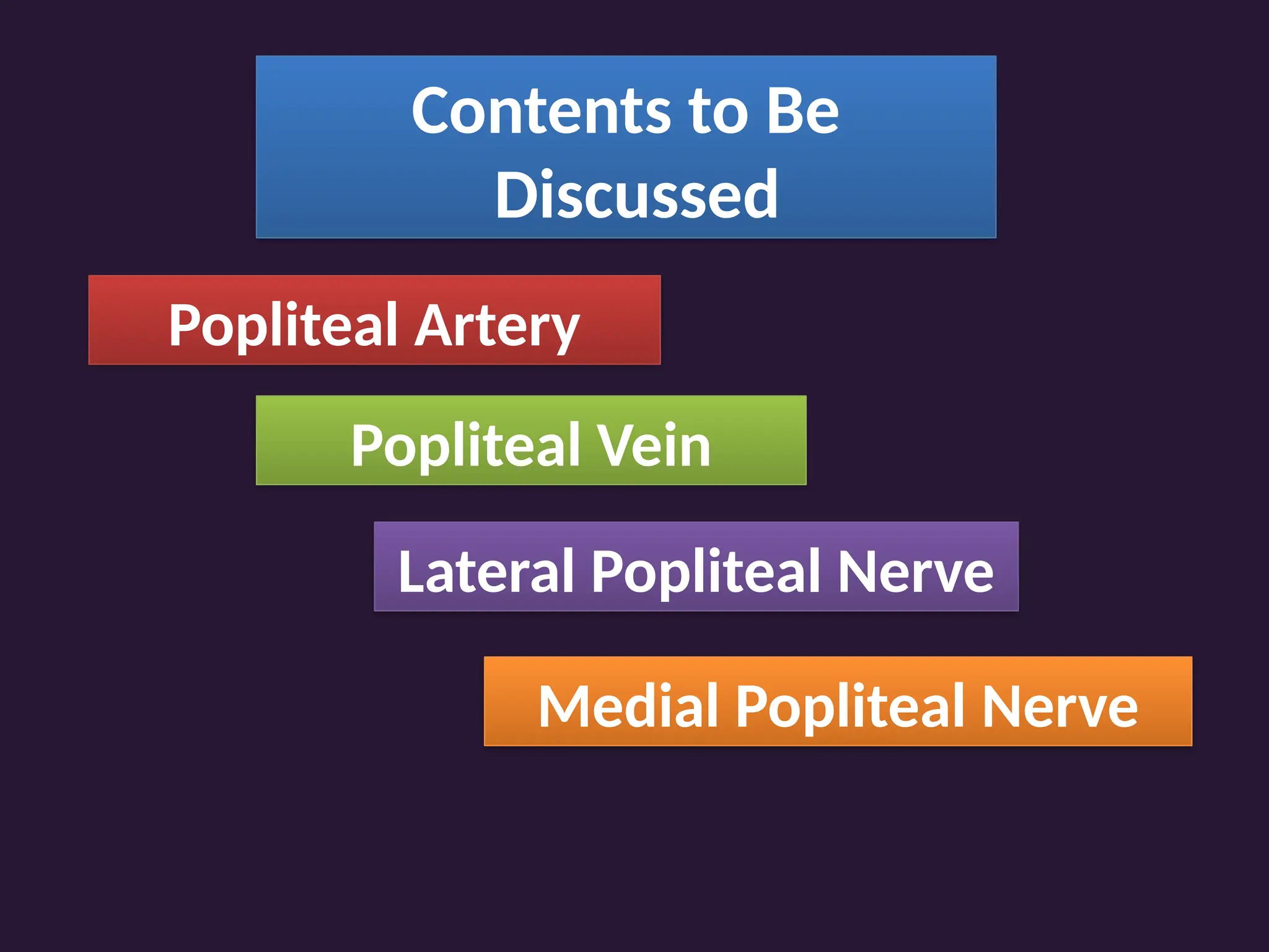
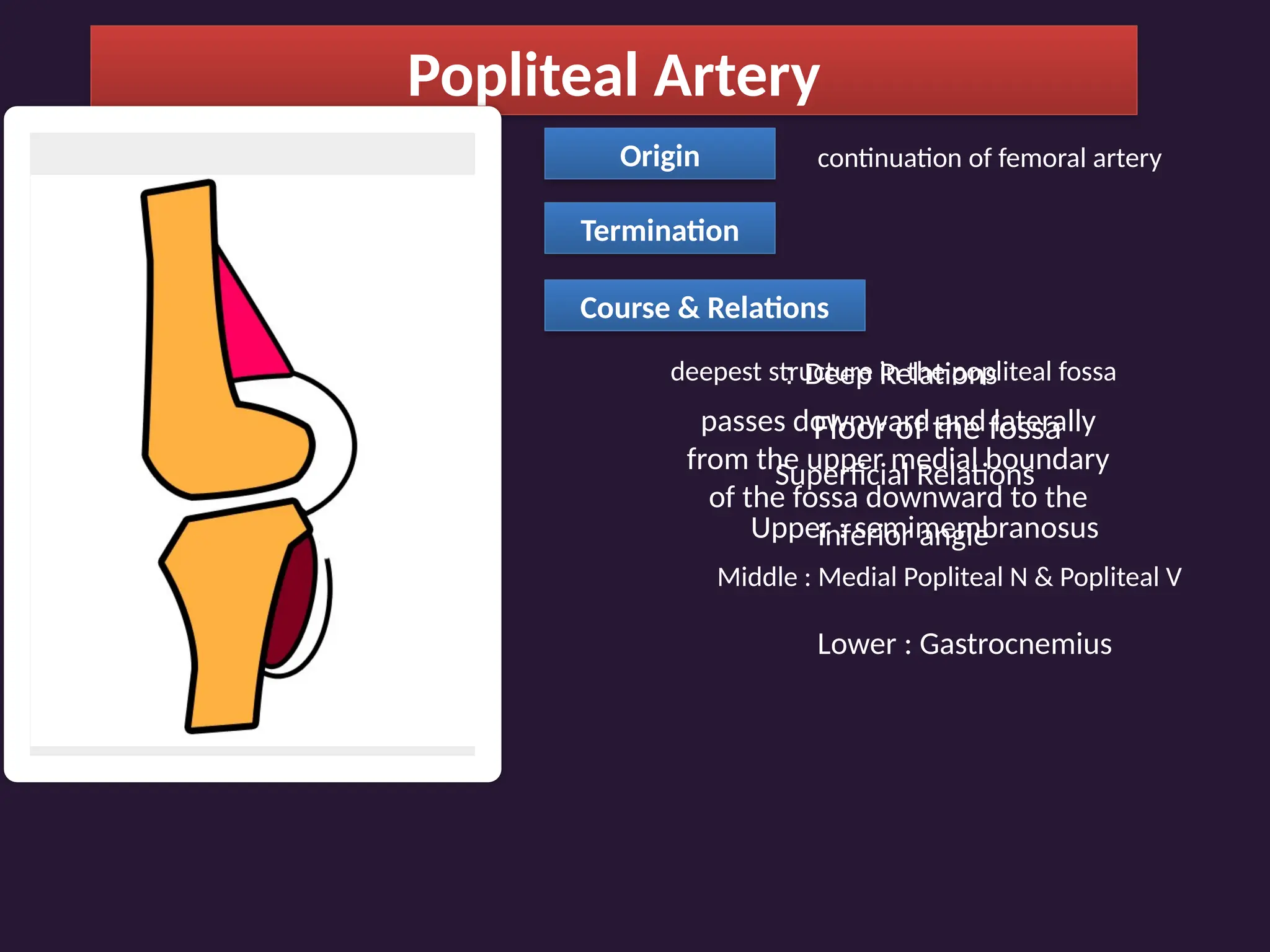
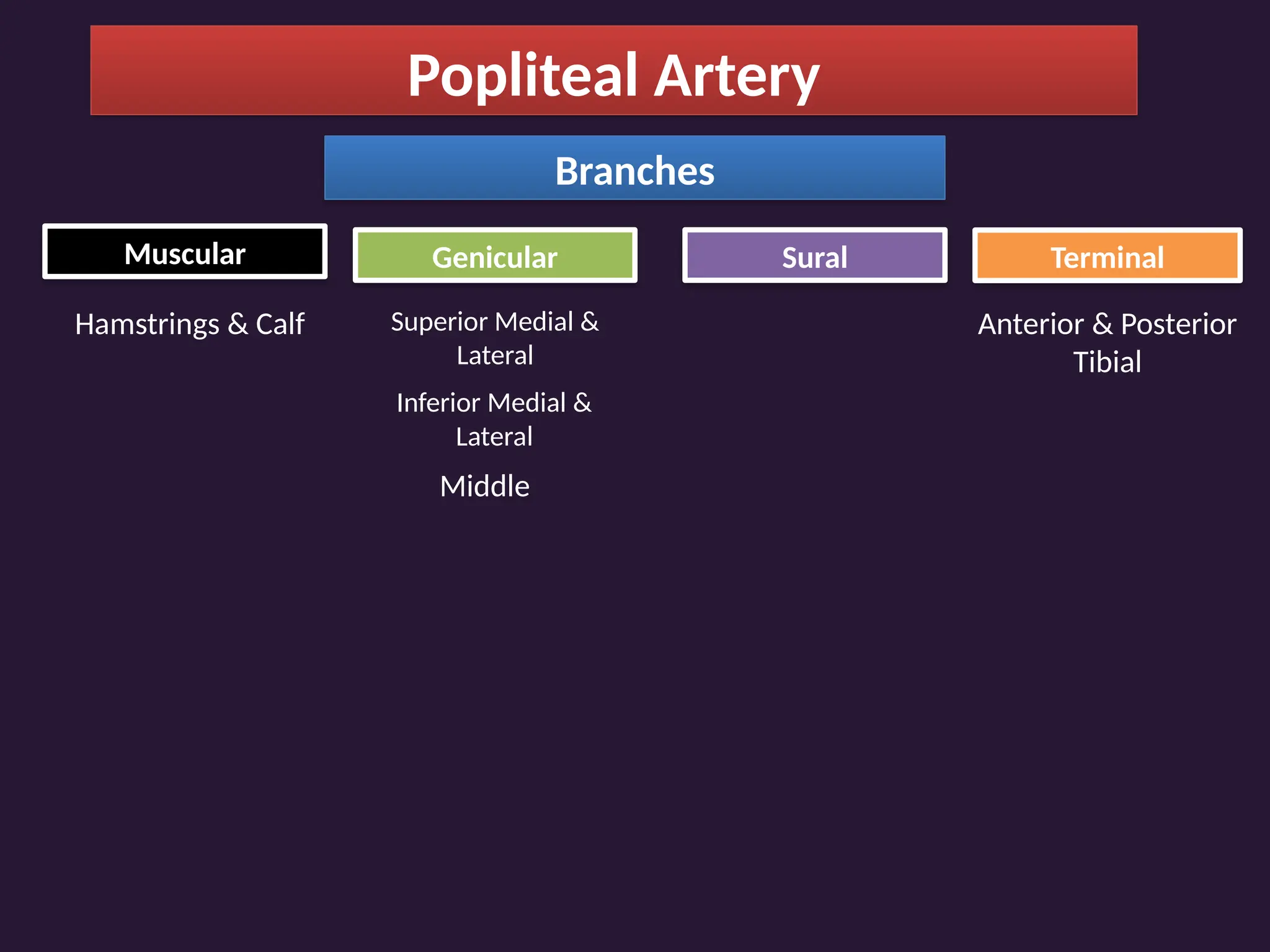
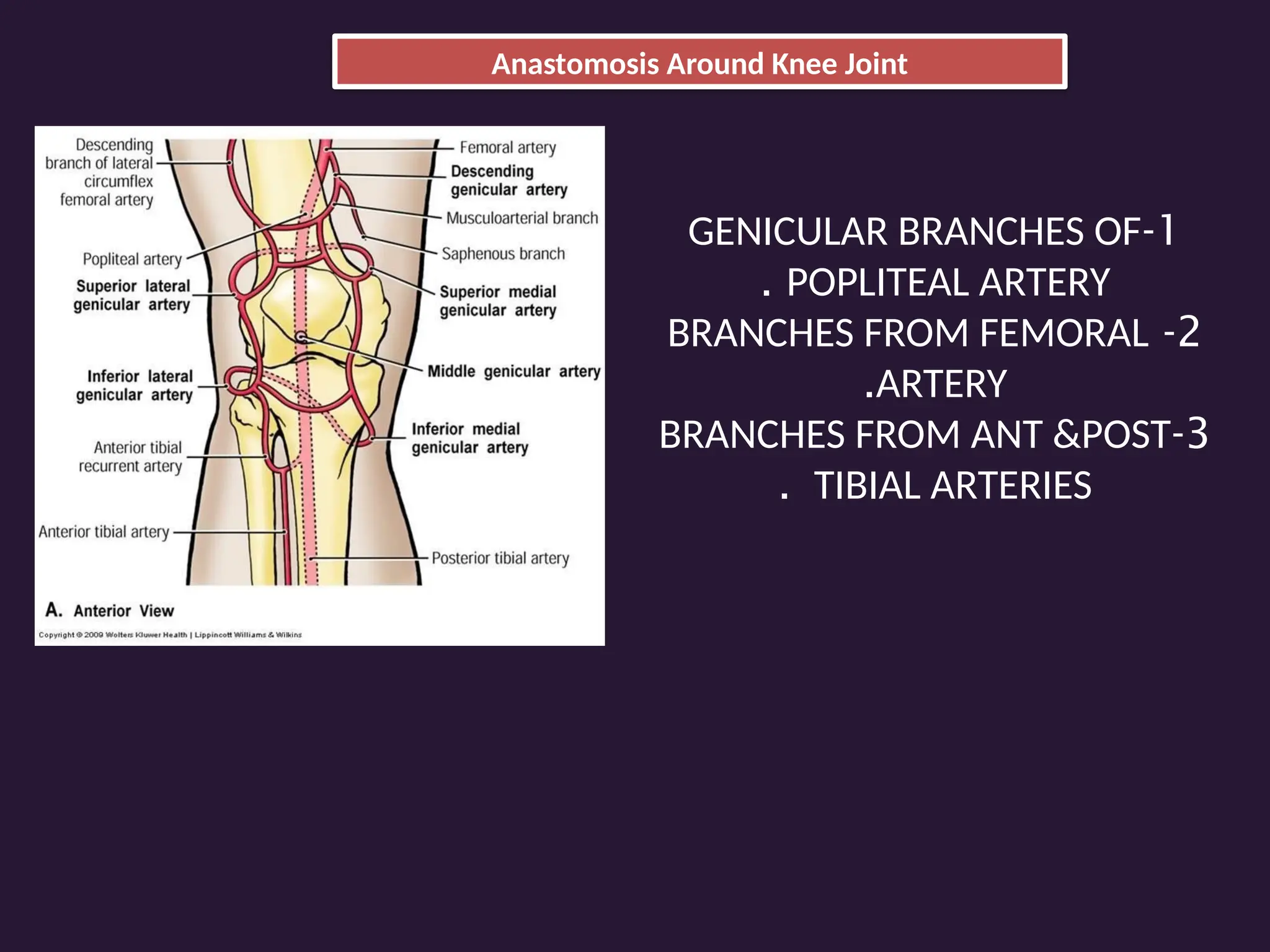
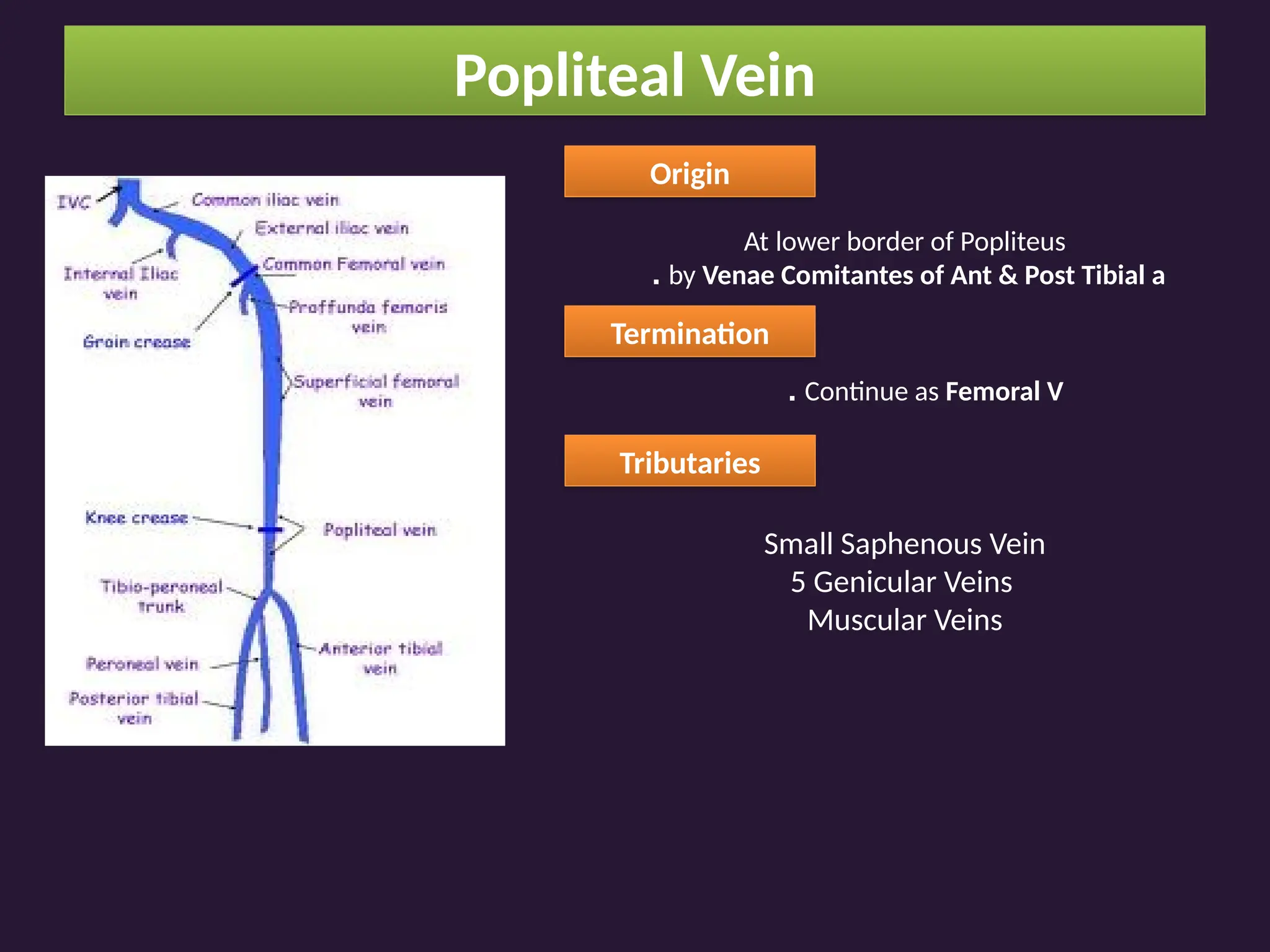
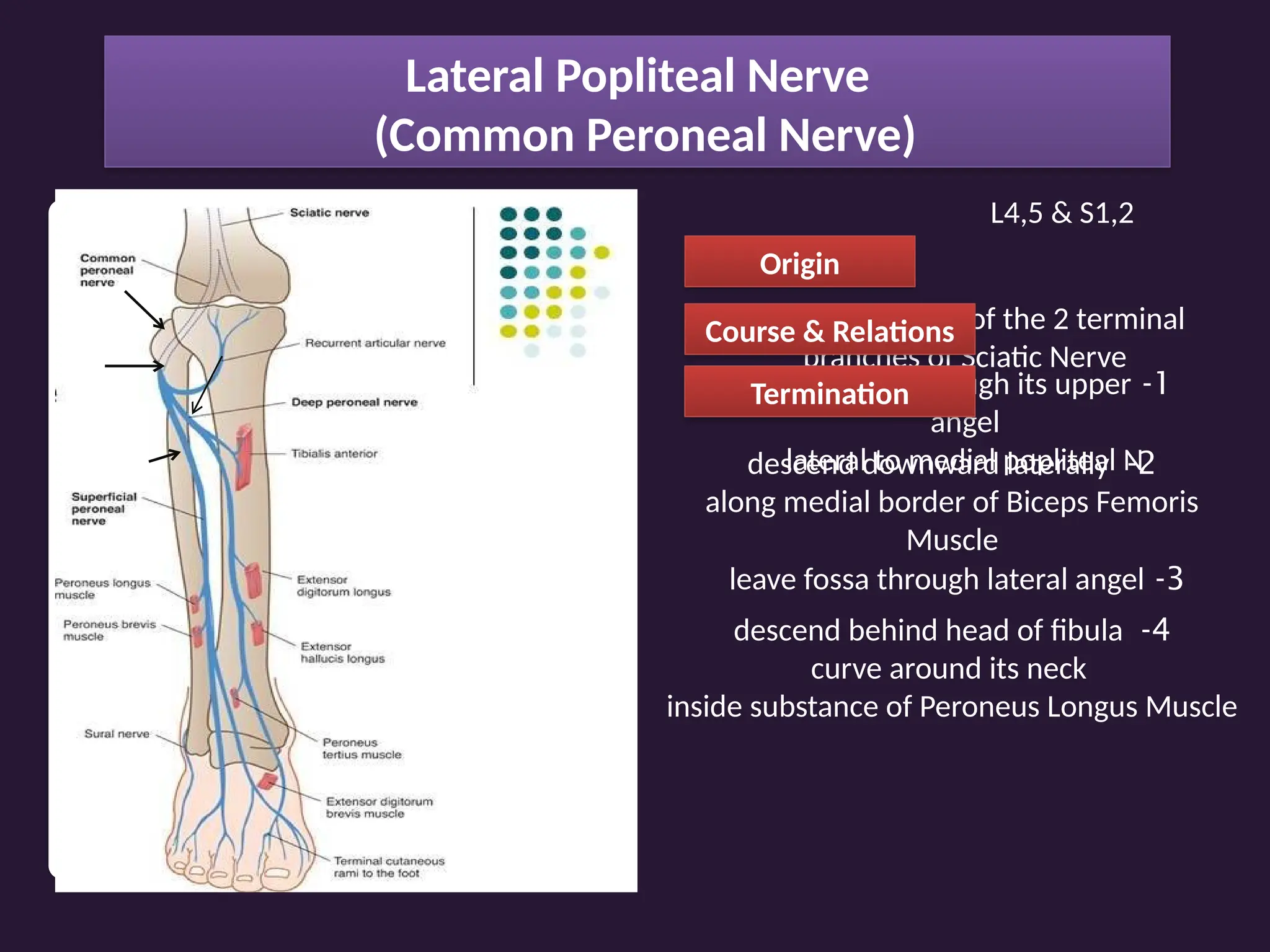
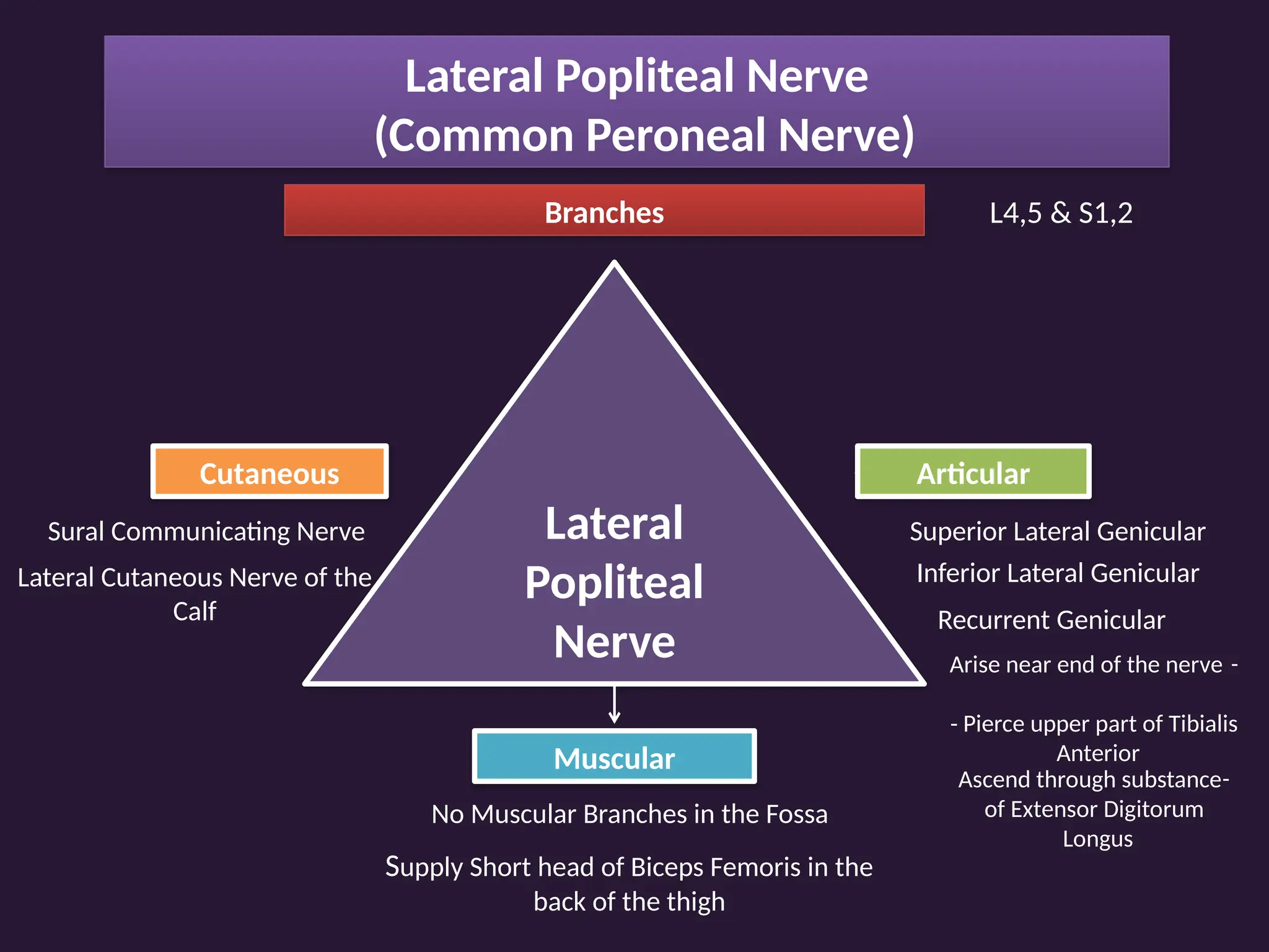
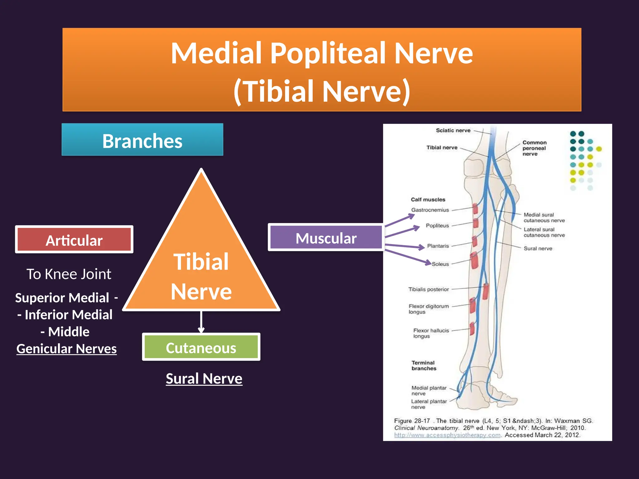
Popliteal Fossa
Presentation by
:
Group (621-630)
Shape Diamond Inter-Muscular Space
Boundaries
1
Upper Lateral
Upper Medial
1
-
Biceps Femoris
2
3
2
-
Semitendinosus
3- Semimembranosus
Lower Medial
4
5
6
4
-
Medial Head of
Gastrocnemius
Lower Lateral 5
-
Plantaris
6- Lateral Head of
Gastrocnemius
Shape
Boundaries
Floor
1
2
3
1
-
Popliteal Surface of
Femur
2
-
Back of the Capsule
of Knee Joint
3
-
Popliteus Muscle &
Fascia
Vertical Sectional View
Shape
Boundaries
Floor
Roof
2
-
Small Saphenous Vein
1 2
Skin
Superficial Fascia
Deep Fascia
*
End of Small Saphenous Vein
*Medial Cutaneous Nerves of Thigh
*Lateral Cutaneous Nerves of Calf
Pierced by
:
1
-
Posterior Cutaneous Nerve of
Thigh
Shape
Boundaries
Floor
Roof
Contents
In Addition to Diagram
*
Genicular Branch of Posterior
Division of Obturator Nerve
*
Popliteal Lymph nodes
*
Popliteal Fat
Contents to Be
Discussed
Popliteal Artery
Popliteal Vein
Lateral Popliteal Nerve
Medial Popliteal Nerve
Contents to Be
Discussed
Popliteal Artery
Popliteal Artery
Origin continuation of femoral artery
Termination
Course & Relations
deepest structure in the popliteal fossa
passes downward and laterally
from the upper medial boundary
of the fossa downward to the
inferior angle
Deep Relations
:
Floor of the fossa
Superficial Relations
Upper : semimembranosus
Middle : Medial Popliteal N & Popliteal V
Lower : Gastrocnemius
Popliteal Artery
Branches
Muscular Genicular Sural Terminal
Hamstrings & Calf Superior Medial &
Lateral
Inferior Medial &
Lateral
Middle
Anterior & Posterior
Tibial
Anastomosis Around Knee Joint
1
-
GENICULAR BRANCHES OF
POPLITEAL ARTERY
.
2
-
BRANCHES FROM FEMORAL
ARTERY
.
3
-
BRANCHES FROM ANT &POST
TIBIAL ARTERIES
.
Contents to Be
Discussed
Popliteal Artery
Popliteal Vein
Popliteal Vein
Origin
At lower border of Popliteus
by Venae Comitantes of Ant & Post Tibial a
.
Termination
Continue as Femoral V
.
Tributaries
Small Saphenous Vein
5 Genicular Veins
Muscular Veins
Contents to Be
Discussed
Popliteal Artery
Popliteal Vein
Lateral Popliteal Nerve
(Common Peroneal Nerve)
L4,5 & S1,2
Origin
Lateral & smaller of the 2 terminal
branches of Sciatic Nerve
Course & Relations
1
-
enter fossa through its upper
angel
lateral to medial popliteal N
2
-
descend downward laterally
along medial border of Biceps Femoris
Muscle
3
-
leave fossa through lateral angel
4
-
descend behind head of fibula
curve around its neck
inside substance of Peroneus Longus Muscle
Termination
Lateral Popliteal Nerve
(Common Peroneal Nerve)
L4,5 & S1,2
Branches
Lateral
Popliteal
Nerve
Muscular
No Muscular Branches in the Fossa
Supply Short head of Biceps Femoris in the
back of the thigh
Cutaneous
Sural Communicating Nerve
Lateral Cutaneous Nerve of the
Calf
Articular
Superior Lateral Genicular
Inferior Lateral Genicular
Recurrent Genicular
-
Arise near end of the nerve
- Pierce upper part of Tibialis
Anterior
-
Ascend through substance
of Extensor Digitorum
Longus
Contents to Be
Discussed
Popliteal Artery
Popliteal Vein
Lateral Popliteal Nerve
Medial Popliteal Nerve
(Tibial Nerve)
L4,5 & S1,2,3
Origin
Medial Terminal Branch of
Sciatic Nerve
Termination
Become Posterior Tibial
Nerve
Course
-
Enter through Upper Angle
-
Most Superficial Structure
-
Leave through Lower Angle
Coverings :
- Upper part :
Hamestring
- Lower Part :
Gastrocnemius
Relation with Vessels :
Lateral
Superficial
Medial
Medial Popliteal Nerve
(Tibial Nerve)
Branches
Tibial
Nerve
Articular
To Knee Joint
-
Superior Medial
- Inferior Medial
- Middle
Genicular Nerves Cutaneous
Sural Nerve
Muscular
Sural Nerve
*
Descends in the groove between 2
heads of Gastrocnemius by short
saphenous nerve
.
*
In the lower 1/3 of the leg, it is
joined by the Sural Communicating
branch of lateral popliteal nerve
*
It runs behind & below the lateral
malleolus then forward along the
lateral border of the foot
.
*
It supplies the skin of the lower 1/3
of the back of the leg & lateral
border of the foot & little toe
.
Popliteal Fossa
Presentation by
:
Group (621-630)

popliteal fossa and its boundaries .ppt

  • 1.
  • 2.
    Shape Diamond Inter-MuscularSpace Boundaries 1 Upper Lateral Upper Medial 1 - Biceps Femoris 2 3 2 - Semitendinosus 3- Semimembranosus Lower Medial 4 5 6 4 - Medial Head of Gastrocnemius Lower Lateral 5 - Plantaris 6- Lateral Head of Gastrocnemius
  • 4.
    Shape Boundaries Floor 1 2 3 1 - Popliteal Surface of Femur 2 - Backof the Capsule of Knee Joint 3 - Popliteus Muscle & Fascia Vertical Sectional View
  • 5.
    Shape Boundaries Floor Roof 2 - Small Saphenous Vein 12 Skin Superficial Fascia Deep Fascia * End of Small Saphenous Vein *Medial Cutaneous Nerves of Thigh *Lateral Cutaneous Nerves of Calf Pierced by : 1 - Posterior Cutaneous Nerve of Thigh
  • 6.
    Shape Boundaries Floor Roof Contents In Addition toDiagram * Genicular Branch of Posterior Division of Obturator Nerve * Popliteal Lymph nodes * Popliteal Fat
  • 7.
    Contents to Be Discussed PoplitealArtery Popliteal Vein Lateral Popliteal Nerve Medial Popliteal Nerve
  • 8.
  • 9.
    Popliteal Artery Origin continuationof femoral artery Termination Course & Relations deepest structure in the popliteal fossa passes downward and laterally from the upper medial boundary of the fossa downward to the inferior angle Deep Relations : Floor of the fossa Superficial Relations Upper : semimembranosus Middle : Medial Popliteal N & Popliteal V Lower : Gastrocnemius
  • 10.
    Popliteal Artery Branches Muscular GenicularSural Terminal Hamstrings & Calf Superior Medial & Lateral Inferior Medial & Lateral Middle Anterior & Posterior Tibial
  • 11.
    Anastomosis Around KneeJoint 1 - GENICULAR BRANCHES OF POPLITEAL ARTERY . 2 - BRANCHES FROM FEMORAL ARTERY . 3 - BRANCHES FROM ANT &POST TIBIAL ARTERIES .
  • 12.
  • 13.
    Popliteal Vein Origin At lowerborder of Popliteus by Venae Comitantes of Ant & Post Tibial a . Termination Continue as Femoral V . Tributaries Small Saphenous Vein 5 Genicular Veins Muscular Veins
  • 14.
    Contents to Be Discussed PoplitealArtery Popliteal Vein Lateral Popliteal Nerve
  • 15.
    Lateral Popliteal Nerve (CommonPeroneal Nerve) L4,5 & S1,2 Origin Lateral & smaller of the 2 terminal branches of Sciatic Nerve Course & Relations 1 - enter fossa through its upper angel lateral to medial popliteal N 2 - descend downward laterally along medial border of Biceps Femoris Muscle 3 - leave fossa through lateral angel 4 - descend behind head of fibula curve around its neck inside substance of Peroneus Longus Muscle Termination
  • 16.
    Lateral Popliteal Nerve (CommonPeroneal Nerve) L4,5 & S1,2 Branches Lateral Popliteal Nerve Muscular No Muscular Branches in the Fossa Supply Short head of Biceps Femoris in the back of the thigh Cutaneous Sural Communicating Nerve Lateral Cutaneous Nerve of the Calf Articular Superior Lateral Genicular Inferior Lateral Genicular Recurrent Genicular - Arise near end of the nerve - Pierce upper part of Tibialis Anterior - Ascend through substance of Extensor Digitorum Longus
  • 17.
    Contents to Be Discussed PoplitealArtery Popliteal Vein Lateral Popliteal Nerve Medial Popliteal Nerve
  • 18.
    Medial Popliteal Nerve (TibialNerve) L4,5 & S1,2,3 Origin Medial Terminal Branch of Sciatic Nerve Termination Become Posterior Tibial Nerve Course - Enter through Upper Angle - Most Superficial Structure - Leave through Lower Angle Coverings : - Upper part : Hamestring - Lower Part : Gastrocnemius Relation with Vessels : Lateral Superficial Medial
  • 19.
    Medial Popliteal Nerve (TibialNerve) Branches Tibial Nerve Articular To Knee Joint - Superior Medial - Inferior Medial - Middle Genicular Nerves Cutaneous Sural Nerve Muscular
  • 20.
    Sural Nerve * Descends inthe groove between 2 heads of Gastrocnemius by short saphenous nerve . * In the lower 1/3 of the leg, it is joined by the Sural Communicating branch of lateral popliteal nerve * It runs behind & below the lateral malleolus then forward along the lateral border of the foot . * It supplies the skin of the lower 1/3 of the back of the leg & lateral border of the foot & little toe .
  • 21.