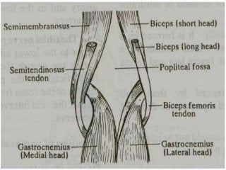
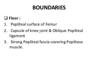
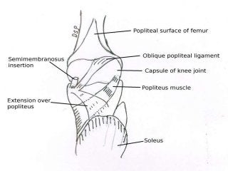
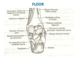
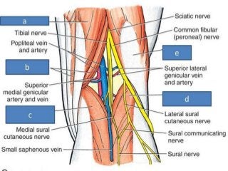
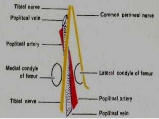
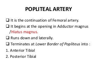
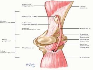
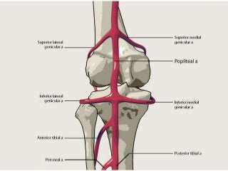
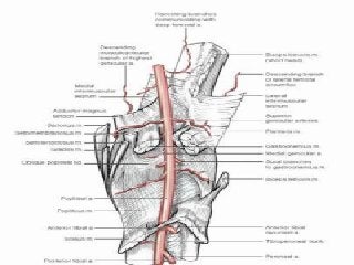
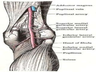
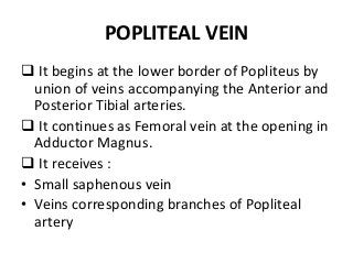
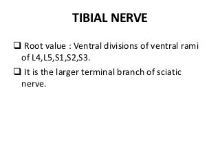
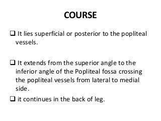
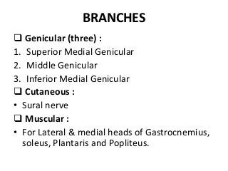
POPLITEAL FOSSA
INTRODUCTION
• It is a diamond shaped depression lying
behind Knee Joint, Lower part of femur and
Upper part of tibia.
BOUNDARIES
 Supero-laterally : Biceps femoris
 Supero-medially : Semitendinosus,
Semimembranosus, Gracilis, Sartorius and Adductor
magnus.
 Infero-laterally : Lateral head of gastrocnemius ,
plantaris.
 Infero -medially : Medial head of gastrocnemius.
BOUNDARIES
 Roof :
 Deep fascia / Popliteal fascia.
 Superficial fascia over the roof contains :
1. Small Saphenous vein and cutaneous nerves.
2. Branches & terminal part of Posterior
Cutaneous nerve of thigh
3. Posterior division of Medial Cutaneous nerve of
thigh
4. Peroneal / Sural Communicating nerve
BOUNDARIES
 Floor :
1. Popliteal surface of Femur
2. Capsule of knee joint & Oblique Popliteal
ligament
3. Strong Popliteal fascia covering Popliteus
muscle.
CONTENTS
1. Popliteal artery and its branches
2. Popliteal vein and its tributaries
3. Tibial nerve and its branches
4. Common Peroneal nerve and its branches
5. Posterior cutaneous nerve of thigh
6. Genicular branch of obturator nerve
7. Popliteal lymph nodes
8. Fat
ARRANGEMENT OF STRUCTURES
 Tibial nerve is most superficial
 Popliteal vein lies deep /anterior to tibial
nerve.
 Popliteal artery lies deep to all.
RELATION OF STRUCTURES
 In upper part :
 Medial to lateral – artery, vein & nerve
 In middle part :
 Behind forwards – nerve, vein & artery.
 In lower part :
 medial to lateral – nerve , vein & artery.
POPLITEAL ARTERY
 It is the continuation of Femoral artery.
 It begins at the opening in Adductor magnus
/Hiatus magnus.
 Runs down and laterally.
 Terminates at Lower Border of Popliteus into :
1. Anterior Tibial
2. Posterior Tibial
RELATIONS
 Anterior / Deep : Above down
1. Popliteal surface of femur
2. Back of knee joint
3. Fascia covering Popliteus
 Posterior / Superficial : Tibial nerve
 Laterally :
Upper part - Biceps femoris & lateral condyle of
femur
Lower part – Plantaris & lateral head of
gastrocnemius
RELATIONS
 Medially :
 Upper part – Semimembranosus & medial
condyle of femur
 Lower part : Tibial nerve, Popliteal Vein & Medial
Head of Gastrocnemius
BRANCHES
 Muscular :
• Upper 2-3 branches – Adductor magnus,
Hamstrings
• Lower branches – Gastrocnemius, Soleus and
Plantaris.
 Cutaneous branches :
• Arise directly from PA or indirectly from
muscular branches.
• One cutaneous branch always accompanies
Small saphenous vein.
BRANCHES
 Genicular :
1. Medial and Lateral Superior Genicular
2. Medial and Lateral Inferior Genicular
3. Middle Genicular
POPLITEAL VEIN
 It begins at the lower border of Popliteus by
union of veins accompanying the Anterior and
Posterior Tibial arteries.
 It continues as Femoral vein at the opening in
Adductor Magnus.
 It receives :
• Small saphenous vein
• Veins corresponding branches of Popliteal
artery
TIBIAL NERVE
 Root value : Ventral divisions of ventral rami
of L4,L5,S1,S2,S3.
 It is the larger terminal branch of sciatic
nerve.
COURSE
 It lies superficial or posterior to the popliteal
vessels.
 It extends from the superior angle to the
inferior angle of the Popliteal fossa crossing
the popliteal vessels from lateral to medial
side.
 it continues in the back of leg.
BRANCHES
 Genicular (three) :
1. Superior Medial Genicular
2. Middle Genicular
3. Inferior Medial Genicular
 Cutaneous :
• Sural nerve
 Muscular :
• For Lateral & medial heads of Gastrocnemius,
soleus, Plantaris and Popliteus.
COMMON PERONEAL NERVE
Root value : Dorsal divisions of Ventral rami of
L4,L5,S1,S2.
It is the smaller terminal branch of Sciatic
nerve.
It lies in the same plane as the Tibial nerve.
COURSE
 It extends from the superior angle of the
fossa to the lateral angle of the Popliteal fossa
along the medial border of the Biceps
Femoris.
 It continues downwards and forwards and
winds round the postero-lateral aspect of the
neck of fibula and divides into Superficial and
Deep Peroneal nerves.
BRANCHES
 Genicular (three) :
• Superior Lateral Genicular
• Inferior Lateral Genicular
• Recurrent Genicular
 Cutaneous :
• Lateral Cutaneous nerve of calf
• Sural Communicating nerve
 Muscular :
• None except a branch to short head of Biceps
Femoris occasionally.
POSTERIOR CUTANEOUS NERVE OF
THIGH
GENICULAR BRANCH OF OBTURATOR
NERVE
ANASTOMOSES AROUND KNEE JOINT
APPLIED ANATOMY
1. Blood pressure in Lower limb is recorded
from Popliteal artery.
2. Constant pulsations of PA against unyielding
tendon of Adductor Magnus leads to vascular
changes in the vessel wall – narrowing and
occlusion of artery.
3. Popliteal artery is fixed to the Capsule of
Knee joint by a fibrous band.
4. Popliteal artery is most prone for aneurysm
than any other artery in body.
APPLIED ANATOMY
5. Most of the muscular branches of Tibial
nerve arise from its lateral side except to
medial head of gastrocnemius. So medial
side is the safe side.
6. Common Peroneal nerve can be palpated
against Postero-lateral side of the neck of the
fibula.
7. It is the most frequently injured nerve in
lower limb.

Popliteal fossa 1.pptx

  • 1.
  • 2.
    INTRODUCTION • It isa diamond shaped depression lying behind Knee Joint, Lower part of femur and Upper part of tibia.
  • 3.
    BOUNDARIES  Supero-laterally :Biceps femoris  Supero-medially : Semitendinosus, Semimembranosus, Gracilis, Sartorius and Adductor magnus.  Infero-laterally : Lateral head of gastrocnemius , plantaris.  Infero -medially : Medial head of gastrocnemius.
  • 5.
    BOUNDARIES  Roof : Deep fascia / Popliteal fascia.  Superficial fascia over the roof contains : 1. Small Saphenous vein and cutaneous nerves. 2. Branches & terminal part of Posterior Cutaneous nerve of thigh 3. Posterior division of Medial Cutaneous nerve of thigh 4. Peroneal / Sural Communicating nerve
  • 8.
    BOUNDARIES  Floor : 1.Popliteal surface of Femur 2. Capsule of knee joint & Oblique Popliteal ligament 3. Strong Popliteal fascia covering Popliteus muscle.
  • 11.
    CONTENTS 1. Popliteal arteryand its branches 2. Popliteal vein and its tributaries 3. Tibial nerve and its branches 4. Common Peroneal nerve and its branches 5. Posterior cutaneous nerve of thigh 6. Genicular branch of obturator nerve 7. Popliteal lymph nodes 8. Fat
  • 14.
    ARRANGEMENT OF STRUCTURES Tibial nerve is most superficial  Popliteal vein lies deep /anterior to tibial nerve.  Popliteal artery lies deep to all.
  • 15.
    RELATION OF STRUCTURES In upper part :  Medial to lateral – artery, vein & nerve  In middle part :  Behind forwards – nerve, vein & artery.  In lower part :  medial to lateral – nerve , vein & artery.
  • 16.
    POPLITEAL ARTERY  Itis the continuation of Femoral artery.  It begins at the opening in Adductor magnus /Hiatus magnus.  Runs down and laterally.  Terminates at Lower Border of Popliteus into : 1. Anterior Tibial 2. Posterior Tibial
  • 20.
    RELATIONS  Anterior /Deep : Above down 1. Popliteal surface of femur 2. Back of knee joint 3. Fascia covering Popliteus  Posterior / Superficial : Tibial nerve  Laterally : Upper part - Biceps femoris & lateral condyle of femur Lower part – Plantaris & lateral head of gastrocnemius
  • 21.
    RELATIONS  Medially : Upper part – Semimembranosus & medial condyle of femur  Lower part : Tibial nerve, Popliteal Vein & Medial Head of Gastrocnemius
  • 23.
    BRANCHES  Muscular : •Upper 2-3 branches – Adductor magnus, Hamstrings • Lower branches – Gastrocnemius, Soleus and Plantaris.  Cutaneous branches : • Arise directly from PA or indirectly from muscular branches. • One cutaneous branch always accompanies Small saphenous vein.
  • 24.
    BRANCHES  Genicular : 1.Medial and Lateral Superior Genicular 2. Medial and Lateral Inferior Genicular 3. Middle Genicular
  • 25.
    POPLITEAL VEIN  Itbegins at the lower border of Popliteus by union of veins accompanying the Anterior and Posterior Tibial arteries.  It continues as Femoral vein at the opening in Adductor Magnus.  It receives : • Small saphenous vein • Veins corresponding branches of Popliteal artery
  • 26.
    TIBIAL NERVE  Rootvalue : Ventral divisions of ventral rami of L4,L5,S1,S2,S3.  It is the larger terminal branch of sciatic nerve.
  • 27.
    COURSE  It liessuperficial or posterior to the popliteal vessels.  It extends from the superior angle to the inferior angle of the Popliteal fossa crossing the popliteal vessels from lateral to medial side.  it continues in the back of leg.
  • 28.
    BRANCHES  Genicular (three): 1. Superior Medial Genicular 2. Middle Genicular 3. Inferior Medial Genicular  Cutaneous : • Sural nerve  Muscular : • For Lateral & medial heads of Gastrocnemius, soleus, Plantaris and Popliteus.
  • 29.
    COMMON PERONEAL NERVE Rootvalue : Dorsal divisions of Ventral rami of L4,L5,S1,S2. It is the smaller terminal branch of Sciatic nerve. It lies in the same plane as the Tibial nerve.
  • 30.
    COURSE  It extendsfrom the superior angle of the fossa to the lateral angle of the Popliteal fossa along the medial border of the Biceps Femoris.  It continues downwards and forwards and winds round the postero-lateral aspect of the neck of fibula and divides into Superficial and Deep Peroneal nerves.
  • 31.
    BRANCHES  Genicular (three): • Superior Lateral Genicular • Inferior Lateral Genicular • Recurrent Genicular  Cutaneous : • Lateral Cutaneous nerve of calf • Sural Communicating nerve  Muscular : • None except a branch to short head of Biceps Femoris occasionally.
  • 32.
  • 33.
    GENICULAR BRANCH OFOBTURATOR NERVE
  • 34.
  • 35.
    APPLIED ANATOMY 1. Bloodpressure in Lower limb is recorded from Popliteal artery. 2. Constant pulsations of PA against unyielding tendon of Adductor Magnus leads to vascular changes in the vessel wall – narrowing and occlusion of artery. 3. Popliteal artery is fixed to the Capsule of Knee joint by a fibrous band. 4. Popliteal artery is most prone for aneurysm than any other artery in body.
  • 36.
    APPLIED ANATOMY 5. Mostof the muscular branches of Tibial nerve arise from its lateral side except to medial head of gastrocnemius. So medial side is the safe side. 6. Common Peroneal nerve can be palpated against Postero-lateral side of the neck of the fibula. 7. It is the most frequently injured nerve in lower limb.