1. POPLITEAL FOSSA
۞ Nabeela Kanwal ۞ Sabrina Yousaf
۞ Sehrish Perwaiz ۞ Samer
۞ Hina ۞ Sara ۞ Urooj
End
Presented by:
2. End
The word Popliteal refers to any of the anatomical structures
located in the back of the knee.
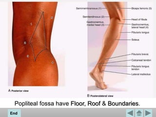
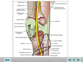
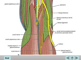
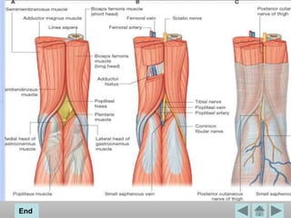
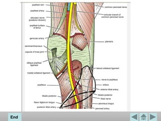
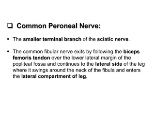
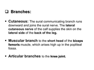
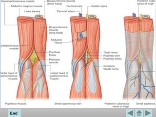
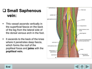
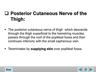
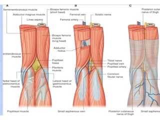
The popliteal fossa is a diamond-shaped intermuscular space
situated at the back of the knee .
The fossa is most prominent when the knee joint is flexed. The
bones of the popliteal fossa are the femur and the tibia. Colloquially, it is
referred to as a "knee pit.“
The popliteal fossa is an important area of transition between the thigh
and leg and is the major route by which structures pass from one region to
the other
4. End
Floor:
The floor is formed by
Popliteal surface of femur
Capsule of knee joint & oblique Popliteal ligament
Strong fascia covering the Popliteal muscle
6. Roof:
Roof is formed by (from superficial to deep).
Skin
Superficial fascia which contains short saphenous vein,
three cutaneous nerves i.e, terminal branch of posterior
cutaneous nerve of thigh, posterior division of medial
cutaneous nerve, and peroneal or sural communicating
nerve
Deep fascia or popliteal fascia
End
9. BOUNDARIES:
The margins of the upper part of the diamond are formed
Medially by the distal ends of the semitendinosus and
semimembranosus muscles and
Laterally by the distal end of the biceps femoris muscle.
The margins of smaller lower part of the space are formed
Medially by the medial head of the gastrocnemius muscle
and
Laterally by the plantaris muscle and the lateral head of the
gastrocnemius muscle.
End
11. CONTENTS:
Popliteal fossa contains
Popliteal artery
Popliteal vein
The tibial and common fibular/ Peroneal nerves
Small saphenous vein
Posterior cutaneous nerve of thigh
Genicular branch of obturator nerve
Popliteal Lymph Nodes
End
12. Popliteal artery:
Popliteal artery is the continuation of the femoral artery in the anterior
compartment of thigh and begins as the femoral artery passes posteriorly
through the adductor hiatus in the adductor magnus muscle.
Popliteal artery appears in the popliteal fossa on the upper medial side
under the margin of the semimembranosus muscle.
Descends obliquely through the fossa with the tibial nerve
Enters the posterior compartment of leg
Ends just lateral to the midline of the leg by dividing into the anterior and
posterior tibial arteries.
Popliteal artery is the deepest of the neurovascular structures in the popliteal
fossa and therefore difficult to palpate, however, a pulse can usually be
detected by deep palpation medial to the midline.
In the popliteal fossa, the popliteal artery gives rise to branches, which supply
adjacent muscles, and to a series of geniculate arteries, which contribute to
vascular anastomoses around the knee.
End
13. Popliteal vein:
The popliteal vein is superficial to and travels
with the popliteal artery. It exits the popliteal
fossa superiorly to become the femoral vein by
passing through the adductor hiatus.
16. Tibial nerve:
Originate proximal to the popliteal fossa as the two major
branches of the sciatic nerve. They are the most
superficial of the neurovascular structures in the
popliteal fossa and enter the region directly from above
under the margin of the biceps femoris muscle.
The tibial nerve descends vertically through the
popliteal fossa and exits deep to the margin of plantaris
muscle to enter the posterior compartment of leg.
End
17. Branches:
Cutaneous: The sural nerve descends between the
two heads of the gastrocnemius muscle and is usually
joined by the sural communicating branch of the
common peroneal nerve. Numerous small branches arise
from the sural nerve to supply the skin of the calf and the
back of the leg. The sural nerve accompanies the small
saphenous vein behind the lateral malleolus and is
distributed to the skin along the lateral border of the foot
and the lateral side of the little toe.
Muscular branches: Supply both heads of the
gastrocnemius, plantaris, soleus, and poplite.
Articular branches: Supply the knee joint.
18. Common Peroneal Nerve:
The smaller terminal branch of the sciatic nerve.
The common fibular nerve exits by following the biceps
femoris tendon over the lower lateral margin of the
popliteal fossa and continues to the lateral side of the leg
where it swings around the neck of the fibula and enters
the lateral compartment of leg.
19. Branches:
Cutaneous: The sural communicating branch runs
downward and joins the sural nerve. The lateral
cutaneous nerve of the calf supplies the skin on the
lateral side of the back of the leg.
Muscular branch to the short head of the biceps
femoris muscle, which arises high up in the popliteal
fossa.
Articular branches to the knee joint.
21. Small Saphenous
vein:
This vessel ascends vertically in
the superficial fascia on the back
of the leg from the lateral side of
the dorsal venous arch in the foot.
It ascends to the back of the knee
where it penetrates deep fascia,
which forms the roof of the
popliteal fossa and joins with the
popliteal vein.
End
22. Posterior Cutaneous Nerve of the
Thigh:
The posterior cutaneous nerve of thigh which descends
through the thigh superficial to the hamstring muscles,
passes through the roof of the popliteal fossa and then
continues inferiorly with the small saphenous vein.
Tereminates by supplying skin over popliteal fossa.
End
23. Genicular branch of Obturator
nerve:
The posterior division of the obturator nerve in the
medial compartment of the thigh.
It leaves the subsartorial canal with the femoral artery
by passing through the opening in the adductor magnus.
The nerve terminates by supplying the knee joint.
End
26. Popliteal Lymph Nodes:
Six lymph nodes are embedded in the fatty connective
tissue of the popliteal fossa.
They receive superficial lymph vessels from lateral side
of the foot and leg.
Accompany the small saphenous vein into popliteal
fossa.
They also receive lymph from knee joint and from deep
lymph vessels.
Accompanying the anterior and posterior tibial arteries.
End