
Recommended
More Related Content
What's hot
What's hot (20)
Similar to adductor canal
Similar to adductor canal (20)
Recently uploaded
Recently uploaded (20)
adductor canal
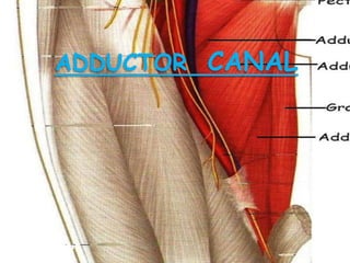
- 2. Anterior superior iliac spine Inguinal ligament Pubic tubercle Adductor longus Adductor canal Sartorius DEFINATION SITUATION EXTENT AND FUNCTION
- 3. HISTORY OF THE ADDUCTOR CANAL Surgeon John Hunter utilized this region as the site for compression of femoral vessels- by applying the tourniquet against the bony resistance of linea aspera to arrest the bleeding in the operation of - aneurysm of popliteal vessels or during the amputation of lower limb below the knee, hence it is called as HUNTERS CANAL.
- 4. Adductor magnus Adductor longus Saphaneous nerve Femoral artery Femoral vein N.to vastus medialis Vastus medialis Fascial roof Sartorius Sub-sartorial nerve plexus BOUNDARIES
- 5. Nerve to vastus medialis Descending genicular artery Saphaneous artery Saphaneous nerve Sub sartorial plexus Sartorius Obturator nerve Med.fem.cut.nerve CONTENTS AND STRUCTURES PIERCING THE ROOF OF ADDUCTOR CANAL
- 6. Femoral vein Femoral artery Saphaneous nerve Saphaneous artery Nerve to vastus medialis CONTENTS AND COURSE OF CONTENTS IN THE CANAL Descending genicular artery
- 7. APPLIED ANATOMY OF ADDUCTOR CANAL • Penetrating injuries at the canal will damage the femoral artery, sectioning of the both the divisions of obturator nerve and saphaneous branch of femoral nerve. Manifestations seen are:- • Loss of blood supply below the knee region. • Loss of cutaneous sensation on medial side of thigh. • Loss of cutaneous sensation on medial border of the foot upto great toe. • Loss of proprioception of knee joint due to sectioning of genicular branch of obturator nerve.
- 8. ANEURYSM OF POPLITEALARTERY • In the aneurysm of popliteal artery, the surgeons ligated the femoral artery in subsartorial canal. • The fibrous roof is incised to enter the canal.
- 10. L1 L2 L3 L4 Obturator nerve Femoral nerve Genitofemoral nerve Ilioinguinal nerve Iliohypogastric nerve Accessory obturator nerve Branch to Lumbosacral trunk Lat.cut.nerve.of.thigh VENTRAL DIVISIONS DORSAL DIVISIONS ANTERIOR PRIMARY RAMI FORMATION-
- 11. Psoas major Sacrum Hip joint Obturator nerve Obturator externus For hip joint Pectineus Gracilis Adductor longus Adductor brevis Ant.division Post. division Adductor magnus To subsartorial nerve plexus Genicular branch L1 L2 L3 L4 L5 COURSE-
- 12. Accessory obturator nerve L3 L4 Obturator internus Superior ramus of pubis Pectineus Hip joint Anterior division of obturator nerve Obturator externus Communicating branch ACCESSORY OBTURATOR NERVE-
- 13. APPLIED ANATOMY- • 1) Referred pain- In the diseases of hip joint, referred pain is there along the medial side of thigh or to knee jt, due to common nerve supply of obturator nerve. • 2) Surgical division of obturator nerve is essential to relieve the spasm of the adductor muscles in spastic paralysis. • 3) In inflammation of ovary, localized peritonitis irritates the obturator nerve. Pain is referred in such condition to the hip, knee or inner side of thigh. • 4) In case of spastic paraplegia obturator nerve is cut in order to ensure better perineal care. • 5) Damage to obturator nerve leads to paralysis of adductor muscles of the medial compartment of thigh and sensory loss on the medial side of the skin of the thigh.