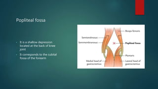
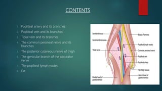
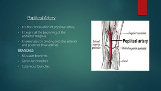
Popliteal Fossa
BY MAZEN BIN KHALID
ROLL NO : 88
Popliteal fossa
• It is a shallow depression
located at the back of knee
joint
• It corresponds to the cubital
fossa of the forearm
Boundaries
• Superolateral: Biceps femoris
• Superomedial:
1. Semitendinosus
2. Semimembranosus
3. Gracilis
4. Adductor Magnus
5. Sartorius
• Inferolateral: Gastrocnemius supplemented
by Plantaris
• Inferomedial: Medial head of
gastrocnemius
CONTENTS
1. Popliteal artery and its branches
2. Popliteal vein and its branches
3. Tibial vein and its branches
4. The common peroneal nerve and its
branches
5. The posterior cutaneous nerve of thigh
6. The genicular branch of the obturator
nerve
7. The popliteal lymph nodes
8. Fat
POPLITEAL FOSSA
Popliteal Artery
• It is the continuation of popliteal artery
• It begins at the beginning of the
adductor magnus
• It terminates by dividing into the anterior
and posterior tibial arteries
BRANCHES
• Muscular branches
• Genicular branches
• Cutaneous branches
Clinical Aspects
• Blood Pressure in Lower limb is recorded
from Popliteal artery
• Popliteal artery is fixed to the capsule of the
knee joint by a fibrous band present just
above the femoral condyles. This may be a
source of continuous traction or stretching
on the artery causing primary thrombosis.
• It is more prone to aneurysm than any other
arteries of the body
Tibial Nerve
• Ventral divisions of ventral rami of L4,
L5, S1, S2, S3.
• This is the larger terminal branch of
the sciatic nerve.
• It lies superficial or posterior to the
popliteal vessels.
BRANCHES
• Three genicular branches
• Cutaneous nerve or sural nerve
• Muscular branches
Clinical Aspects
 Most of the muscular branches of tibial nerve arise from its lateral side
except to medial head of gastrocnemius
 Damage to the tibial nerve causes motor and sensory loss
a) Motor Loss: Superficial and deep muscles of calf and intrinsic muscles of
sole
b) Sensory loss: Loss of sensation on whole of sole of foot, plantar aspect
of digits and nail beds on dorsum of foot.

Popliteal fossa

  • 1.
    Popliteal Fossa BY MAZENBIN KHALID ROLL NO : 88
  • 2.
    Popliteal fossa • Itis a shallow depression located at the back of knee joint • It corresponds to the cubital fossa of the forearm
  • 3.
    Boundaries • Superolateral: Bicepsfemoris • Superomedial: 1. Semitendinosus 2. Semimembranosus 3. Gracilis 4. Adductor Magnus 5. Sartorius • Inferolateral: Gastrocnemius supplemented by Plantaris • Inferomedial: Medial head of gastrocnemius
  • 4.
    CONTENTS 1. Popliteal arteryand its branches 2. Popliteal vein and its branches 3. Tibial vein and its branches 4. The common peroneal nerve and its branches 5. The posterior cutaneous nerve of thigh 6. The genicular branch of the obturator nerve 7. The popliteal lymph nodes 8. Fat
  • 5.
  • 6.
    Popliteal Artery • Itis the continuation of popliteal artery • It begins at the beginning of the adductor magnus • It terminates by dividing into the anterior and posterior tibial arteries BRANCHES • Muscular branches • Genicular branches • Cutaneous branches
  • 7.
    Clinical Aspects • BloodPressure in Lower limb is recorded from Popliteal artery • Popliteal artery is fixed to the capsule of the knee joint by a fibrous band present just above the femoral condyles. This may be a source of continuous traction or stretching on the artery causing primary thrombosis. • It is more prone to aneurysm than any other arteries of the body
  • 8.
    Tibial Nerve • Ventraldivisions of ventral rami of L4, L5, S1, S2, S3. • This is the larger terminal branch of the sciatic nerve. • It lies superficial or posterior to the popliteal vessels. BRANCHES • Three genicular branches • Cutaneous nerve or sural nerve • Muscular branches
  • 9.
    Clinical Aspects  Mostof the muscular branches of tibial nerve arise from its lateral side except to medial head of gastrocnemius  Damage to the tibial nerve causes motor and sensory loss a) Motor Loss: Superficial and deep muscles of calf and intrinsic muscles of sole b) Sensory loss: Loss of sensation on whole of sole of foot, plantar aspect of digits and nail beds on dorsum of foot.