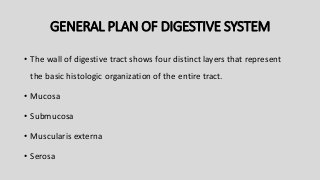
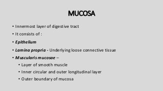
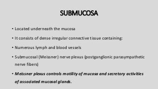
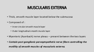
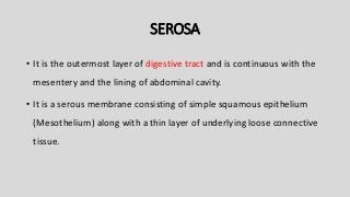
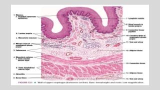
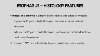
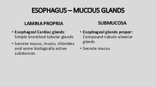
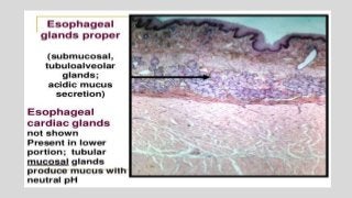
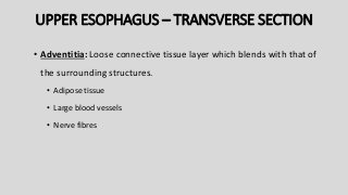
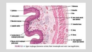
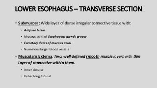
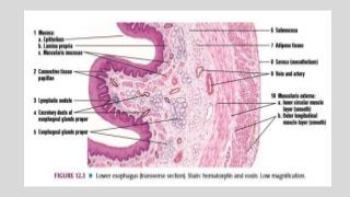
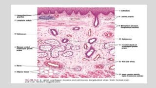
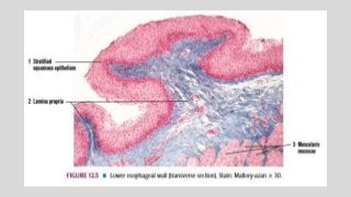
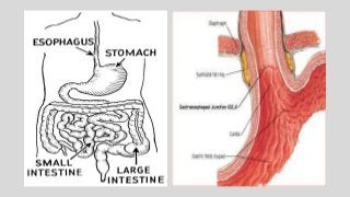
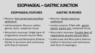
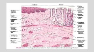
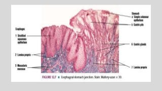
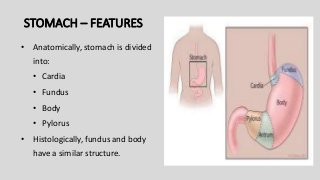
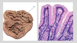
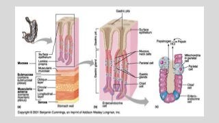
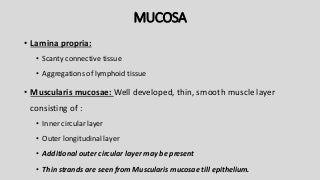
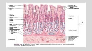
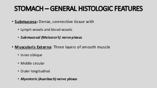
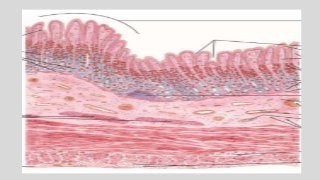
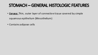
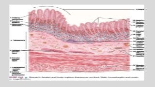
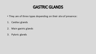
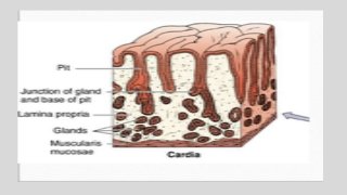
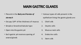
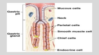
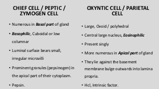
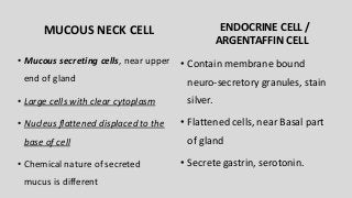
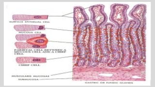
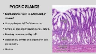
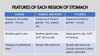
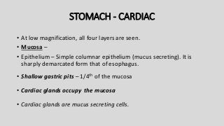
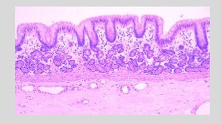
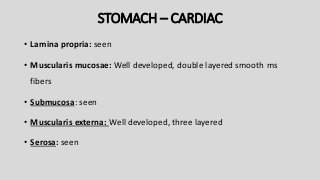
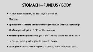
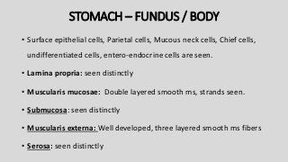
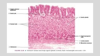
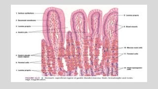
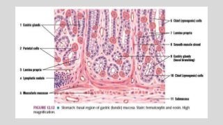
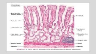
DIGESTIVE SYSTEM - 2
ESOPHAGUS
AND
STOMACH
COMPONENTS
• Oral cavity
• Pharynx
• Esophagus
• Stomach
• Small intestine
• Large intestine
• Rectum
• Anal canal
ACCESSORY DIGESTIVE ORGANS
• Salivary glands
• Liver
• Pancreas
GENERAL PLAN OF DIGESTIVE SYSTEM
• The wall of digestive tract shows four distinct layers that represent
the basic histologic organization of the entire tract.
• Mucosa
• Submucosa
• Muscularis externa
• Serosa
MUCOSA
• Innermost layer of digestive tract
• It consists of :
• Epithelium
• Lamina propria - Underlying loose connective tissue
• Muscularis mucosae –
• Layer of smooth muscle
• Inner circular and outer longitudinal layer
• Outer boundary of mucosa
SUBMUCOSA
• Located underneath the mucosa
• It consists of dense irregular connective tissue containing:
• Numerous lymph and blood vessels
• Submucosal (Meissner) nerve plexus (postganglionic parasympathetic
nerve fibers)
• Meissner plexus controls motility of mucosa and secretory activities
of associated mucosal glands.
MUSCULARIS EXTERNA
• Thick, smooth muscle layer located below the submucosa
• Composed of :
• Inner circular smooth muscle layer
• Outer longitudinal smooth muscle layer
• Myenteric (Auerbach) nerve plexus – present between the two layers
• Contain post ganglionic parasympathetic nerve fibers controlling the
motility of smooth muscles of muscularis externa.
SEROSA
• It is the outermost layer of digestive tract and is continuous with the
mesentery and the lining of abdominal cavity.
• It is a serous membrane consisting of simple squamous epithelium
(Mesothelium) along with a thin layer of underlying loose connective
tissue.
ADVENTITIA
• When the viscera and part of digestive tract is not covered by
mesothelium, the outermost layer is formed by connective tissue
layer only called Adventitia.
ESOPHAGUS
ESOPHAGUS – PARTS
• Esophagus is divided into Thoracic and Abdominal part
• Histologic features of Abdominal part of esophagus varies in:
• Upper one third
• Middle one third
• Lower one third
ESOPHAGUS – HISTOLOGY FEATURES
• Mucosa: In empty state, mucosa shows numerous temporary
longitudinal folds.
i. Epithelium : Moist, stratified non keratinized epithelium
ii. Lamina propria: Connective tissue and glands
iii. Muscularis mucosa:
• Submucosa: Connective tissue and glands
ESOPHAGUS – HISTOLOGY FEATURES
• Muscularis externa: Contains both skeletal and smooth muscles
i. Upper 1/3rd part – Both the layers contain striated skeletal
muscles
ii. Middle 1/3rd part – Both the layers contain both striated skeletal
and smooth muscles
iii. Lower 1/3rd part – Both the layers contain smooth muscles
UPPER ESOPHAGUS – TRANSVERSE SECTION
 Mucosa: Numerous temporary longitudinal folds
• Epithelium – Stratified squamous non keratinized epithelium
• Lamina propria – Underlying thin layer of connective tissue
• Connective tissue papillae
• Smaller blood vessels
• Diffuse lymphatic tissue & solitary lymphatic nodule
• Mucous secreting glands – Esophageal Cardiac glands
• Excretory ducts
• Muscularis mucosae: Longitudinal layer of smooth muscle fibres
UPPER ESOPHAGUS – TRANSVERSE SECTION
• Submucosa: Wide layer of dense irregular connective tissue with:
• Adipose tissue
• Mucous acini of Esophageal glands proper
• Excretory ducts of mucous acini
• Numerous larger blood vessels
• Muscularis Externa: Two, well defined striated muscle layers with thin
layer of connective within them.
• Inner circular
• Outer longitudinal
ESOPHAGUS – MUCOUS GLANDS
LAMINA PROPRIA
• Esophageal Cardiac glands:
Simple branched tubular glands
• Secrete mucus, mucin, chlorides
and some biologically active
substances.
SUBMUCOSA
• Esophageal glands proper:
Compound tubulo-alveolar
glands
• Secrete mucus
UPPER ESOPHAGUS – TRANSVERSE SECTION
• Adventitia: Loose connective tissue layer which blends with that of
the surrounding structures.
• Adipose tissue
• Large blood vessels
• Nerve fibres
LOWER ESOPHAGUS – TRANSVERSE SECTION
 Mucosa: Numerous temporary longitudinal folds
• Epithelium – Stratified squamous non keratinized epithelium
• Lamina propria – Underlying thin layer of connective tissue
• Connective tissue papillae
• Smaller blood vessels
• Diffuse lymphatic tissue & solitary lymphatic nodule
• Mucous secreting glands – Esophageal Cardiac glands
• Excretory ducts
• Muscularis mucosae: Longitudinal layer of smooth muscle fibres
LOWER ESOPHAGUS – TRANSVERSE SECTION
• Submucosa: Wide layer of dense irregular connective tissue with:
• Adipose tissue
• Mucous acini of Esophageal glands proper
• Excretory ducts of mucous acini
• Numerous larger blood vessels
• Muscularis Externa: Two, well defined smooth muscle layers with thin
layer of connective within them.
• Inner circular
• Outer longitudinal
LOWER ESOPHAGUS – TRANSVERSE SECTION
• Serosa: Loose connective tissue layer lined by a simple squamous
layer (Mesothelium).
• Adipose tissue
• Large blood vessels
• Nerve fibers
• Upper 1/3rd of esophagus contains lesser amount of loose
connective tissue at all sites.
UPPER ESOPHAGUS :
LONGITUDINAL SECTION
UPPER ESOPHAGUS – LONGITUDINAL SECTION
 Mucosa: Numerous temporary longitudinal folds
• Epithelium – Stratified squamous non keratinized epithelium with
different layers
• Lamina propria – Underlying thin connective tissue with papillae,
blood vessels, Diffuse and aggregated lymphatic tissue, Esophageal
Cardiac glands and excretory ducts
• Muscularis mucosae: Longitudinally sectioned bundles of smooth
muscle fibers
UPPER ESOPHAGUS – LONGITUDINAL SECTION
• Submucosa: Wide layer of dense irregular connective tissue
containing adipose tissue, mucous acini of Esophageal glands
proper, excretory ducts of mucous acini and numerous larger blood
vessels.
• Muscularis Externa: Inner circular muscle layer is seen sectioned
transversely.
ESOPHAGEAL- GASTRIC
JUNCTION
ESOPHAGEAL – GASTRIC JUNCTION
ESOPHAGEAL FEATURES
• Mucosa: Non keratinized stratified
epithelium
• Lamina propria: Mucous cardiac
glands, ducts, lymphatic tissue
• Muscularis mucosae: Single layer of
longitudinal smooth muscle fibers
• Submucosa and Muscularis Externa:
Similar in structure and continuous
with that of stomach
GASTRIC FEATURES
• Mucosa: Simple columnar
epithelium
• Lamina propria: Filled with gastric
glands, gastric pits, lymphoid tissue.
• Muscularis mucosae: Double layer of
longitudinal smooth muscle fibers
• Submucosa and Muscularis Externa:
Similar in structure and continuous
with that of esophagus.
STOMACH
STOMACH – FEATURES
• Anatomically, stomach is divided
into:
• Cardia
• Fundus
• Body
• Pylorus
• Histologically, fundus and body
have a similar structure.
STOMACH – HISTOLOGIC FEATURES
• Histologically, stomach has three regions – Cardia, Fundus / Body and
Pylorus.
• Stomach wall exhibits four basic layers:
• Mucosa
• Submucosa
• Muscularis externa
• Serosa
MUCOSA
• Rugae: Numerous temporary folds
• Epithelium – Simple columnar epithelium (mucus secreting)
• Gastric pits – Tubular infoldings of the epithelium into lamina propria.
• Lamina propria – Present underneath the epithelium that fills spaces
between the gastric glands.
• Gastric glands- Deep to the pits, the mucous membrane is packed
with numerous gastric glands. Empty into the bottom of gastric pit.
MUCOSA
• Lamina propria:
• Scanty connective tissue
• Aggregations of lymphoid tissue
• Muscularis mucosae: Well developed, thin, smooth muscle layer
consisting of :
• Inner circular layer
• Outer longitudinal layer
• Additional outer circular layer may be present
• Thin strands are seen from Muscularis mucosae till epithelium.
STOMACH – GENERAL HISTOLOGIC FEATURES
• Submucosa: Dense, connective tissue with
• Lymph vessels and blood vessels
• Submucosal (Meissner’s) nerve plexus
• Muscularis Externa: Three layers of smooth muscle
• Inner oblique
• Middle circular
• Outer longitudinal
• Myenteric (Auerbach) nerve plexus
STOMACH – GENERAL HISTOLOGIC FEATURES
• Serosa: Thin, outer layer of connective tissue covered by simple
squamous epithelium (Mesothelium)
• Contains adipose cells
GASTRIC GLANDS
• They are of three types depending on their site of presence:
1. Cardiac glands
2. Main gastric glands
3. Pyloric glands
CARDIAC GLANDS
• Present in the Cardiac part of
stomach
• Simple tubular or compound
tubulo-alveolar
• Secrete mucus
• An occasional oxyntic or peptic cell
may be present
MAIN GASTRIC GLANDS
• Present in the Body and Fundus of
stomach
• Occupy 3/4th of the thickness of mucosa
• Simple or branched tubular type
• Open into the gastric pit
• Each gastric pit receives opening of
several glands
• Various types of cells present in the
epithelium lining the gastric glands are:
1. Chief cells
2. Oxyntic cells
3. Mucous neck cells
4. Endocrine cells
5. Stem cells
CHIEF CELL / PEPTIC /
ZYMOGEN CELL
• Numerous in Basal part of gland
• Basophilic, Cuboidal or low
columnar
• Luminal surface bears small,
irregular microvilli
• Prominent granules (pepsinogen) in
the apical part of their cytoplasm.
• Pepsin.
OXYNTIC CELL / PARIETAL
CELL
• Large, Ovoid / polyhedral
• Central large nucleus, Eosinophilic
• Present singly
• More numerous in Apical part of gland
• They lie against the basement
membrane bulge outwards into lamina
propria.
• Hcl, Intrinsic factor.
MUCOUS NECK CELL
• Mucous secreting cells, near upper
end of gland
• Large cells with clear cytoplasm
• Nucleus flattened displaced to the
base of cell
• Chemical nature of secreted
mucus is different
ENDOCRINE CELL /
ARGENTAFFIN CELL
• Contain membrane bound
neuro-secretory granules, stain
silver.
• Flattened cells, near Basal part
of gland
• Secrete gastrin, serotonin.
STEM CELL
• Undifferentiated cells
• Multiply to replace other cells
PYLORIC GLANDS
• Short glands present in pyloric part of
stomach
• Occupy deeper 1/3rd of the mucosa
• Simple or branched tubular glands, coiled.
• Lined by mucus secreting cells
• Occasionally oxyntic and argentaffin cells
are present.
• Gastrin
FEATURES OF EACH REGION OF STOMACH
CARDIA FUNDUS AND BODY PYLORUS
Presence of Cardiac
glands – mucus
Presence of Gastric
glands – hcl, pepsin,
etc.
Presence of Pyloric
glands – mucus
Shallow gastric pits Shallow gastric pits,
1/4th of mucosa
Deep gastric pits, 2/3rd
of mucosa
Change of epithelium
seen
Simple tall columnar Simple tall columnar
Circular muscle is thick-
pyloric sphincter
STOMACH - CARDIAC
• At low magnification, all four layers are seen.
• Mucosa –
• Epithelium – Simple columnar epithelium (mucus secreting). It is
sharply demarcated form that of esophagus.
• Shallow gastric pits – 1/4th of the mucosa
• Cardiac glands occupy the mucosa
• Cardiac glands are mucus secreting cells.
STOMACH – CARDIAC
• Lamina propria: seen
• Muscularis mucosae: Well developed, double layered smooth ms
fibers
• Submucosa: seen
• Muscularis externa: Well developed, three layered
• Serosa: seen
STOMACH – FUNDUS / BODY
• At low magnification, all four layers are seen.
• Mucosa:
• Epithelium – Simple tall columnar epithelium (mucus secreting)
• Shallow gastric pits – 1/4th of the mucosa
• Tubular gastric glands occupy – 3/4th of the thickness of mucosa
• In Deeper part- gastric glands branch, hence
• Each gland shows three regions: Isthmus, Neck and basal part.
STOMACH – FUNDUS / BODY
• Surface epithelial cells, Parietal cells, Mucous neck cells, Chief cells,
undifferentiated cells, entero-endocrine cells are seen.
• Lamina propria: seen distinctly
• Muscularis mucosae: Double layered smooth ms, strands seen.
• Submucosa: seen distinctly
• Muscularis externa: Well developed, three layered smooth ms fibers
• Serosa: seen distinctly
STOMACH – PYLORUS
• At low magnification, all four layers are seen.
• Mucosa:
• Epithelium – Simple tall columnar epithelium (mucus secreting)
• Deeper gastric pits – 2/3rd of the mucosa
• Short Pyloric glands (coiled) occupy – 1/3rd of the thickness of mucosa
• In Deeper part- gastric glands branch, hence
• Only one type of cell is found in epithelium of Pyloric glands - Tall
columnar, mucus secreting cell.
STOMACH – PYLORUS
• Entero-endocrine cells can be seen by special stains.
• Lamina propria: seen
• Muscularis mucosae: Well developed, double layered smooth ms
fibers
• Submucosa: seen
• Muscularis externa: Well developed, three layered
• Serosa: seen
SUMMARY

Oesophagus and Stomach.pptx

  • 1.
    DIGESTIVE SYSTEM -2 ESOPHAGUS AND STOMACH
  • 2.
    COMPONENTS • Oral cavity •Pharynx • Esophagus • Stomach • Small intestine • Large intestine • Rectum • Anal canal
  • 3.
    ACCESSORY DIGESTIVE ORGANS •Salivary glands • Liver • Pancreas
  • 4.
    GENERAL PLAN OFDIGESTIVE SYSTEM • The wall of digestive tract shows four distinct layers that represent the basic histologic organization of the entire tract. • Mucosa • Submucosa • Muscularis externa • Serosa
  • 5.
    MUCOSA • Innermost layerof digestive tract • It consists of : • Epithelium • Lamina propria - Underlying loose connective tissue • Muscularis mucosae – • Layer of smooth muscle • Inner circular and outer longitudinal layer • Outer boundary of mucosa
  • 6.
    SUBMUCOSA • Located underneaththe mucosa • It consists of dense irregular connective tissue containing: • Numerous lymph and blood vessels • Submucosal (Meissner) nerve plexus (postganglionic parasympathetic nerve fibers) • Meissner plexus controls motility of mucosa and secretory activities of associated mucosal glands.
  • 7.
    MUSCULARIS EXTERNA • Thick,smooth muscle layer located below the submucosa • Composed of : • Inner circular smooth muscle layer • Outer longitudinal smooth muscle layer • Myenteric (Auerbach) nerve plexus – present between the two layers • Contain post ganglionic parasympathetic nerve fibers controlling the motility of smooth muscles of muscularis externa.
  • 8.
    SEROSA • It isthe outermost layer of digestive tract and is continuous with the mesentery and the lining of abdominal cavity. • It is a serous membrane consisting of simple squamous epithelium (Mesothelium) along with a thin layer of underlying loose connective tissue.
  • 9.
    ADVENTITIA • When theviscera and part of digestive tract is not covered by mesothelium, the outermost layer is formed by connective tissue layer only called Adventitia.
  • 10.
  • 11.
    ESOPHAGUS – PARTS •Esophagus is divided into Thoracic and Abdominal part • Histologic features of Abdominal part of esophagus varies in: • Upper one third • Middle one third • Lower one third
  • 12.
    ESOPHAGUS – HISTOLOGYFEATURES • Mucosa: In empty state, mucosa shows numerous temporary longitudinal folds. i. Epithelium : Moist, stratified non keratinized epithelium ii. Lamina propria: Connective tissue and glands iii. Muscularis mucosa: • Submucosa: Connective tissue and glands
  • 14.
    ESOPHAGUS – HISTOLOGYFEATURES • Muscularis externa: Contains both skeletal and smooth muscles i. Upper 1/3rd part – Both the layers contain striated skeletal muscles ii. Middle 1/3rd part – Both the layers contain both striated skeletal and smooth muscles iii. Lower 1/3rd part – Both the layers contain smooth muscles
  • 15.
    UPPER ESOPHAGUS –TRANSVERSE SECTION  Mucosa: Numerous temporary longitudinal folds • Epithelium – Stratified squamous non keratinized epithelium • Lamina propria – Underlying thin layer of connective tissue • Connective tissue papillae • Smaller blood vessels • Diffuse lymphatic tissue & solitary lymphatic nodule • Mucous secreting glands – Esophageal Cardiac glands • Excretory ducts • Muscularis mucosae: Longitudinal layer of smooth muscle fibres
  • 16.
    UPPER ESOPHAGUS –TRANSVERSE SECTION • Submucosa: Wide layer of dense irregular connective tissue with: • Adipose tissue • Mucous acini of Esophageal glands proper • Excretory ducts of mucous acini • Numerous larger blood vessels • Muscularis Externa: Two, well defined striated muscle layers with thin layer of connective within them. • Inner circular • Outer longitudinal
  • 17.
    ESOPHAGUS – MUCOUSGLANDS LAMINA PROPRIA • Esophageal Cardiac glands: Simple branched tubular glands • Secrete mucus, mucin, chlorides and some biologically active substances. SUBMUCOSA • Esophageal glands proper: Compound tubulo-alveolar glands • Secrete mucus
  • 19.
    UPPER ESOPHAGUS –TRANSVERSE SECTION • Adventitia: Loose connective tissue layer which blends with that of the surrounding structures. • Adipose tissue • Large blood vessels • Nerve fibres
  • 21.
    LOWER ESOPHAGUS –TRANSVERSE SECTION  Mucosa: Numerous temporary longitudinal folds • Epithelium – Stratified squamous non keratinized epithelium • Lamina propria – Underlying thin layer of connective tissue • Connective tissue papillae • Smaller blood vessels • Diffuse lymphatic tissue & solitary lymphatic nodule • Mucous secreting glands – Esophageal Cardiac glands • Excretory ducts • Muscularis mucosae: Longitudinal layer of smooth muscle fibres
  • 22.
    LOWER ESOPHAGUS –TRANSVERSE SECTION • Submucosa: Wide layer of dense irregular connective tissue with: • Adipose tissue • Mucous acini of Esophageal glands proper • Excretory ducts of mucous acini • Numerous larger blood vessels • Muscularis Externa: Two, well defined smooth muscle layers with thin layer of connective within them. • Inner circular • Outer longitudinal
  • 23.
    LOWER ESOPHAGUS –TRANSVERSE SECTION • Serosa: Loose connective tissue layer lined by a simple squamous layer (Mesothelium). • Adipose tissue • Large blood vessels • Nerve fibers • Upper 1/3rd of esophagus contains lesser amount of loose connective tissue at all sites.
  • 25.
  • 26.
    UPPER ESOPHAGUS –LONGITUDINAL SECTION  Mucosa: Numerous temporary longitudinal folds • Epithelium – Stratified squamous non keratinized epithelium with different layers • Lamina propria – Underlying thin connective tissue with papillae, blood vessels, Diffuse and aggregated lymphatic tissue, Esophageal Cardiac glands and excretory ducts • Muscularis mucosae: Longitudinally sectioned bundles of smooth muscle fibers
  • 27.
    UPPER ESOPHAGUS –LONGITUDINAL SECTION • Submucosa: Wide layer of dense irregular connective tissue containing adipose tissue, mucous acini of Esophageal glands proper, excretory ducts of mucous acini and numerous larger blood vessels. • Muscularis Externa: Inner circular muscle layer is seen sectioned transversely.
  • 30.
  • 32.
    ESOPHAGEAL – GASTRICJUNCTION ESOPHAGEAL FEATURES • Mucosa: Non keratinized stratified epithelium • Lamina propria: Mucous cardiac glands, ducts, lymphatic tissue • Muscularis mucosae: Single layer of longitudinal smooth muscle fibers • Submucosa and Muscularis Externa: Similar in structure and continuous with that of stomach GASTRIC FEATURES • Mucosa: Simple columnar epithelium • Lamina propria: Filled with gastric glands, gastric pits, lymphoid tissue. • Muscularis mucosae: Double layer of longitudinal smooth muscle fibers • Submucosa and Muscularis Externa: Similar in structure and continuous with that of esophagus.
  • 35.
  • 36.
    STOMACH – FEATURES •Anatomically, stomach is divided into: • Cardia • Fundus • Body • Pylorus • Histologically, fundus and body have a similar structure.
  • 37.
    STOMACH – HISTOLOGICFEATURES • Histologically, stomach has three regions – Cardia, Fundus / Body and Pylorus. • Stomach wall exhibits four basic layers: • Mucosa • Submucosa • Muscularis externa • Serosa
  • 38.
    MUCOSA • Rugae: Numeroustemporary folds • Epithelium – Simple columnar epithelium (mucus secreting) • Gastric pits – Tubular infoldings of the epithelium into lamina propria. • Lamina propria – Present underneath the epithelium that fills spaces between the gastric glands. • Gastric glands- Deep to the pits, the mucous membrane is packed with numerous gastric glands. Empty into the bottom of gastric pit.
  • 41.
    MUCOSA • Lamina propria: •Scanty connective tissue • Aggregations of lymphoid tissue • Muscularis mucosae: Well developed, thin, smooth muscle layer consisting of : • Inner circular layer • Outer longitudinal layer • Additional outer circular layer may be present • Thin strands are seen from Muscularis mucosae till epithelium.
  • 43.
    STOMACH – GENERALHISTOLOGIC FEATURES • Submucosa: Dense, connective tissue with • Lymph vessels and blood vessels • Submucosal (Meissner’s) nerve plexus • Muscularis Externa: Three layers of smooth muscle • Inner oblique • Middle circular • Outer longitudinal • Myenteric (Auerbach) nerve plexus
  • 45.
    STOMACH – GENERALHISTOLOGIC FEATURES • Serosa: Thin, outer layer of connective tissue covered by simple squamous epithelium (Mesothelium) • Contains adipose cells
  • 47.
    GASTRIC GLANDS • Theyare of three types depending on their site of presence: 1. Cardiac glands 2. Main gastric glands 3. Pyloric glands
  • 48.
    CARDIAC GLANDS • Presentin the Cardiac part of stomach • Simple tubular or compound tubulo-alveolar • Secrete mucus • An occasional oxyntic or peptic cell may be present
  • 50.
    MAIN GASTRIC GLANDS •Present in the Body and Fundus of stomach • Occupy 3/4th of the thickness of mucosa • Simple or branched tubular type • Open into the gastric pit • Each gastric pit receives opening of several glands • Various types of cells present in the epithelium lining the gastric glands are: 1. Chief cells 2. Oxyntic cells 3. Mucous neck cells 4. Endocrine cells 5. Stem cells
  • 52.
    CHIEF CELL /PEPTIC / ZYMOGEN CELL • Numerous in Basal part of gland • Basophilic, Cuboidal or low columnar • Luminal surface bears small, irregular microvilli • Prominent granules (pepsinogen) in the apical part of their cytoplasm. • Pepsin. OXYNTIC CELL / PARIETAL CELL • Large, Ovoid / polyhedral • Central large nucleus, Eosinophilic • Present singly • More numerous in Apical part of gland • They lie against the basement membrane bulge outwards into lamina propria. • Hcl, Intrinsic factor.
  • 54.
    MUCOUS NECK CELL •Mucous secreting cells, near upper end of gland • Large cells with clear cytoplasm • Nucleus flattened displaced to the base of cell • Chemical nature of secreted mucus is different ENDOCRINE CELL / ARGENTAFFIN CELL • Contain membrane bound neuro-secretory granules, stain silver. • Flattened cells, near Basal part of gland • Secrete gastrin, serotonin.
  • 56.
    STEM CELL • Undifferentiatedcells • Multiply to replace other cells
  • 57.
    PYLORIC GLANDS • Shortglands present in pyloric part of stomach • Occupy deeper 1/3rd of the mucosa • Simple or branched tubular glands, coiled. • Lined by mucus secreting cells • Occasionally oxyntic and argentaffin cells are present. • Gastrin
  • 58.
    FEATURES OF EACHREGION OF STOMACH CARDIA FUNDUS AND BODY PYLORUS Presence of Cardiac glands – mucus Presence of Gastric glands – hcl, pepsin, etc. Presence of Pyloric glands – mucus Shallow gastric pits Shallow gastric pits, 1/4th of mucosa Deep gastric pits, 2/3rd of mucosa Change of epithelium seen Simple tall columnar Simple tall columnar Circular muscle is thick- pyloric sphincter
  • 59.
    STOMACH - CARDIAC •At low magnification, all four layers are seen. • Mucosa – • Epithelium – Simple columnar epithelium (mucus secreting). It is sharply demarcated form that of esophagus. • Shallow gastric pits – 1/4th of the mucosa • Cardiac glands occupy the mucosa • Cardiac glands are mucus secreting cells.
  • 61.
    STOMACH – CARDIAC •Lamina propria: seen • Muscularis mucosae: Well developed, double layered smooth ms fibers • Submucosa: seen • Muscularis externa: Well developed, three layered • Serosa: seen
  • 62.
    STOMACH – FUNDUS/ BODY • At low magnification, all four layers are seen. • Mucosa: • Epithelium – Simple tall columnar epithelium (mucus secreting) • Shallow gastric pits – 1/4th of the mucosa • Tubular gastric glands occupy – 3/4th of the thickness of mucosa • In Deeper part- gastric glands branch, hence • Each gland shows three regions: Isthmus, Neck and basal part.
  • 63.
    STOMACH – FUNDUS/ BODY • Surface epithelial cells, Parietal cells, Mucous neck cells, Chief cells, undifferentiated cells, entero-endocrine cells are seen. • Lamina propria: seen distinctly • Muscularis mucosae: Double layered smooth ms, strands seen. • Submucosa: seen distinctly • Muscularis externa: Well developed, three layered smooth ms fibers • Serosa: seen distinctly
  • 68.
    STOMACH – PYLORUS •At low magnification, all four layers are seen. • Mucosa: • Epithelium – Simple tall columnar epithelium (mucus secreting) • Deeper gastric pits – 2/3rd of the mucosa • Short Pyloric glands (coiled) occupy – 1/3rd of the thickness of mucosa • In Deeper part- gastric glands branch, hence • Only one type of cell is found in epithelium of Pyloric glands - Tall columnar, mucus secreting cell.
  • 69.
    STOMACH – PYLORUS •Entero-endocrine cells can be seen by special stains. • Lamina propria: seen • Muscularis mucosae: Well developed, double layered smooth ms fibers • Submucosa: seen • Muscularis externa: Well developed, three layered • Serosa: seen
  • 71.