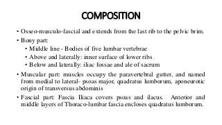
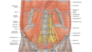
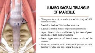
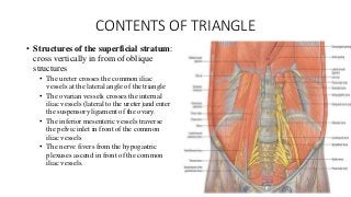
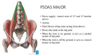
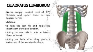
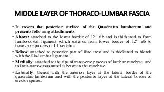
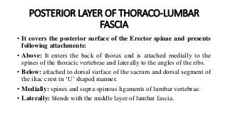
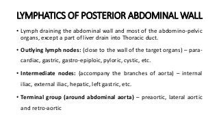
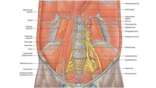
POSTERIOR
ABDOMINAL WALL
DR. SUNDIP CHARMODE
ASSOCIATE PROFESSOR
DEPARTMENT OF ANATOMY
AIIMS RAJKOT
COMPOSITION
• Osseo-musculo-fascial and extends from the last rib to the pelvic brim.
• Bony part:
• Middle line - Bodies of five lumbar vertebrae
• Above and laterally: inner surface of lower ribs
• Below and laterally: iliac fossae and ale of sacrum
• Muscular part: muscles occupy the paravertebral gutter, and named
from medial to lateral- psoas major, quadratus lumborum, aponeurotic
origin of transversus abdominis
• Fascial part: Fascia Iliaca covers psoas and ilacus. Anterior and
middle layers of Thoraco-lumbar fascia encloses quadratus lumborum.
LUMBO-SACRAL TRIANGLE
OF MARCILLE
• Triangular interval on each side of the body of fifth
lumbar vertebra.
• Medially: body of fifth lumbar vertebra
• Laterally: medial border of psoas major muscle
• Apex: directed above and below by junction of psoas
and body of fifth lumbar vertebra
• Base: upper surface of lateral mass or ala of the
sacrum
• Floor or posterior wall: transverse process of fifth
lumbar vertebra and ilio-lumbar ligament.
CONTENTS OF TRIANGLE
• Structures of the deep stratum: run
vertically over the ala of sacrum
• Ganglionated sympathetic trunk
• Lumbo-sacral trunk
• Ilio-lumbar artery
• Obturator nerve
• Structures of the Intermediate
stratum: pass obliquely downwards
and laterally
• Common iliac artery dividing into
external and internal iliac arteries
• Common iliac vein formed by union of
external and internal iliac veins
CONTENTS OF TRIANGLE
• Structures of the superficial stratum:
cross vertically in from of oblique
structures
• The ureter crosses the common iliac
vessels at the lateral angle of the triangle
• The ovarian vessels crosses the internal
iliac vessels (lateral to the ureter)and enter
the suspensory ligament of the ovary.
• The inferior mesenteric vessels traverse
the pelvic inlet in front of the common
iliac vessels
• The nerve fivers from the hypogastric
plexuses ascend in front of the common
iliac vessels.
PSOAS MAJOR
• Long fusiform muscle
• Covered together with the iliacus by the fascia
iliaca.
• Origin:
• Anterior surfaces and lower borders of
transverse processes of all lumbar vertebrae
• Five fleshy slips from the bodies and
intervertebral discs of all lumbar vertebra
• Four tendinous arches bridging across the
sides of the bodies of lumbar vertebrae
• Insertion: anterior surface of the tip of lesser
trochanter of the femur
PSOAS MAJOR
• Nerve supply: ventral rami of 2nd and 3rd lumbar
nerves
• Action:
• Chief flexor of hip joint (acting from above)
• Flexes the trunk at the hip joint
• When the foot is on ground, it acts as a medial
rotator of hip joint
• When the foot is off the ground, it acts as a lateral
rotator of hip joint
FASCIA ILIACA
• It is a strong sheet of fascia which covers
the iliacus and psoas major and encloses
them in a closed osseo-fascial pocket.
Two parts:
• Iliac part – which covers both the iliacus
and psoas major below the iliac crest
• Psoas part – covers only the psoas major
above the iliac crest
FASCIA ILIACA
ATTACHMENT OF ILIAC PART
• Laterally: Inner lip of ventral
segment of iliac crest where it
blends with fascia transversalis
• Medially: Alae of sacrum and
pelvic brim up to ilio-pubic
eminence
• Above: Continuous with psoas part
• Below:
ATTACHMENT OF PSOAS PART
• Above: thickened to form medial
arcuate ligament
• Laterally: it is attached to the
anterior surface of transverse
processes of lumbar vertebrae,
where it blends with thoraco-
lumbar fascia
• Medially: bodies and discs of
lumbar vertebra
FASCIA ILIACA
STRUCTURES PIERCING FASCIA ILIACA
• In front: Genito-femoral nerve
• Laterally: Ilio-hypogastric and ilio-
inguinal nerves and the fourth
lumbar artery
• Medially: Lumbo-sacral trunk, ilio-
lumbar artery and obturator nerve
CLINICAL CORRELATES
• An abscess may collect beneath
the fascia iliaca from the
tuberculous disease of the lumbar
vertebrae.
QUADRATUS LUMBORUM
• Quadrilateral muscle
• Fills up the gap between the last rib and the iliac
crest
• It is enclosed between anterior and middle
layers of the thoraco-lumbar fascia.
• Origin:
• Posterior one-third of inner lip of ventral segment of
iliac crest
• Ilio-lumbar ligament
• Insertion:
• Fleshy fibers, attached to lower border and
anterior surface of medial part of 12th rib
• Tendinous fibers attached to the anterior surface of
transverse processes of upper four lumbar
vertebrae.
QUADRATUS LUMBORUM
• Nerve supply: ventral rami of 12th
thoracic and upper three or four
lumbar nerves
• Actions:
• It fixes the last rib and helps the
diaphragm during inspiration.
• Acting on one side it acts as lateral
flexor of trunk
• Acting on both sides they produce
extension of the vertebral column.
THORACO-LUMBAR FASCIA
In lumbar region, the fascia
consists of three layers: Anterior,
Middle and posterior.
The layers enclose 2 spaces.
Quadratus lumborum between
anterior and middle layers.
Erector spinae between middle
and posterior layers.
ANTERIOR LAYER OF THORACO-LUMBAR
FASCIA
• It covers the anterior surface of the Quadratus lumborum and
presents following attachments:
• Above: thickened to form lateral arcuate ligament
• Below: attached to posterior part of iliac crest and is thickened to form
the ilio-lumbar ligament
• Medially: attached to a vertical ridge on the anterior surface of
transverse process of lumbar vertebrae where it blends with the psoas
fascia.
• Laterally: blends with the middle layer of lumbar fascia at the lateral
border of the quadratus lumborum
MIDDLE LAYER OF THORACO-LUMBAR FASCIA
• It covers the posterior surface of the Quadratus lumborum and
presents following attachments:
• Above: attached to the lower border of 12th rib and is thickened to form
lumbo-costal ligament which extends from lower border of 12th rib to
transverse process of L1 vertebra.
• Below: attached to posterior part of iliac crest and is thickened to blends
with the ilio-lumbar ligament
• Medially: attached to the tips of transverse process of lumbar vertebrae and
to inter-transversus muscles between the vertebrae.
• Laterally: blends with the anterior layer at the lateral border of the
quadratus lumborum and with the posterior layer at the lateral border of
erector spinae.
POSTERIOR LAYER OF THORACO-LUMBAR
FASCIA
• It covers the posterior surface of the Erector spinae and presents
following attachments:
• Above: It enters the back of thorax and is attached medially to the
spines of the thoracic vertebrae and laterally to the angles of the ribs.
• Below: attached to dorsal surface of the sacrum and dorsal segment of
the iliac crest in ‘U’ shaped manner.
• Medially: spines and supra-spinous ligaments of lumbar vertebrae.
• Laterally: blends with the middle layer of lumbar fascia.
MUSCLES ATTACHED TO THE THORACO-
LUMBAR FASCIA
1. From the anterior layer: a few fibers of the diaphragm.
2. From the fusion of the anterior and middle layers – transversus
abdominis and internal oblique
3. From posterior layer – latissimus Dorsii, serratus posterior inferior,
a few fibers of gluteus maximus
LYMPHATICS OF POSTERIOR ABDOMINAL WALL
• Lymph draining the abdominal wall and most of the abdomino-pelvic
organs, except a part of liver drain into Thoracic duct.
• Outlying lymph nodes: (close to the wall of the target organs) – para-
cardiac, gastric, gastro-epiploic, pyloric, cystic, etc.
• Intermediate nodes: (accompany the branches of aorta) – internal
iliac, external iliac, hepatic, left gastric, etc.
• Terminal group (around abdominal aorta) – preaortic, lateral aortic
and retro-aortic
NERVES OF POSTERIOR ABDOMINAL WALL
• The innervations of the abdomen are derived from the somatic spinal
nerves and the autonomic nerves.
THANK YOU
Posterior abdominal wall.pptx

Posterior abdominal wall.pptx

  • 1.
    POSTERIOR ABDOMINAL WALL DR. SUNDIPCHARMODE ASSOCIATE PROFESSOR DEPARTMENT OF ANATOMY AIIMS RAJKOT
  • 2.
    COMPOSITION • Osseo-musculo-fascial andextends from the last rib to the pelvic brim. • Bony part: • Middle line - Bodies of five lumbar vertebrae • Above and laterally: inner surface of lower ribs • Below and laterally: iliac fossae and ale of sacrum • Muscular part: muscles occupy the paravertebral gutter, and named from medial to lateral- psoas major, quadratus lumborum, aponeurotic origin of transversus abdominis • Fascial part: Fascia Iliaca covers psoas and ilacus. Anterior and middle layers of Thoraco-lumbar fascia encloses quadratus lumborum.
  • 5.
    LUMBO-SACRAL TRIANGLE OF MARCILLE •Triangular interval on each side of the body of fifth lumbar vertebra. • Medially: body of fifth lumbar vertebra • Laterally: medial border of psoas major muscle • Apex: directed above and below by junction of psoas and body of fifth lumbar vertebra • Base: upper surface of lateral mass or ala of the sacrum • Floor or posterior wall: transverse process of fifth lumbar vertebra and ilio-lumbar ligament.
  • 6.
    CONTENTS OF TRIANGLE •Structures of the deep stratum: run vertically over the ala of sacrum • Ganglionated sympathetic trunk • Lumbo-sacral trunk • Ilio-lumbar artery • Obturator nerve • Structures of the Intermediate stratum: pass obliquely downwards and laterally • Common iliac artery dividing into external and internal iliac arteries • Common iliac vein formed by union of external and internal iliac veins
  • 7.
    CONTENTS OF TRIANGLE •Structures of the superficial stratum: cross vertically in from of oblique structures • The ureter crosses the common iliac vessels at the lateral angle of the triangle • The ovarian vessels crosses the internal iliac vessels (lateral to the ureter)and enter the suspensory ligament of the ovary. • The inferior mesenteric vessels traverse the pelvic inlet in front of the common iliac vessels • The nerve fivers from the hypogastric plexuses ascend in front of the common iliac vessels.
  • 8.
    PSOAS MAJOR • Longfusiform muscle • Covered together with the iliacus by the fascia iliaca. • Origin: • Anterior surfaces and lower borders of transverse processes of all lumbar vertebrae • Five fleshy slips from the bodies and intervertebral discs of all lumbar vertebra • Four tendinous arches bridging across the sides of the bodies of lumbar vertebrae • Insertion: anterior surface of the tip of lesser trochanter of the femur
  • 9.
    PSOAS MAJOR • Nervesupply: ventral rami of 2nd and 3rd lumbar nerves • Action: • Chief flexor of hip joint (acting from above) • Flexes the trunk at the hip joint • When the foot is on ground, it acts as a medial rotator of hip joint • When the foot is off the ground, it acts as a lateral rotator of hip joint
  • 12.
    FASCIA ILIACA • Itis a strong sheet of fascia which covers the iliacus and psoas major and encloses them in a closed osseo-fascial pocket. Two parts: • Iliac part – which covers both the iliacus and psoas major below the iliac crest • Psoas part – covers only the psoas major above the iliac crest
  • 13.
    FASCIA ILIACA ATTACHMENT OFILIAC PART • Laterally: Inner lip of ventral segment of iliac crest where it blends with fascia transversalis • Medially: Alae of sacrum and pelvic brim up to ilio-pubic eminence • Above: Continuous with psoas part • Below: ATTACHMENT OF PSOAS PART • Above: thickened to form medial arcuate ligament • Laterally: it is attached to the anterior surface of transverse processes of lumbar vertebrae, where it blends with thoraco- lumbar fascia • Medially: bodies and discs of lumbar vertebra
  • 14.
    FASCIA ILIACA STRUCTURES PIERCINGFASCIA ILIACA • In front: Genito-femoral nerve • Laterally: Ilio-hypogastric and ilio- inguinal nerves and the fourth lumbar artery • Medially: Lumbo-sacral trunk, ilio- lumbar artery and obturator nerve CLINICAL CORRELATES • An abscess may collect beneath the fascia iliaca from the tuberculous disease of the lumbar vertebrae.
  • 15.
    QUADRATUS LUMBORUM • Quadrilateralmuscle • Fills up the gap between the last rib and the iliac crest • It is enclosed between anterior and middle layers of the thoraco-lumbar fascia. • Origin: • Posterior one-third of inner lip of ventral segment of iliac crest • Ilio-lumbar ligament • Insertion: • Fleshy fibers, attached to lower border and anterior surface of medial part of 12th rib • Tendinous fibers attached to the anterior surface of transverse processes of upper four lumbar vertebrae.
  • 16.
    QUADRATUS LUMBORUM • Nervesupply: ventral rami of 12th thoracic and upper three or four lumbar nerves • Actions: • It fixes the last rib and helps the diaphragm during inspiration. • Acting on one side it acts as lateral flexor of trunk • Acting on both sides they produce extension of the vertebral column.
  • 17.
    THORACO-LUMBAR FASCIA In lumbarregion, the fascia consists of three layers: Anterior, Middle and posterior. The layers enclose 2 spaces. Quadratus lumborum between anterior and middle layers. Erector spinae between middle and posterior layers.
  • 18.
    ANTERIOR LAYER OFTHORACO-LUMBAR FASCIA • It covers the anterior surface of the Quadratus lumborum and presents following attachments: • Above: thickened to form lateral arcuate ligament • Below: attached to posterior part of iliac crest and is thickened to form the ilio-lumbar ligament • Medially: attached to a vertical ridge on the anterior surface of transverse process of lumbar vertebrae where it blends with the psoas fascia. • Laterally: blends with the middle layer of lumbar fascia at the lateral border of the quadratus lumborum
  • 19.
    MIDDLE LAYER OFTHORACO-LUMBAR FASCIA • It covers the posterior surface of the Quadratus lumborum and presents following attachments: • Above: attached to the lower border of 12th rib and is thickened to form lumbo-costal ligament which extends from lower border of 12th rib to transverse process of L1 vertebra. • Below: attached to posterior part of iliac crest and is thickened to blends with the ilio-lumbar ligament • Medially: attached to the tips of transverse process of lumbar vertebrae and to inter-transversus muscles between the vertebrae. • Laterally: blends with the anterior layer at the lateral border of the quadratus lumborum and with the posterior layer at the lateral border of erector spinae.
  • 20.
    POSTERIOR LAYER OFTHORACO-LUMBAR FASCIA • It covers the posterior surface of the Erector spinae and presents following attachments: • Above: It enters the back of thorax and is attached medially to the spines of the thoracic vertebrae and laterally to the angles of the ribs. • Below: attached to dorsal surface of the sacrum and dorsal segment of the iliac crest in ‘U’ shaped manner. • Medially: spines and supra-spinous ligaments of lumbar vertebrae. • Laterally: blends with the middle layer of lumbar fascia.
  • 21.
    MUSCLES ATTACHED TOTHE THORACO- LUMBAR FASCIA 1. From the anterior layer: a few fibers of the diaphragm. 2. From the fusion of the anterior and middle layers – transversus abdominis and internal oblique 3. From posterior layer – latissimus Dorsii, serratus posterior inferior, a few fibers of gluteus maximus
  • 22.
    LYMPHATICS OF POSTERIORABDOMINAL WALL • Lymph draining the abdominal wall and most of the abdomino-pelvic organs, except a part of liver drain into Thoracic duct. • Outlying lymph nodes: (close to the wall of the target organs) – para- cardiac, gastric, gastro-epiploic, pyloric, cystic, etc. • Intermediate nodes: (accompany the branches of aorta) – internal iliac, external iliac, hepatic, left gastric, etc. • Terminal group (around abdominal aorta) – preaortic, lateral aortic and retro-aortic
  • 24.
    NERVES OF POSTERIORABDOMINAL WALL • The innervations of the abdomen are derived from the somatic spinal nerves and the autonomic nerves.
  • 34.