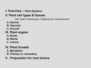
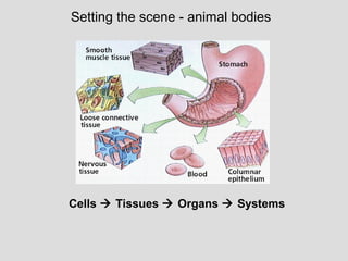
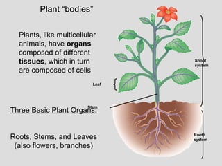
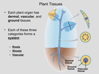
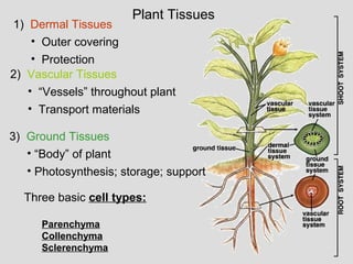
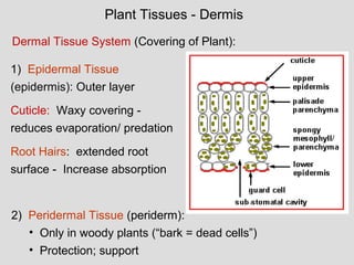
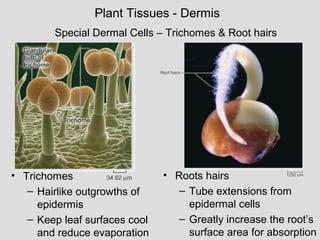
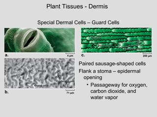
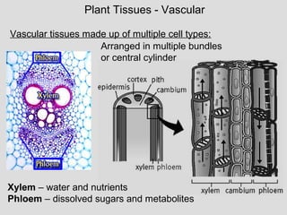
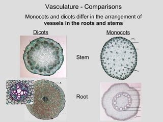
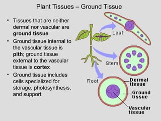
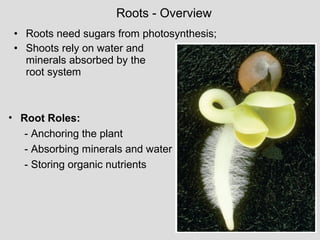
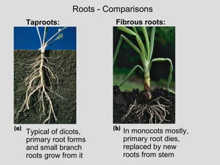
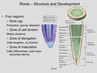
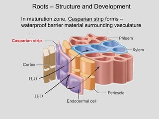
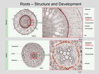
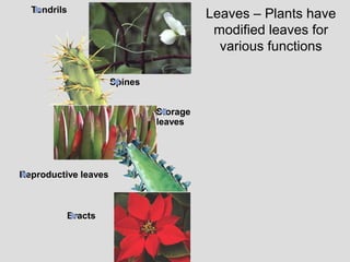
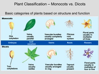
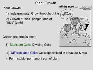
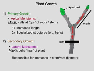
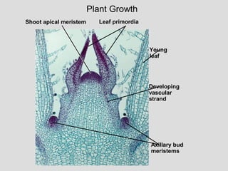
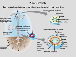
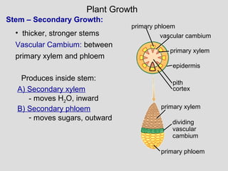
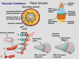
This document provides an overview of plant systems and structures. It discusses the three basic plant organs of roots, stems, and leaves. It describes the tissues that make up plants, including dermal tissue, ground tissue, and vascular tissue. It explains that plants grow through cell division at meristems and differentiate cells. Primary growth increases length while secondary growth increases thickness. Meristems are dividing cells that allow for growth at tips and girth.
In this presentation, concept of xerophytes, types of xerophytes and adaptations (morphological, anatomical and physiological) developed in them are explained.
Presentation on Gymnosperms. Prepared by Rahmat Alam Puniyali, Student of BS IV at Karakoram International University Gilgit, Pakistan. Photos of related plants are taken by the creator at KIU (Karakoram International University) campus.
(Some of the pictures and diagrams are taken from the websites of their resembling organizations (The McGraw-Hill Companies))
Gymnosperm is from the Greek “gymnos” naked, and “sperma” seeds. They are groups of vascular plants that reproduce by means of an exposed seeds or ovules. They are phanerogams according to A. W. Eichler.
Discussion of the functions of leaves, focusing on Photosynthesis and the process. Also covers transpiration, O2 CO2 transfer, germination. Appropriate for high school level students.
The living plant cell
What is the main differences between plant cell and animal cell??
Cell wall: Formed of cellulose.
Chloroplast: Responsible for photosynthesis.
Vacuole: much larger in plant cells, store any nutrients and waste products .
Nutraceutical market, scope and growth: Herbal drug technologyLokesh Patil
As consumer awareness of health and wellness rises, the nutraceutical market—which includes goods like functional meals, drinks, and dietary supplements that provide health advantages beyond basic nutrition—is growing significantly. As healthcare expenses rise, the population ages, and people want natural and preventative health solutions more and more, this industry is increasing quickly. Further driving market expansion are product formulation innovations and the use of cutting-edge technology for customized nutrition. With its worldwide reach, the nutraceutical industry is expected to keep growing and provide significant chances for research and investment in a number of categories, including vitamins, minerals, probiotics, and herbal supplements.
Seminar of U.V. Spectroscopy by SAMIR PANDASAMIR PANDA
Spectroscopy is a branch of science dealing the study of interaction of electromagnetic radiation with matter.
Ultraviolet-visible spectroscopy refers to absorption spectroscopy or reflect spectroscopy in the UV-VIS spectral region.
Ultraviolet-visible spectroscopy is an analytical method that can measure the amount of light received by the analyte.
Observation of Io’s Resurfacing via Plume Deposition Using Ground-based Adapt...Sérgio Sacani
Since volcanic activity was first discovered on Io from Voyager images in 1979, changes
on Io’s surface have been monitored from both spacecraft and ground-based telescopes.
Here, we present the highest spatial resolution images of Io ever obtained from a groundbased telescope. These images, acquired by the SHARK-VIS instrument on the Large
Binocular Telescope, show evidence of a major resurfacing event on Io’s trailing hemisphere. When compared to the most recent spacecraft images, the SHARK-VIS images
show that a plume deposit from a powerful eruption at Pillan Patera has covered part
of the long-lived Pele plume deposit. Although this type of resurfacing event may be common on Io, few have been detected due to the rarity of spacecraft visits and the previously low spatial resolution available from Earth-based telescopes. The SHARK-VIS instrument ushers in a new era of high resolution imaging of Io’s surface using adaptive
optics at visible wavelengths.
(May 29th, 2024) Advancements in Intravital Microscopy- Insights for Preclini...Scintica Instrumentation
Intravital microscopy (IVM) is a powerful tool utilized to study cellular behavior over time and space in vivo. Much of our understanding of cell biology has been accomplished using various in vitro and ex vivo methods; however, these studies do not necessarily reflect the natural dynamics of biological processes. Unlike traditional cell culture or fixed tissue imaging, IVM allows for the ultra-fast high-resolution imaging of cellular processes over time and space and were studied in its natural environment. Real-time visualization of biological processes in the context of an intact organism helps maintain physiological relevance and provide insights into the progression of disease, response to treatments or developmental processes.
In this webinar we give an overview of advanced applications of the IVM system in preclinical research. IVIM technology is a provider of all-in-one intravital microscopy systems and solutions optimized for in vivo imaging of live animal models at sub-micron resolution. The system’s unique features and user-friendly software enables researchers to probe fast dynamic biological processes such as immune cell tracking, cell-cell interaction as well as vascularization and tumor metastasis with exceptional detail. This webinar will also give an overview of IVM being utilized in drug development, offering a view into the intricate interaction between drugs/nanoparticles and tissues in vivo and allows for the evaluation of therapeutic intervention in a variety of tissues and organs. This interdisciplinary collaboration continues to drive the advancements of novel therapeutic strategies.
Richard's entangled aventures in wonderlandRichard Gill
Since the loophole-free Bell experiments of 2020 and the Nobel prizes in physics of 2022, critics of Bell's work have retreated to the fortress of super-determinism. Now, super-determinism is a derogatory word - it just means "determinism". Palmer, Hance and Hossenfelder argue that quantum mechanics and determinism are not incompatible, using a sophisticated mathematical construction based on a subtle thinning of allowed states and measurements in quantum mechanics, such that what is left appears to make Bell's argument fail, without altering the empirical predictions of quantum mechanics. I think however that it is a smoke screen, and the slogan "lost in math" comes to my mind. I will discuss some other recent disproofs of Bell's theorem using the language of causality based on causal graphs. Causal thinking is also central to law and justice. I will mention surprising connections to my work on serial killer nurse cases, in particular the Dutch case of Lucia de Berk and the current UK case of Lucy Letby.
Cancer cell metabolism: special Reference to Lactate PathwayAADYARAJPANDEY1
Normal Cell Metabolism:
Cellular respiration describes the series of steps that cells use to break down sugar and other chemicals to get the energy we need to function.
Energy is stored in the bonds of glucose and when glucose is broken down, much of that energy is released.
Cell utilize energy in the form of ATP.
The first step of respiration is called glycolysis. In a series of steps, glycolysis breaks glucose into two smaller molecules - a chemical called pyruvate. A small amount of ATP is formed during this process.
Most healthy cells continue the breakdown in a second process, called the Kreb's cycle. The Kreb's cycle allows cells to “burn” the pyruvates made in glycolysis to get more ATP.
The last step in the breakdown of glucose is called oxidative phosphorylation (Ox-Phos).
It takes place in specialized cell structures called mitochondria. This process produces a large amount of ATP. Importantly, cells need oxygen to complete oxidative phosphorylation.
If a cell completes only glycolysis, only 2 molecules of ATP are made per glucose. However, if the cell completes the entire respiration process (glycolysis - Kreb's - oxidative phosphorylation), about 36 molecules of ATP are created, giving it much more energy to use.
IN CANCER CELL:
Unlike healthy cells that "burn" the entire molecule of sugar to capture a large amount of energy as ATP, cancer cells are wasteful.
Cancer cells only partially break down sugar molecules. They overuse the first step of respiration, glycolysis. They frequently do not complete the second step, oxidative phosphorylation.
This results in only 2 molecules of ATP per each glucose molecule instead of the 36 or so ATPs healthy cells gain. As a result, cancer cells need to use a lot more sugar molecules to get enough energy to survive.
introduction to WARBERG PHENOMENA:
WARBURG EFFECT Usually, cancer cells are highly glycolytic (glucose addiction) and take up more glucose than do normal cells from outside.
Otto Heinrich Warburg (; 8 October 1883 – 1 August 1970) In 1931 was awarded the Nobel Prize in Physiology for his "discovery of the nature and mode of action of the respiratory enzyme.
WARNBURG EFFECT : cancer cells under aerobic (well-oxygenated) conditions to metabolize glucose to lactate (aerobic glycolysis) is known as the Warburg effect. Warburg made the observation that tumor slices consume glucose and secrete lactate at a higher rate than normal tissues.
The increased availability of biomedical data, particularly in the public domain, offers the opportunity to better understand human health and to develop effective therapeutics for a wide range of unmet medical needs. However, data scientists remain stymied by the fact that data remain hard to find and to productively reuse because data and their metadata i) are wholly inaccessible, ii) are in non-standard or incompatible representations, iii) do not conform to community standards, and iv) have unclear or highly restricted terms and conditions that preclude legitimate reuse. These limitations require a rethink on data can be made machine and AI-ready - the key motivation behind the FAIR Guiding Principles. Concurrently, while recent efforts have explored the use of deep learning to fuse disparate data into predictive models for a wide range of biomedical applications, these models often fail even when the correct answer is already known, and fail to explain individual predictions in terms that data scientists can appreciate. These limitations suggest that new methods to produce practical artificial intelligence are still needed.
In this talk, I will discuss our work in (1) building an integrative knowledge infrastructure to prepare FAIR and "AI-ready" data and services along with (2) neurosymbolic AI methods to improve the quality of predictions and to generate plausible explanations. Attention is given to standards, platforms, and methods to wrangle knowledge into simple, but effective semantic and latent representations, and to make these available into standards-compliant and discoverable interfaces that can be used in model building, validation, and explanation. Our work, and those of others in the field, creates a baseline for building trustworthy and easy to deploy AI models in biomedicine.
Bio
Dr. Michel Dumontier is the Distinguished Professor of Data Science at Maastricht University, founder and executive director of the Institute of Data Science, and co-founder of the FAIR (Findable, Accessible, Interoperable and Reusable) data principles. His research explores socio-technological approaches for responsible discovery science, which includes collaborative multi-modal knowledge graphs, privacy-preserving distributed data mining, and AI methods for drug discovery and personalized medicine. His work is supported through the Dutch National Research Agenda, the Netherlands Organisation for Scientific Research, Horizon Europe, the European Open Science Cloud, the US National Institutes of Health, and a Marie-Curie Innovative Training Network. He is the editor-in-chief for the journal Data Science and is internationally recognized for his contributions in bioinformatics, biomedical informatics, and semantic technologies including ontologies and linked data.
Earliest Galaxies in the JADES Origins Field: Luminosity Function and Cosmic ...Sérgio Sacani
We characterize the earliest galaxy population in the JADES Origins Field (JOF), the deepest
imaging field observed with JWST. We make use of the ancillary Hubble optical images (5 filters
spanning 0.4−0.9µm) and novel JWST images with 14 filters spanning 0.8−5µm, including 7 mediumband filters, and reaching total exposure times of up to 46 hours per filter. We combine all our data
at > 2.3µm to construct an ultradeep image, reaching as deep as ≈ 31.4 AB mag in the stack and
30.3-31.0 AB mag (5σ, r = 0.1” circular aperture) in individual filters. We measure photometric
redshifts and use robust selection criteria to identify a sample of eight galaxy candidates at redshifts
z = 11.5 − 15. These objects show compact half-light radii of R1/2 ∼ 50 − 200pc, stellar masses of
M⋆ ∼ 107−108M⊙, and star-formation rates of SFR ∼ 0.1−1 M⊙ yr−1
. Our search finds no candidates
at 15 < z < 20, placing upper limits at these redshifts. We develop a forward modeling approach to
infer the properties of the evolving luminosity function without binning in redshift or luminosity that
marginalizes over the photometric redshift uncertainty of our candidate galaxies and incorporates the
impact of non-detections. We find a z = 12 luminosity function in good agreement with prior results,
and that the luminosity function normalization and UV luminosity density decline by a factor of ∼ 2.5
from z = 12 to z = 14. We discuss the possible implications of our results in the context of theoretical
models for evolution of the dark matter halo mass function.
2. I. Overview – Plant Systems
II. Plant cell types & tissues
Cell Types: Parenchyma, Collenchyma, Sclerenchyma
A. Dermal
B. Vascular
C. Ground
III. Plant organs
A. Roots
B. Stems
C. Leaves
IV. Plant Growth
A. Meristems
B. Primary vs. secondary
V. Preparation for next lecture
3. Plant Structure, Growth, Development
Plants are notably different from animals:
1. SA:V ratio
2. Mobility
3. Growth
4. Response to environment
5. Cell structure
5. Plant “bodies”
Three Basic Plant Organs:
Roots, Stems, and Leaves
(also flowers, branches)
Plants, like multicellular
animals, have organs
composed of different
tissues, which in turn
are composed of cells
Shoot
system
Leaf
Stem
Root
system
6. • Each plant organ has
dermal, vascular, and
ground tissues
• Each of these three
categories forms a
system
– Roots
– Shoots
– Vascular
Plant Tissues
Dermal
tissue
Ground
tissue Vascular
tissue
7. 1) Dermal Tissues
• Outer covering
• Protection
3) Ground Tissues
• “Body” of plant
• Photosynthesis; storage; support
2) Vascular Tissues
• “Vessels” throughout plant
• Transport materials
Plant Tissues
Three basic cell types:
Parenchyma
Collenchyma
Sclerenchyma
8. What type of tissue transports fluids in plants?
A. Dermal
B. Roots
C. Vascular
D. Stems
E. Ground
10. Plant Cell Types
1) ParenchymaParenchyma (most abundant):
• plant metabolism:
Photosynthesis;
hormone secretion;
sugar storage
Flexible, thin-walled cells; living
renchyma cells in
odea leaf,(w/chloroplasts) • thin wall permeable to gasses
• large central vacuole
• able to divide and differentiate
11. 2) CollenchymaCollenchyma:
Thick-walled (uneven); living
• Offers support
(flexible & strong)
• Able to elongate
• Grouped in
strands, lack
secondary wall
Collenchyma cells sunflower
Plant Cell Types
12. 3) SclerenchymaSclerenchyma: Thick, hard-walled; Dead
• Offer support (e.g. hemp
fibers; nut shells)
• Thick secondary walls
with lignin
• Rigid (cannot elongate)
• Two types –
sclereids and fibers
Sclereid cells
in pear (LM)
Fiber cells in ash tree
l wall
Plant Cell Types
13. Which is a plant cell type?
A. secondary
B. vascular
C. ground
D. collenchyma
E. leaves
14. Dermal Tissue System (Covering of Plant):
1) Epidermal Tissue
(epidermis): Outer layer
Cuticle: Waxy covering -
reduces evaporation/ predation
Root Hairs: extended root
surface - Increase absorption
Plant Tissues - Dermis
2) Peridermal Tissue (periderm):
• Only in woody plants (“bark = dead cells”)
• Protection; support
15. Plant Tissues - Dermis
Special Dermal Cells – Trichomes & Root hairs
• Trichomes
– Hairlike outgrowths of
epidermis
– Keep leaf surfaces cool
and reduce evaporation
• Roots hairs
– Tube extensions from
epidermal cells
– Greatly increase the root’s
surface area for absorption
16. Guard cells
Stoma
Epidermal cell
Guard cells
Stomata
Epidermal cell
Guard cells
Stoma
Epidermal cell
Guard cells
Stomata
Epidermal cell
4 µm 200 µm
71 µm
a. c.
b.
Plant Tissues - Dermis
Paired sausage-shaped cells
Flank a stoma – epidermal
opening
• Passageway for oxygen,
carbon dioxide, and
water vapor
Special Dermal Cells – Guard Cells
17. Vascular tissues made up of multiple cell types:
Plant Tissues - Vascular
Arranged in multiple bundles
or central cylinder
Xylem – water and nutrients
Phloem – dissolved sugars and metabolites
18. 1) Xylem (dead at maturity): water and minerals roots to shoots
Plant Tissues - Vascular
A) Tracheids: Narrow, tube-like cells
B) Vessel Elements: Wide, tube-like cells
C) Fibers
19. 1) Xylem:
Plant Tissues - Vascular
Tracheids:
- Most vascular plants
- Long, thin, tapered ends, lignified
secondary walls
- Water moves cell to cell through pits
Vessel elements:
- Wider and shorter
- Perforation plates ends of vessel
elements
- water flows freely though perforation
plates
20. A) Sieve Tubes: Wide, tube-like cells
B) Companion Cells: support and regulate sieve tubes
2) Phloem (living at maturity) cells:
Plant Tissues - Vascular
21. - Moves water, sugar, amino
acids & hormones
2) Phloem (living at maturity)
Plant Tissues - Vascular
Sieve tube elements/members
• Living parenchyma
• Long narrow cells stack end to end
• Pores in end walls (sieve plates)
• Lack most cellular structures including:
• Distinct vacuole, Some cytoskeletal
elements, Nucleus, Ribosomes
Companion Cells:
• Adjacent to every sieve tube
element
• Non-conducting.
• Regulate both cells
• Connected by numerous
plasmodesmata
22. Dicots Monocots
Vasculature - Comparisons
Monocots and dicots differ in the arrangement of
vessels in the roots and stems
Root
Stem
23. Plant Tissues – Ground Tissue
• Tissues that are neither
dermal nor vascular are
ground tissue
• Ground tissue internal to
the vascular tissue is
pith; ground tissue
external to the vascular
tissue is cortex
• Ground tissue includes
cells specialized for
storage, photosynthesis,
and support
24. • Roots need sugars from photosynthesis;
• Shoots rely on water and
minerals absorbed by the
root system
Roots - Overview
• Root Roles:
- Anchoring the plant
- Absorbing minerals and water
- Storing organic nutrients
25. Taproots: Fibrous roots:
Typical of dicots,
primary root forms
and small branch
roots grow from it
In monocots mostly,
primary root dies,
replaced by new
roots from stem
Roots - Comparisons
26. Roots – Structure and Development
• Four regions:
– Root cap
Protection, gravity detection
– Zone of cell division
Mitotic divisions
– Zone of elongation
Cells lengthen, no division
– Zone of maturation
Cells differentiate, outer layer
becomes dermis
27. Roots – Structure and Development
In maturation zone, Casparian strip forms –
waterproof barrier material surrounding vasculature
30. Stem: an organ made of
– An alternating system
of nodes, points at
which leaves attach
– Internodes, stem length
between nodes
Stems - Overview
• Axillary bud - structure
that can form a lateral
shoot, or branch
• Apical/terminal bud -
located near the shoot
tip, lengthens a shoot
• Apical dominance
maintains dormancy in
most nonapical buds
Apical bud
Node
Internode
Apical
bud
Shoot
system
Vegetative
shoot
Axillary
bud
Stem
31. Phloem Xylem
Sclerenchyma
(fiber cells)
Ground tissue
connecting
pith to cortex
Pith
Cortex
1 mm
Epidermis
Vascular
bundle
Cross section of stem with vascular bundles forming
a ring (typical of eudicots)
a)
Key
to labels
Dermal
Ground
Vascular
Cross section of stem with scattered vascular bundles
(typical of monocots)
(b)
1 mm
Epidermis
Vascular
bundles
Ground
tissue
• In most monocot stems, the vascular bundles are scattered
throughout the ground tissue, rather than forming a ring
Vasculature - Stems
Dicot Monocot
32. Stems – Structure and Development
• Stems have all three types of
plant tissue
• Grow by division at meristems
– Develop into leaves, other
shoots, and even flowers
• Leaves may be arranged in
one of three ways
34. The leaf is the main photosynthetic
organ of most vascular plants
Leaves - Overview
Shoot
system
Leaf
Blade
Petiole
Leaves generally have
a flattened blade
and a stalk called the
petiole, which joins the leaf
to a node of the stem
35. Leaves – Structure and Development
• Leaves are
several layers
thick – each
with different
cell types
36. Leaves – Structure and Development
• Most dicots have 2
types of mesophyll
– Palisade mesophyll
high photosynthesis
– Spongy mesophyll
air spaces for gas
& water exchange
• Monocot leaves have 1
type of mesophyll
37. Leaves
• Leaf epidermis contains stomata - allow CO2 exchange
• Stomata flanked by two guard cells, control open vs. closed
Key
to labels
Dermal
Ground
Vascular
Cuticle Sclerenchyma
fibers
Stoma
undle-
heath
ll
Xylem
Phloem
a) Cutaway drawing of leaf tissues
Guard
cells
Vein
Cuticle
Lower
epidermis
Spongy
mesophyll
Palisade
mesophyll
Upper
epidermis
Guard
cells
Stomatal
pore
Surface view of a spiderwort
(Tradescantia) leaf (LM)
Epidermal
cell
(b)
50µm100µm
Vein Air spaces Guard cells
Cross section of a lilac
(Syringa)) leaf (LM)
(c)
38. Most dicots have
branch-like veins and
palmate leaf shape
Monocots have parallel
leaf veins and longer,
slender blades
Leaves - Comparisons
Monocots and dicots differ in the arrangement of veins,
the vascular tissue of leaves
40. Plant Classification – Monocots vs. Dicots
Basic categories of plants based on structure and function
41. Plant Growth:
1) Indeterminate: Grow throughout life
2) Growth at “tips” (length) and at
“hips” (girth)
Growth patterns in plant:
1) Meristem Cells: Dividing Cells
2) Differentiated Cells: Cells specialized in structure & role
• Form stable, permanent part of plant
Plant Growth
42. 1) Primary Growth:
1) Increased length
2) Specialized structures (e.g. fruits)
2) Secondary Growth:
Responsible for increases in stem/root diameter
• Apical Meristems:
Mitotic cells at “tips” of roots / stems
• Lateral Meristems:
Mitotic cells “hips” of plant
Plant Growth
girth
length
47. A cross section of what tissue is pictured?
A. Monocot root
B. Dicot root
C. Monocot stem
D. Dicot stem
48. Things To Do After Lecture 5…
Reading and Preparation:
1. Re-read today’s lecture, highlight all vocabulary you do not
understand, and look up terms.
2. Ch. 35 Self-Quiz: #1, 3, 6, 7 (correct answers in back of book)
3. Read chapter 35, focus on material covered in lecture (terms,
concepts, and figures!)
4. Skim next lecture.
“HOMEWORK” (NOT COLLECTED – but things to think about for studying):
1. Compare and contrast monocots and dicots.
2. List the different types of plant cells and describe which tissues and
organs they make up, including roles for each organ.
3. Explain the different between apical and lateral meristems and how
growth occurs.
4. Discuss the composition of bark and it’s function for plants (do all plants
have this tissue?)
Editor's Notes
FIGURE 42-5 The structure of ground tissue
(a) Parenchyma cells are living and serve many functions. They have thin, flexible primary cell walls. These parenchyma cells are used for starch storage in a potato. (b) Collenchyma cells are living and have thickened, but somewhat flexible, primary walls. They help support the plant body (as seen in this celery stalk). (c) Sclerenchyma cells have thick, rigid secondary cell walls and die after they differentiate. Illustrated are &quot;stone cells&quot; that give pear fruit its slightly gritty texture.
Figure 35.4 Modified roots
Figure 35.17 Organization of primary tissues in young stems
Figure 35.5 Modified stems
Figure 35.17 Organization of primary tissues in young stems
Figure 35.7 Modified leaves
Figure 35.16 The shoot tip
Figure 35.11 An overview of primary and secondary growth